Home
Images
Vectors
Videos
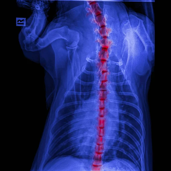
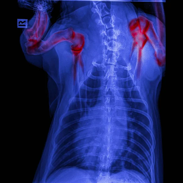
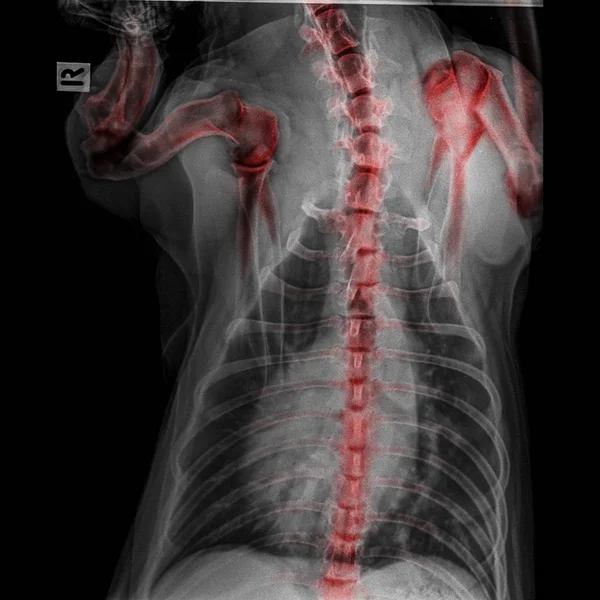
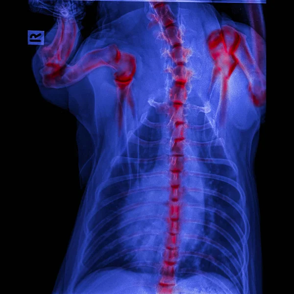
Stock image X-ray of dog posterior view closed up thorax and chest red highlight foreleg bone in shoulder joint and neck bone to back bone-degenerative joint disease in dog- Veterinary medicine-Veterinary anatomy
Published:
Oct.29, 2018 18:54:52
Author:
k.intarapong.gmail.com
Views:
44
Downloads:
1
File type:
image
/
jpg
File size:
6.55 MB
Orginal size:
5500
x
5500
px
Available sizes:
S
(500 x 500)
M
(1000 x 1000)
L
(2000 x 2000)
XL
(5500 x 5500)
EL0
(5500 x 5500)
Level:
beginner
Download Image
Image topics:
Health
Animal
Illness
Medical
Pet
Back
Dog
Emergency
Bone
Body
Neck
Disease
Broken
Leg
Shoulder
Chest
Pain
Elbow
Spine
Joint
Rib
Check
Skeleton
Fracture
Diagnosis
Anatomy
Surgery
Trauma
Accident
Injury
Scan
Veterinary
Vet
Veterinarian
Orthopedic
Thorax
Arthritis
Foreleg
Degeneration
Vertebral
Cartilage
Humerus
Health Care
Intervertebral
X Ray
Arched Back
Cervical Vertebrae
Osteosarcoma
Rickets
Posterior View
Similar stock images
X-ray Of Dog Posterior View Closed Up In Thorax Standard And Chest With Red Highlight In Neck Bone To Back Bone Pain Areas Or Spinal Disease In Dog- Veterinary Medicine- Veterinary Anatomy Concept
5500 × 5500
X-ray Of Dog Posterior View Closed Up Thorax And Chest Red Highlight Foreleg Bone In Shoulder Joint Or Scapula Humerus Joint-degenerative Joint Disease In Dogs-Veterinary Medicine-Veterinary Anatomy
5500 × 5500
Film X-ray T-L Spine(Thoracic-Lumbar Spine) Show : Human's Thoracic-lumbar Spine And Inflammation At Spine
3456 × 5095
Film X-ray T-L Spine(Thoracic-Lumbar Spine) Show : Human's Thoracic-lumbar Spine And Inflammation At Lumbar Spine
3456 × 5095
Rheumatic Heart Disease , Valvular Heart Disease ( Film X-ray Body Of Child With Red Area At Heart ) ( Medical , Science And Healthcare Concept )
3113 × 4174
X-ray Of Dog Posterior View Closed Up Thorax And Chest Red Highlight Foreleg Bone In Shoulder Joint And Neck Bone To Back Bone-degenerative Joint Disease In Dog- Veterinary Medicine-Veterinary Anatomy
5500 × 5500
Scoliosis Film X-ray Show Spinal Bend In Teenager Patient
2200 × 2941
X-Ray Scan Human
2999 × 3734