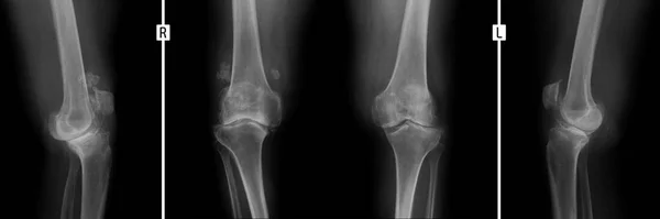
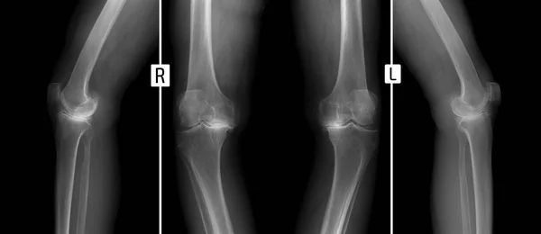
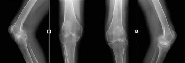
Stock image X-ray of knee joints. Deforming osteoarthritis.
Published: May.03, 2017 05:44:06
Author: vanzittoo
Views: 239
Downloads: 5
File type: image / jpg
File size: 5.62 MB
Orginal size: 6865 x 2984 px
Available sizes:
Level: beginner
Similar stock images
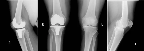
X-ray Of The Knee Joints. Shows The Arthritis Progressiva Deformans Of Knee Joints. Man 38 Years Old.
6754 × 2314