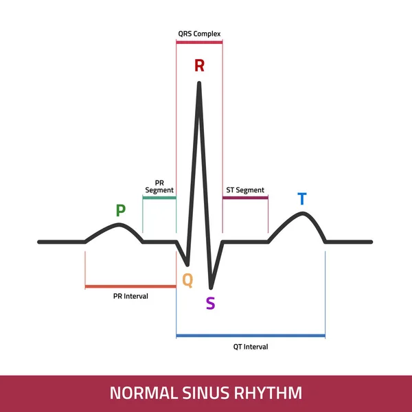
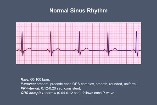
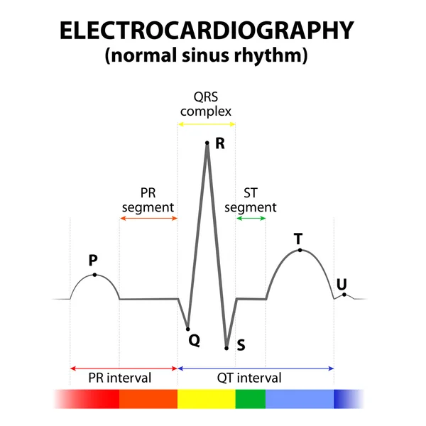
Stock vector ECG of a heart in normal sinus rhythm
Published: Dec.28, 2015 11:30:12
Author: edesignua
Views: 587
Downloads: 10
File type: vector / eps
File size: 0.67 MB
Orginal size: 4103 x 4103 px
Available sizes:
Level: silver