Home
Images
Vectors
Videos
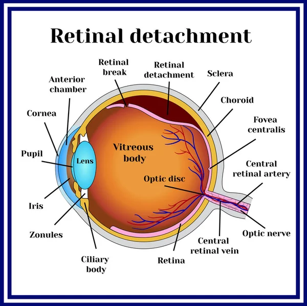
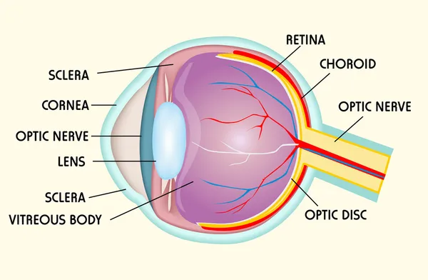
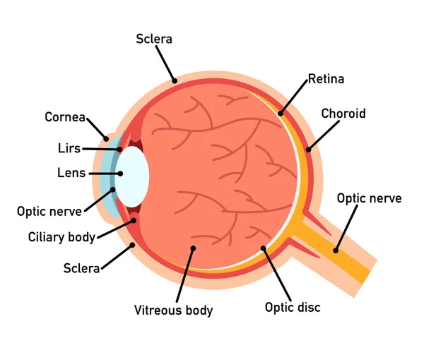
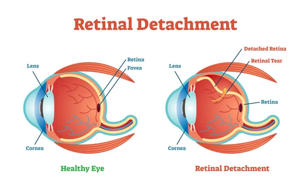
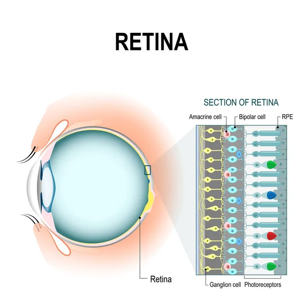
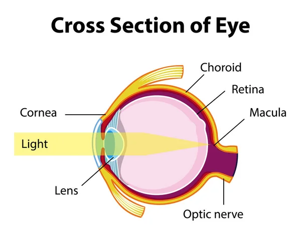
Stock vector human eye anatomy showcasing retinal detachment, including cornea, lens, and optic nerve structure diagram hand drawn schematic vector illustration. Medical science educational illustration
Published:
Apr.17, 2024 19:07:34
Author:
AlexanderPokusay
Views:
5
Downloads:
2
File type:
vector
/
eps
File size:
0.49 MB
Orginal size:
4000
x
4000
px
Available sizes:
S
(500 x 500)
M
(1000 x 1000)
L
(2000 x 2000)
XL
(4000 x 4000)
VECT
(4000 x 4000)
EL0
(4000 x 4000)
Level:
silver
Download Vector
Vector topics:
Vector
Colorful
Illustration
Isolated
Human
Health
Healthy
Medicine
Structure
Eye
Body
Drawing
Education
Lens
Biology
Science
Vision
Organ
Iris
Poster
Educational
Anatomy
Diagram
Pupil
Optometry
Ophthalmology
Cornea
Anatomical
Retina
Sclera
Macula
White Background
Human Eye
Choroid
Medical Illustration
Optic Nerve
Eye Health
Medical Diagram
Medical Education
Eye Disease
Retinal Detachment
Vision Problems
Eye Diagram
Patient Information
Eye Anatomy
Ciliary Body
Anatomy Chart
Eye Structure
Eye Conditions
Ocular Anatomy
Similar stock vectors
Retinal Detachment. Disease.
5306 × 5292
Eye Structure
3300 × 2161
Eye Anatomy Diagram,illustration.
6900 × 5819
Retinal Detachment Vector Illustration Diagram, Anatomical Scheme.
6036 × 3700
Retinal Cells: Rod And Cone Cells
4594 × 4594
Macula, The Central Part Of The Retina In The Human Eye
5333 × 4000
Diabetic Retinopathy
5688 × 4064
Human Eye Anatomy With Cross Section Of Eye Diagram Illustration
5327 × 4172