Home
Images
Vectors
Videos
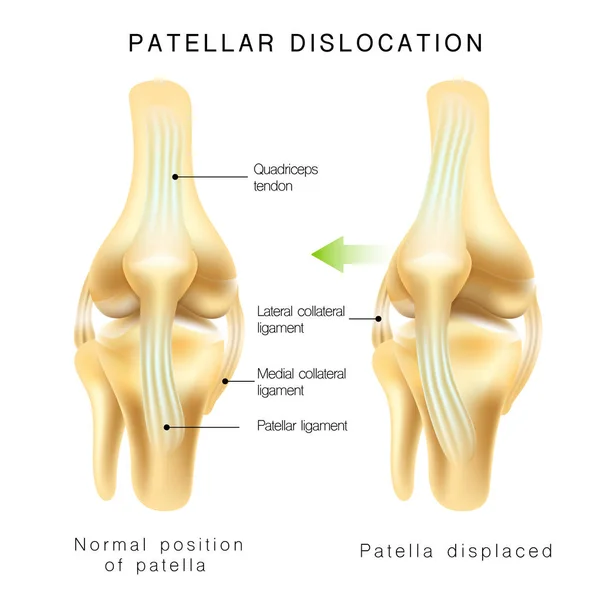
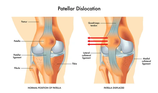
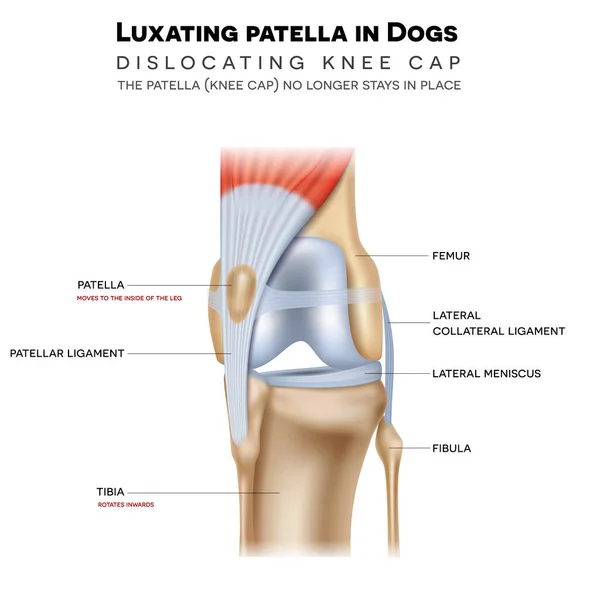
Stock vector Luxating patella in dogs, it shifts either towards the inner or outer knee. Anatomy of the canine (dog's) knee joint colorful design, medical info poster illustration.
Published:
Sep.01, 2020 14:37:13
Author:
megija
Views:
165
Downloads:
14
File type:
vector
/
eps
File size:
12.18 MB
Orginal size:
5000
x
5000
px
Available sizes:
S
(500 x 500)
M
(1000 x 1000)
L
(2000 x 2000)
XL
(5000 x 5000)
VECT
(5000 x 5000)
EL0
(5000 x 5000)
Level:
silver
Download Vector
Vector topics:
Graphic
Health
Healthy
Animal
Medical
Care
Structure
Canine
Dog
Bone
Body
Treatment
Biology
Science
Problem
Cap
Leg
Repair
Pain
Dogs
Knee
Joint
Damage
Poster
Skeleton
Anatomy
Limb
Info
Diagram
Surgery
Trauma
Injury
Veterinary
Ligament
Anatomical
Arthritis
Extremity
Arthritic
Tibia
Femur
Cartilage
Meniscus
Patella
Luxation
Ligaments
Kneecap
3d Illustration
Dislocated
Patellar
Vector Eps10
Similar stock vectors
Normal Position Of Kneecap And Patella Displaced
4380 × 4381
Bursitis. Prepatellar Bursitis. Inflammation Of Sacs With Synovial Fluid. Front And Side View Of Human Knee Joint. Lateral And Anterior Section Of The Knee. Vector Illustration.
5162 × 4000
Bursitis Inflammation Concept
4763 × 3647
Knee Dislocations - Medical Vector Illustration Diagrams. Anatomical Knee Injury Types Scheme.
4528 × 4891
Patellar Dislocation Vector Illustration
4016 × 2364
Flexor Hallucis Longus Muscle With Human Leg And Foot Bones Outline Diagram
4000 × 6667
Knee Bursae. Synovial Pockets Or Sacs That Surround The Knee Joint Cavity. Synovial Joint Anatomy. Frontal And Side View Of Human Knee Joint. Vector Illustration
5000 × 3599
Arthritis Of The Human Knee Joint
5860 × 3754