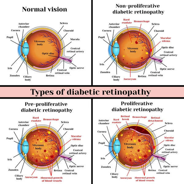
Stock vector Types of diabetic retinopathy

Published: Feb.14, 2017 10:21:09
Author: mrs.bazilio.gmail.com
Views: 605
Downloads: 11
File type: vector / eps
File size: 3.3 MB
Orginal size: 5315 x 5315 px
Available sizes:
Level: beginner