
Stock image Avulsion Fracture

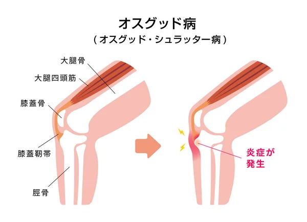
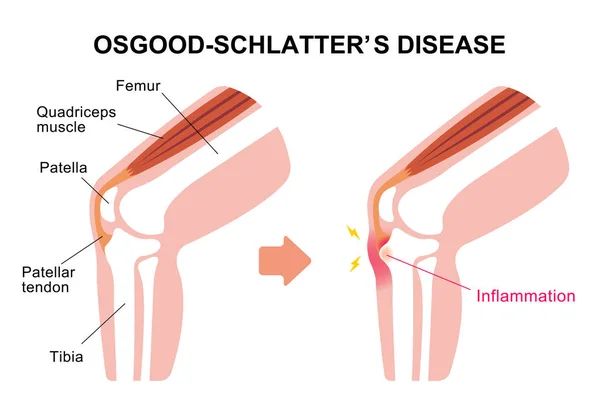
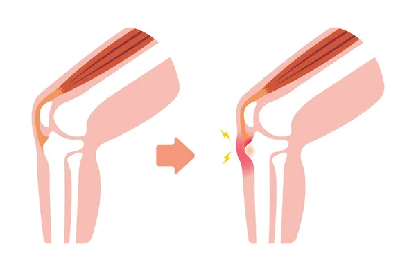
Osgood-schlatter Disease (knee Joint Disease) Illustration (Japanese)
Vector, 1.47MB, 6250 × 4529 eps

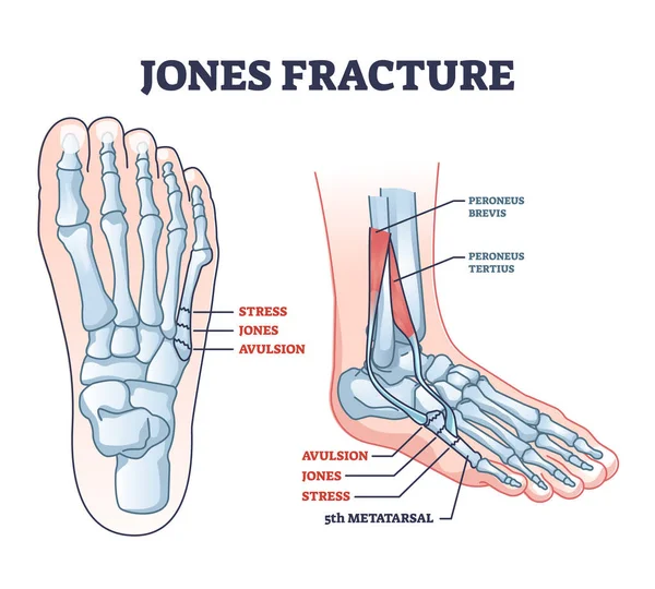
Jones Fracture And Foot Pinky Finger Bone Broken Damage Outline Diagram. Labeled Educational Scheme With Bone Stress Or Avulsion Sections Vetor Illustration. Peroneus Brevis And Tertius Muscle Anatomy
Vector, 6.06MB, 4300 × 3870 eps

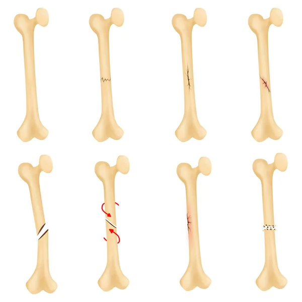
Avulsion Fracture Bone. Infographics. Vector Illustration On A Lined Background.
Vector, 0.87MB, 5000 × 5000 eps
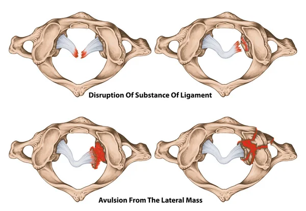
Classification Of Transverse Ligament Injuries. Disruptions If The Midportion, Periosteal Insertion Laterally, Comminuted Fracture And Avulsion Fracture Of Atlas, First Cervical Vertebra.
Image, 3.95MB, 5906 × 4075 jpg
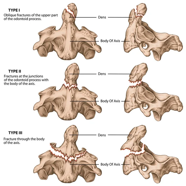
Three Types Of Odontoid Fractures. Fractures Of The Axis. Oblique Avulsion Fractures, Fractures At The Junctions Of The Dens With The Body Of The Axis, Fractures Extend Into The Body Of The Axis.
Image, 6.09MB, 5906 × 5906 jpg
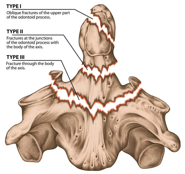
Three Types Of Odontoid Fractures. Fractures Of The Axis. Oblique Avulsion Fractures, Fractures At The Junctions Of The Dens With The Body Of The Axis, Fractures Extend Into The Body Of The Axis.
Image, 5.27MB, 5906 × 5543 jpg

Classification Of Fractures Of The Atlas. Jefferson Fracture. Bone Fracture Of The Arches Of The C1 Vertebra. Posterior, Anterior Arch Fracture, Semi Ring, Transverse Process And Lateral Mass Fracture.
Image, 4.99MB, 5906 × 5036 jpg


Fifth Metatarsal Or Foot Little Finger Fracture After Injury Outline Diagram. Labeled Educational Scheme With Feet Trauma After Twisting Motion Vector Illustration. Anatomical Skeletal Bone Zones.
Vector, 5.93MB, 4348 × 4000 eps
Page 1 >> Next