
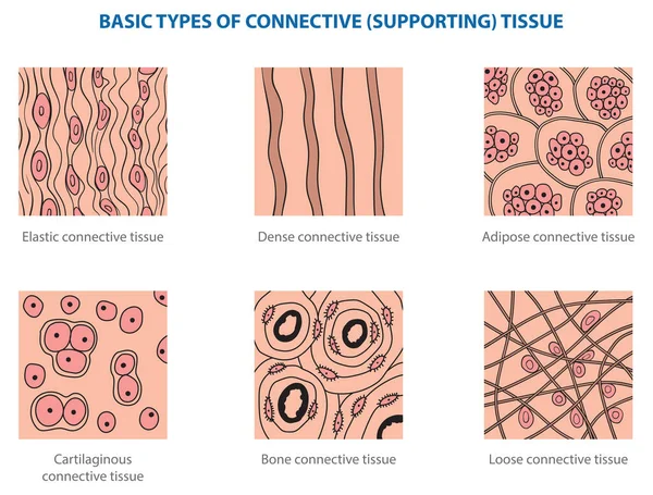
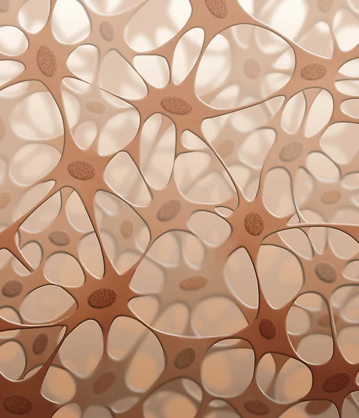
Stock image Connective Tissue


Fibroblast. Cell Structure And Anatomy. Collagen Fibers And Skin Cell. Vector Illustration
Vector, 10.12MB, 4444 × 4306 eps
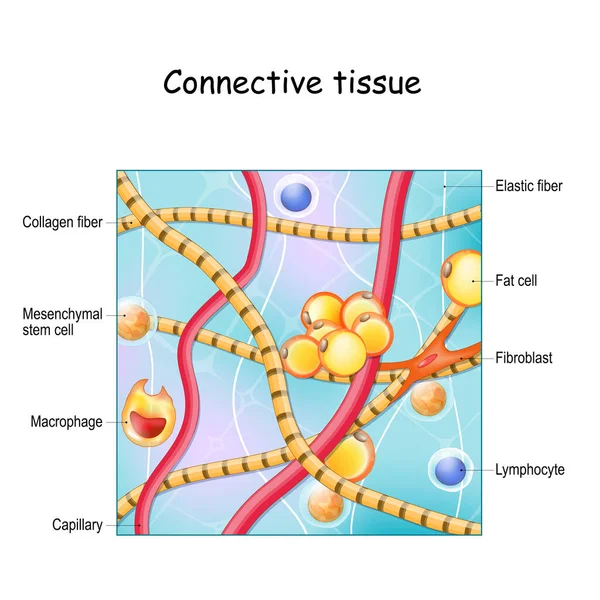
Connective Tissue. Structure And Anatomy. Extracellular Matrix, Elastic And Collagen Fibers, Blood Vessel And Cells: Lymphocyte, Fibroblast, Mesenchymal Stem Cell And Macrophage. Vector Illustration
Vector, 9.23MB, 4444 × 4444 eps
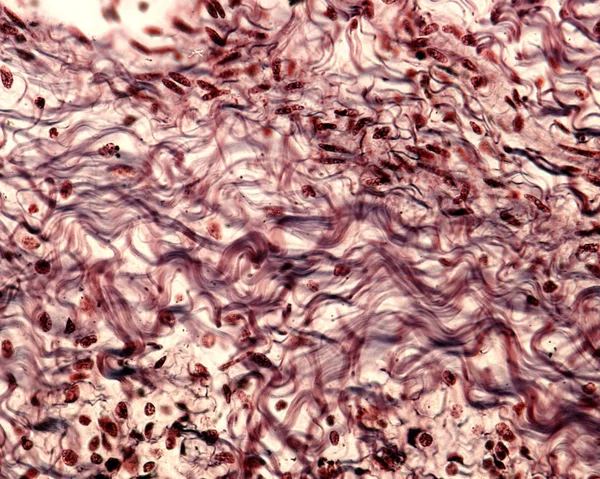
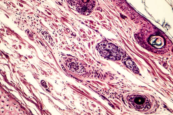
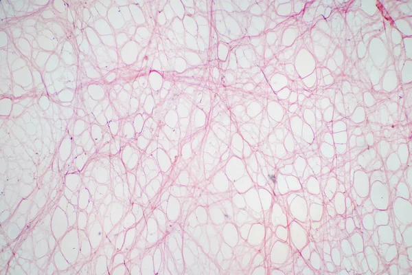
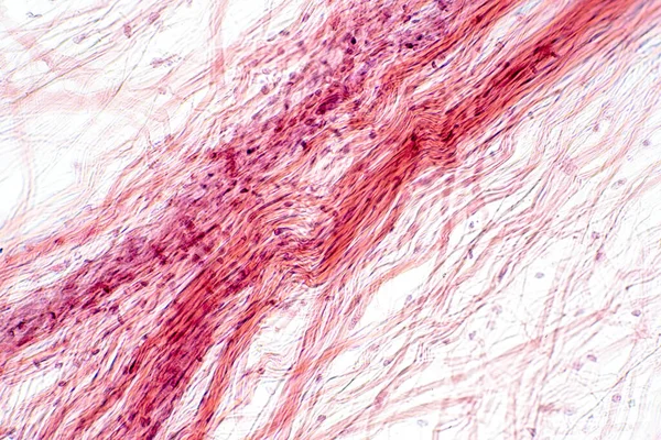
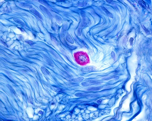
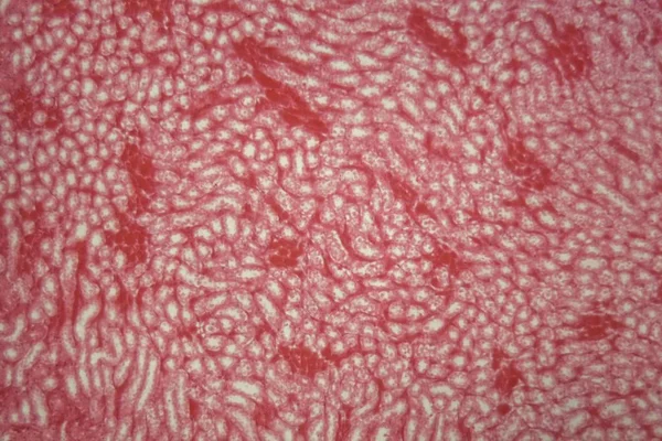
Areolar Connective Tissue Under The Microscope View. Histological For Human Physiology.
Image, 9.18MB, 4412 × 2942 jpg
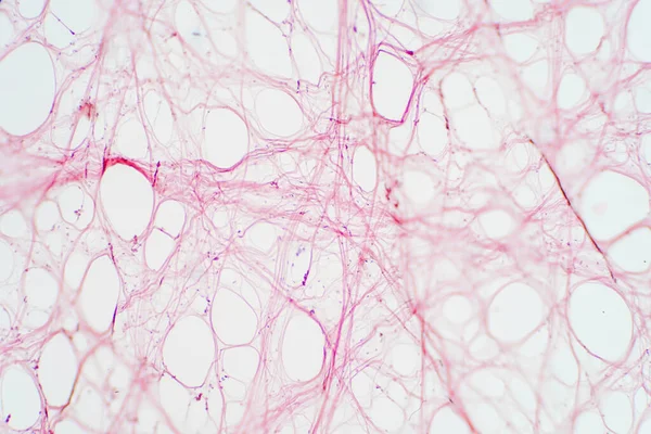
Areolar Connective Tissue Under The Microscope View. Histological For Human Physiology.
Image, 9.51MB, 6000 × 4000 jpg
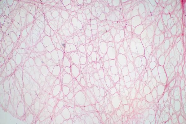
Areolar Connective Tissue Under The Microscope View. Histological For Human Physiology.
Image, 10.06MB, 6000 × 4000 jpg
Areolar Connective Tissue Under The Microscope View. Histological For Human Physiology.
Image, 11.47MB, 6000 × 4000 jpg

Skeletal Muscle Flat Illustration. Fascia, Endomysium, Fiber. Connective Tissue That Separates Muscles And Internal Organs. Can Be Used For Topics Like Anatomy, Human Body, Myofascial Release, Massage
Vector, 5.55MB, 10418 × 10418 eps
Areolar Connective Tissue Under The Microscope View. Histological For Human Physiology.
Image, 10.52MB, 6000 × 4000 jpg
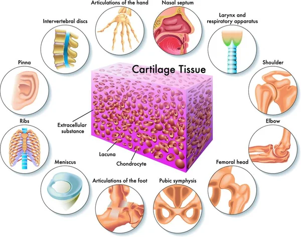
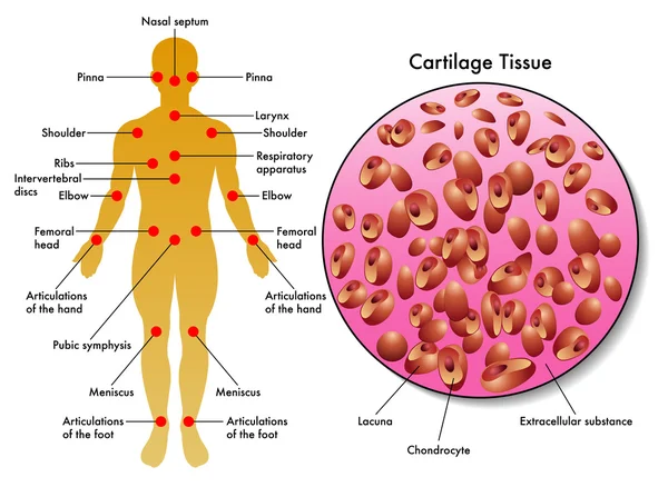
Medical Illustration Of Cartilage Tissue And Its Position In The Human Body
Vector, 0MB, 5144 × 4048 zip
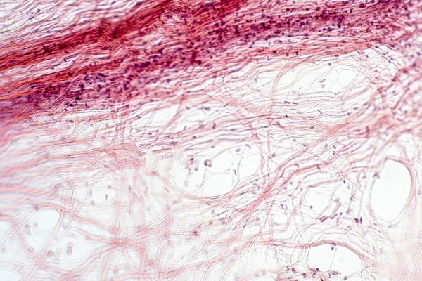
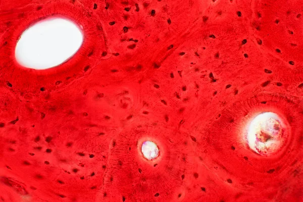
Areolar Connective Tissue Under The Microscope View. Histological For Human Physiology.
Image, 11.28MB, 6000 × 4000 jpg

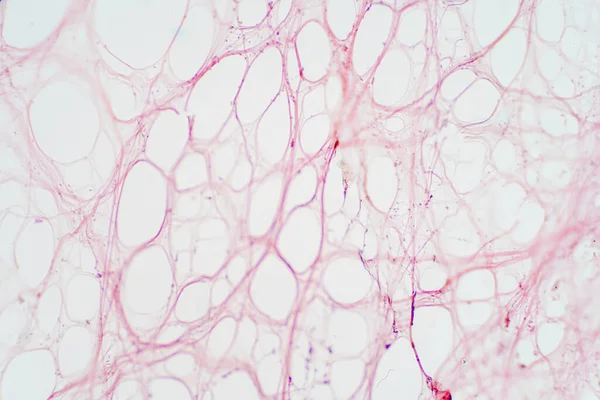
Areolar Connective Tissue Under The Microscope View. Histological For Human Physiology.
Image, 14.13MB, 6000 × 4000 jpg
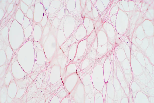
Areolar Connective Tissue Under The Microscope View. Histological For Human Physiology.
Image, 17.1MB, 6000 × 4000 jpg


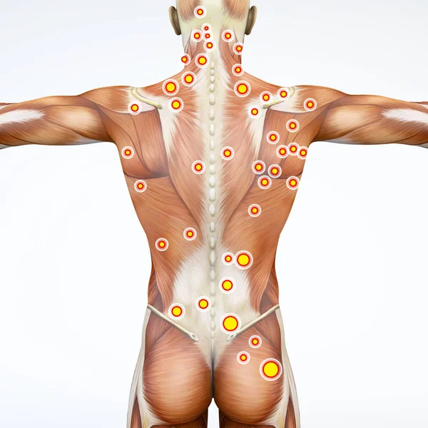
Back View Of A Man And His Trigger Points. Anatomy Muscles. 3d Rendering. Myofascial Trigger Points, Are Described As Hyperirritable Spots In The Fascia Surrounding Skeletal Muscle. Palpable Nodules In Taut Bands Of Muscle Fibers
Image, 11.53MB, 5511 × 5511 jpg

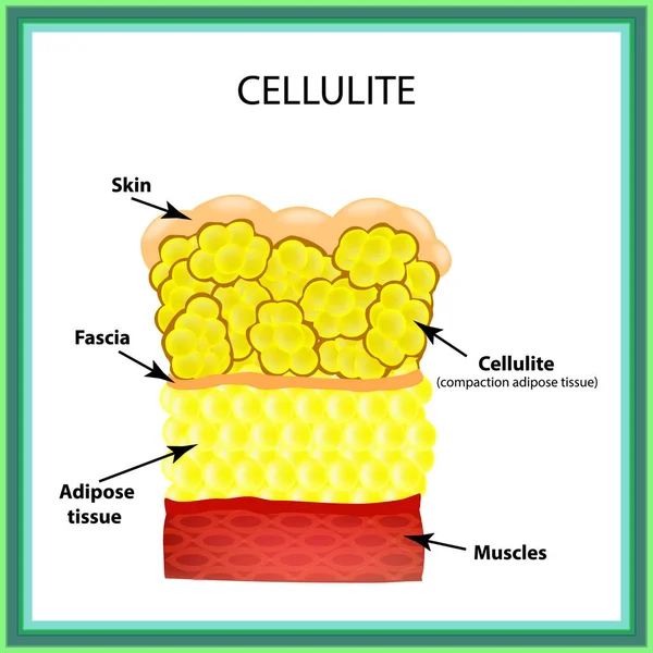
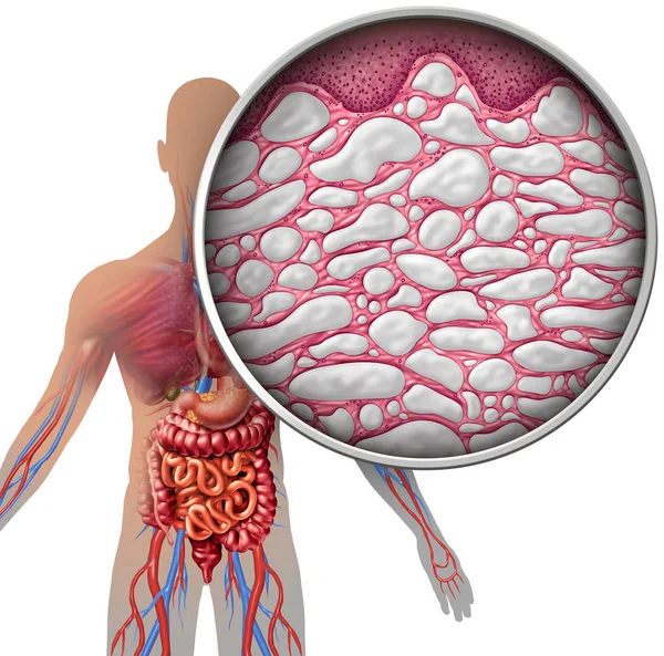
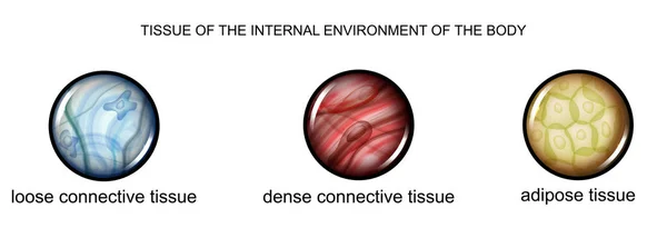
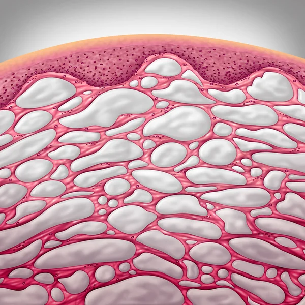
Cellulite. The Anatomical Structure Of The Adipose Tissue. Infographics. Vector Illustration On Isolated Background
Vector, 5.16MB, 5000 × 5000 eps


A Doctor In Medical Gloves Holds An X-ray Of The Foot And Examines A Sore Leg With A Heel Spur On A Woman, Close-up, Osteophytes And Heel, Fascia
Image, 7.23MB, 4860 × 3396 jpg
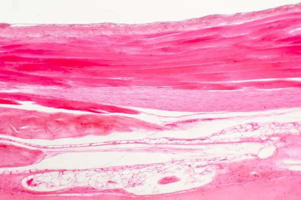
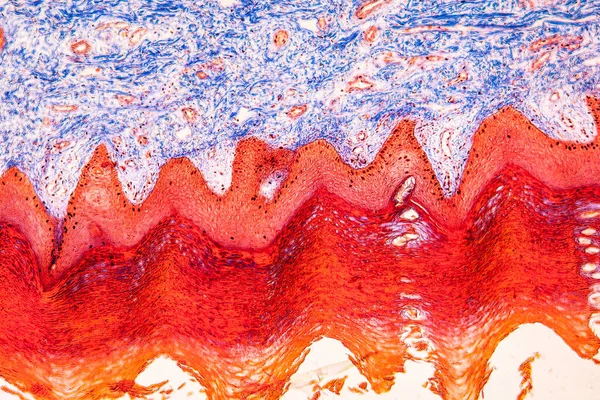
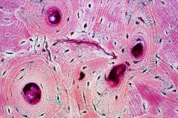
Histology Of Human Compact Bone Tissue Under Microscope View For Education, Muscle Bone Connection And Connective Tissue
Image, 13.74MB, 5786 × 3857 jpg
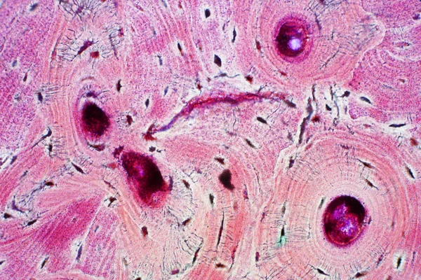
Histology Of Human Compact Bone Tissue Under Microscope View For Education, Muscle Bone Connection And Connective Tissue
Image, 18.53MB, 6000 × 4000 jpg
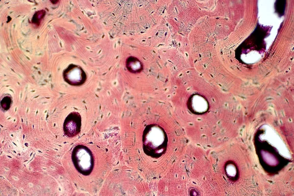
Histology Of Human Compact Bone Tissue Under Microscope View For Education, Muscle Bone Connection And Connective Tissue
Image, 18.19MB, 6000 × 4000 jpg
Histology Of Human Compact Bone Tissue Under Microscope View For Education, Muscle Bone Connection And Connective Tissue
Image, 17.36MB, 6000 × 4000 jpg


Histology Of Human Compact Bone Tissue Under Microscope View For Education, Muscle Bone Connection And Connective Tissue
Image, 20.09MB, 6000 × 4000 jpg
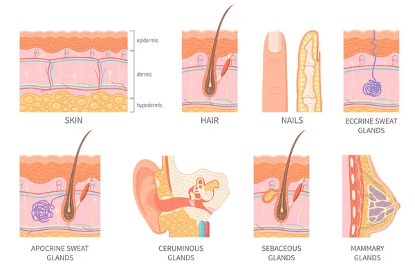
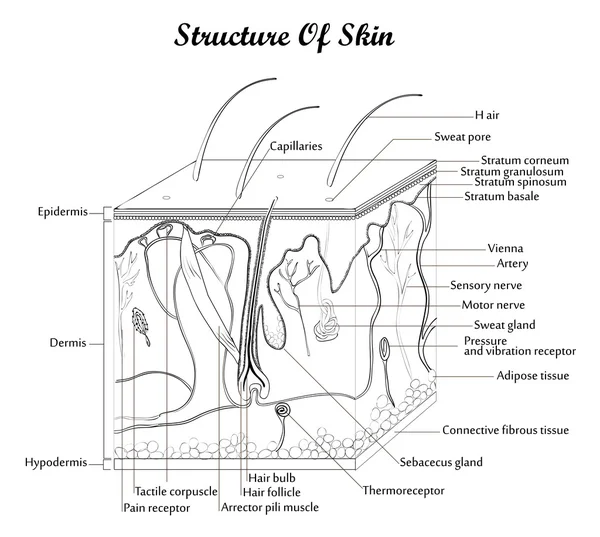
Human Epidermis Layer Structure Cross Section With Hair Follicle Blood Vessels And Glands Isolated Icons Flat Vector Illustration
Vector, 5.78MB, 5000 × 3371 eps


Page 1 >> Next