
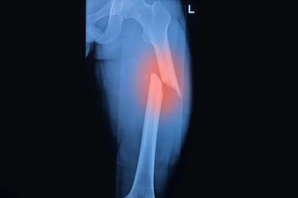
Stock image Femur Fracture page 2


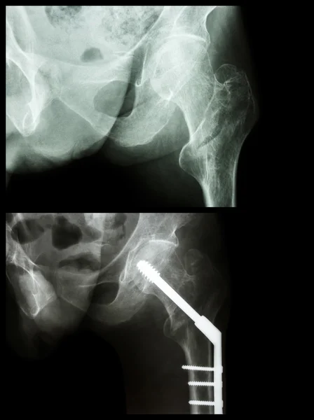
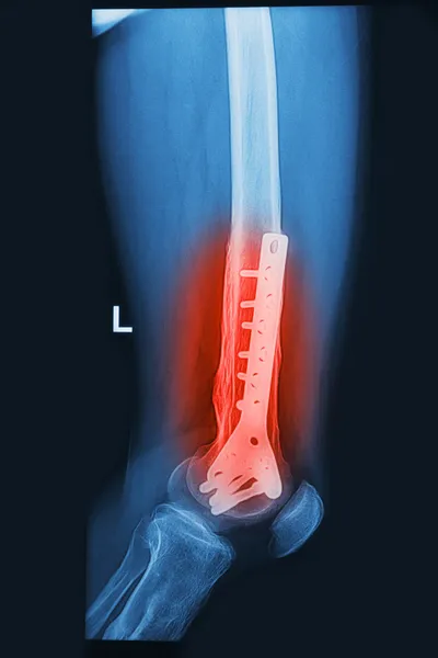
Intertrochanteric Fracture Left Femur (fracture Thigh's Bone). It Was Operated And Insert Intramedullary Nail.
Image, 12.41MB, 5384 × 7212 jpg


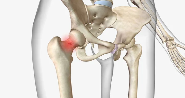
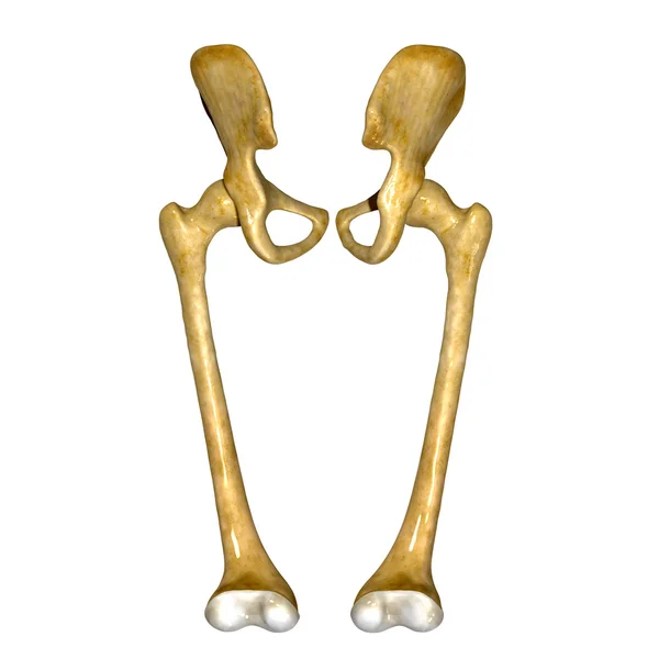
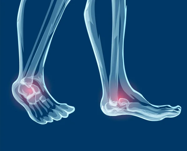
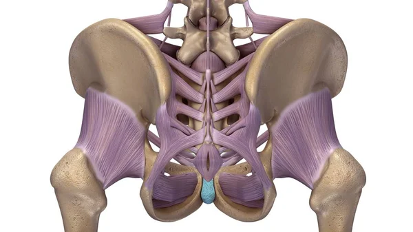
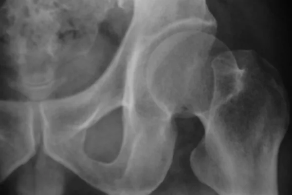
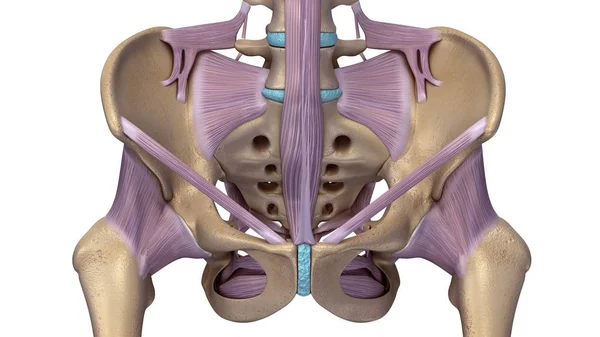
A Femoral Neck Fracture Is A Type Of Hip Fracture That Occurs In The Section Of The Femur Closest To The Pelvis.3D Rendering
Image, 2.83MB, 7340 × 3884 jpg


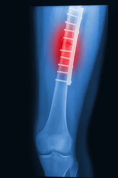
Intertrochanteric Fracture Left Femur (fracture Thigh's Bone). It Was Operated And Insert Intramedullary Nail.
Image, 15.99MB, 5384 × 7212 jpg


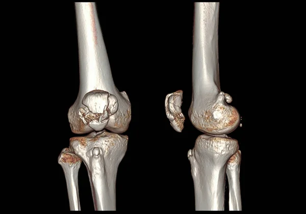
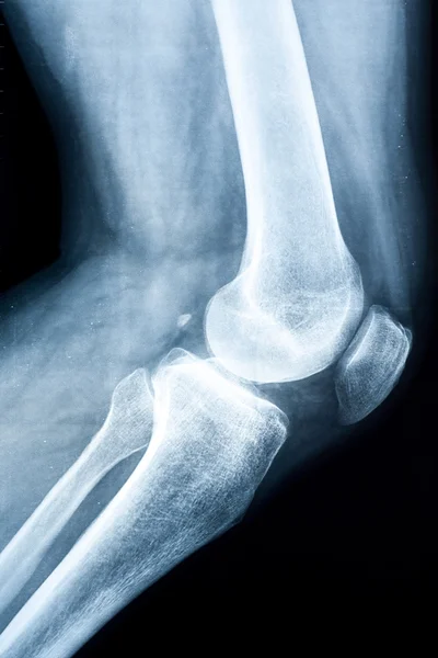
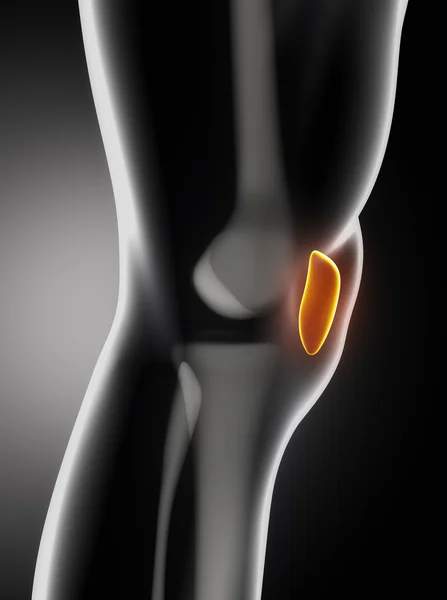
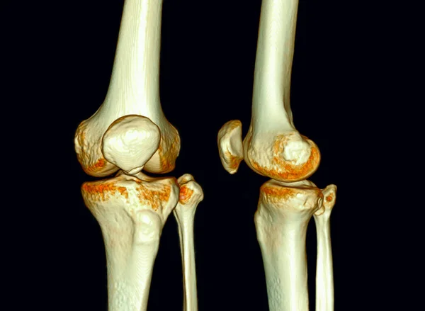
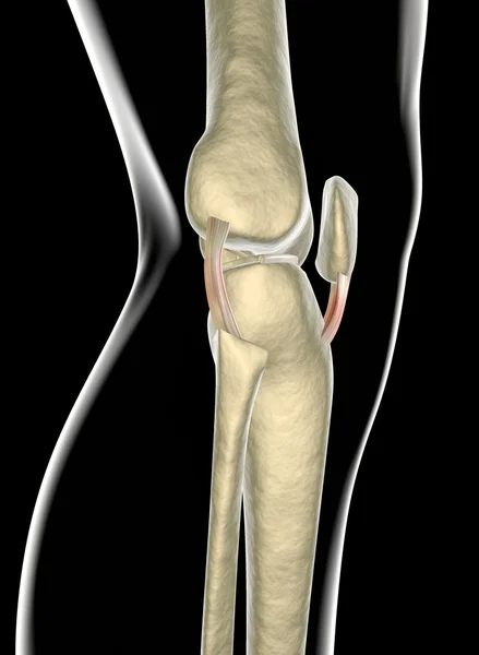
CT Knee 3D Rendering Image AP And Lateral View Isolated On Black Background Showing Fracture Patella.
Image, 8.38MB, 9570 × 6729 jpg
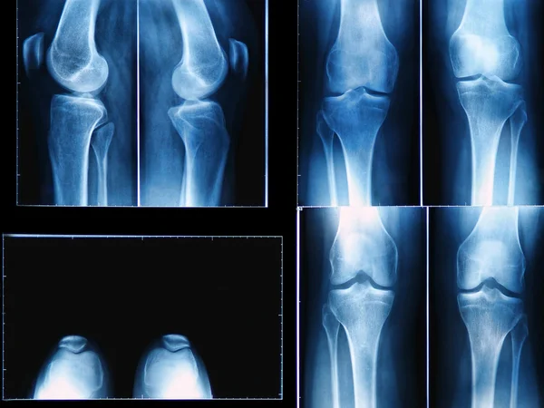
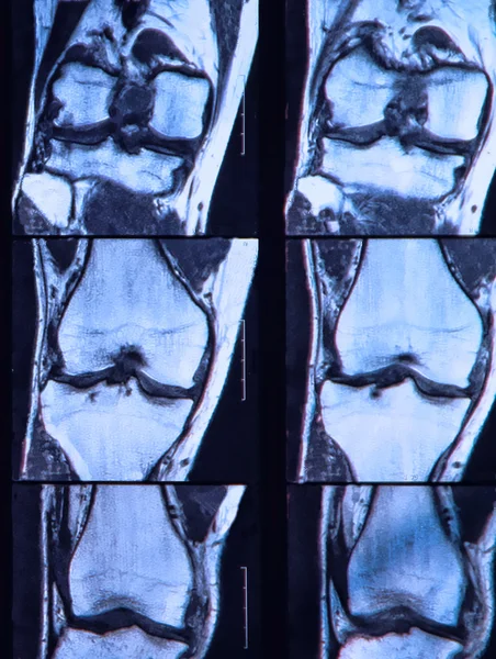

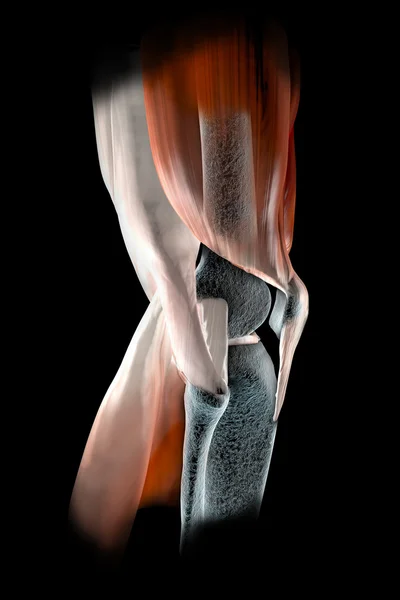
CT Scan Of Knee 3D Rendering Image Isolated On Black Background For Diagnosis Knee Fracture.
Image, 2.34MB, 3840 × 3036 jpg

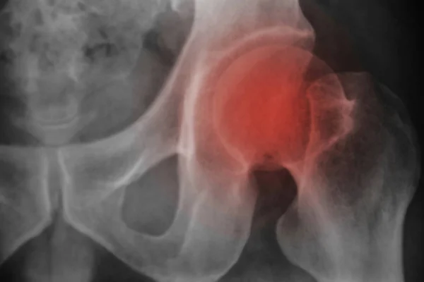
Doctor Holds X-ray Picture On The Background Of A Girl With A Sore Hip Joint And Intervertebral Hernia, Fibromyalgia, Close-up
Image, 4.57MB, 5184 × 3456 jpg


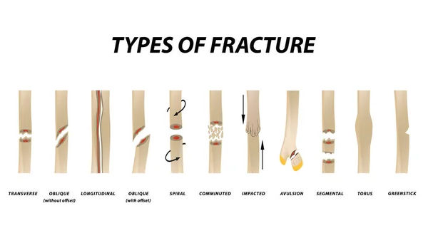
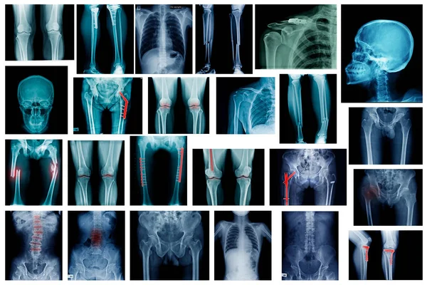
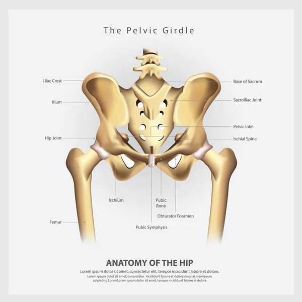
Types Of Fracture. Fracture Bone Set. Infographics. Vector Illustration On Isolated Background.
Vector, 2.09MB, 5000 × 2847 eps
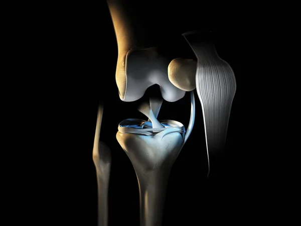
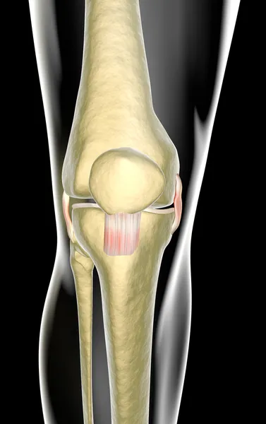
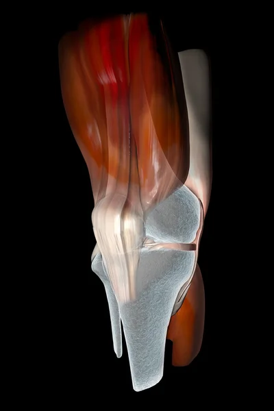
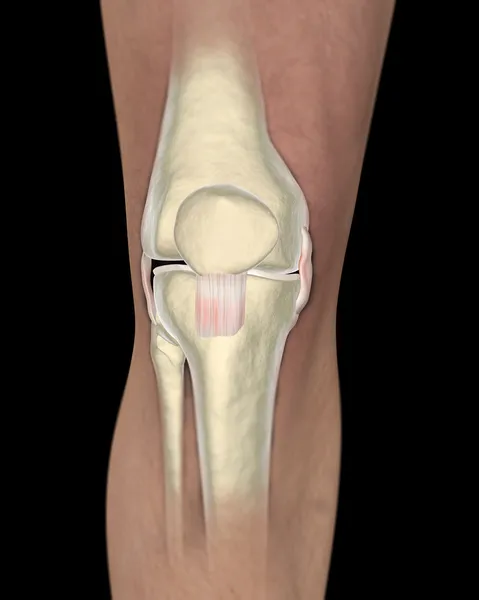
3D Illustration Showing Knee Joint With Ligaments, Meniscus, Articular Cartilage, Fibula And Tibia.
Image, 4.39MB, 6666 × 5000 jpg
Previous << Page 2 >> Next