
Stock image Langerhans
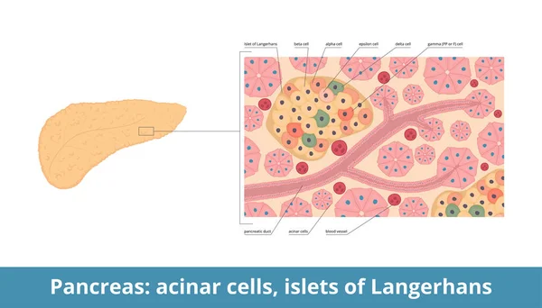
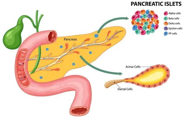
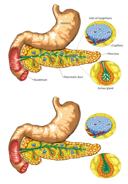
Islets Of Langerhans. Pancreatic Islets Contain Endocrine Cells: Alpha, Beta, Delta, PP Or Gamma, Epsilon Cells. Pancreas Histology (tissue) With Islets And Acinar Cells.
Vector, 7.97MB, 7292 × 4167 eps
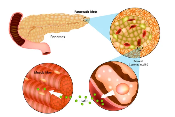
Illustration Of Insulin Production In The Pancreas. Metabolic Actions Of Insulin In Striated Muscle.
Vector, 4.66MB, 5000 × 3334 eps

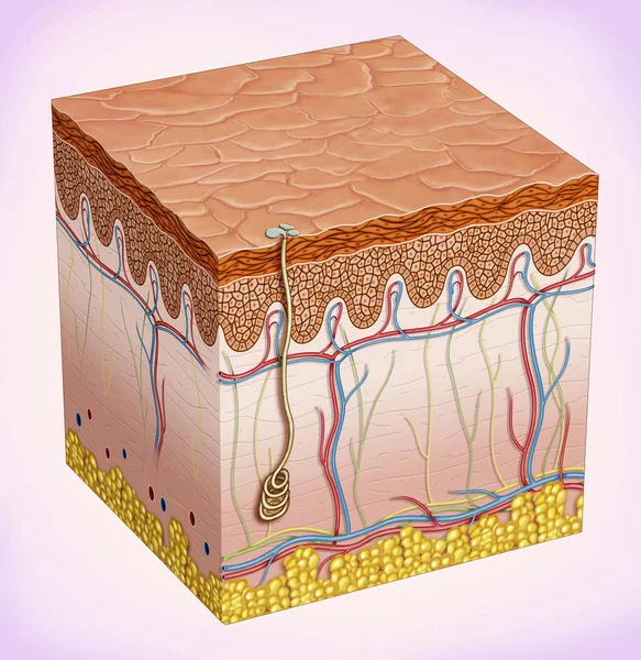
Schematic Illustration Of The Skin. It Provides Protection And Isolation Of The Environmental Organism. There Are Three Main Layers That TheThey Make Up: Epidermis, Dermis, Subcutaneous Tissue And Skin Accessories Such As Hair, Sebaceous Glands And
Image, 5.89MB, 3149 × 3236 jpg

Dendritic Cells Vector Illustration. Anatomical Labeled Closeup Scheme With Progenitor, Immature, Nucleus And Membrane Extensions. Antigen, Receptor And T Cell Diagram.
Vector, 4.58MB, 4800 × 3386 eps
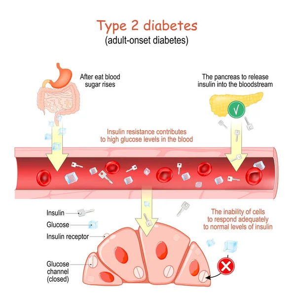
Type 2 Diabetes. Adult-onset Diabetes. Insulin Resistance Contributes To High Glucose Levels In The Blood. The Inability Of Cells To Respond Adequately To Normal Levels Of Insulin. Vector Poster For Educational And Medical Use
Vector, 10.2MB, 4444 × 4444 eps


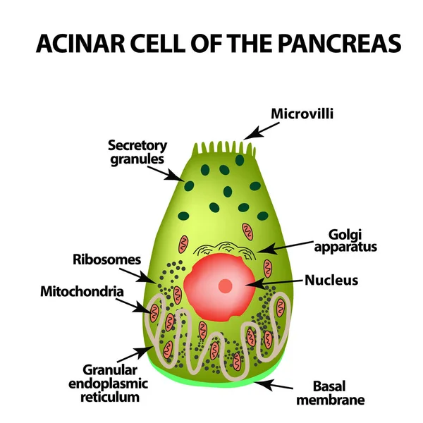
Acinar Cell Of The Pancreas. Acinus. Infographics. Vector Illustration On Isolated Background
Vector, 1.08MB, 5000 × 5000 eps


Insulin Regulates The Metabolism And Is The Key That Unlocks The Cell's Glucose Channel, 3d 2d Graphic, Render, Illustration
Image, 3.15MB, 6000 × 4500 jpg

Melanin Structural Chemical Formula With Blue Liquid Fluid Gradient Shape With Copy Space On White Background
Vector, 1.62MB, 5833 × 4167 eps
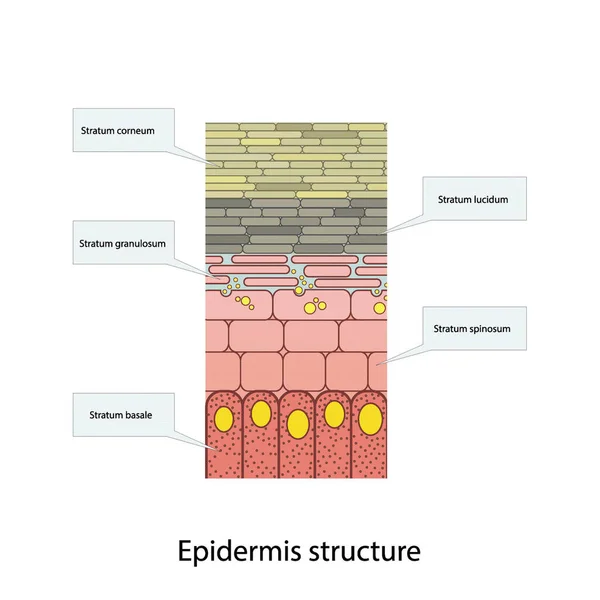
Histological Structure Of Epidermis - Skin Layers Shcematic Vector Illustration Showing Stratum Basale, Spinosum, Granulosum, Lucidum And Corneum
Vector, 5.89MB, 3090 × 3090 eps
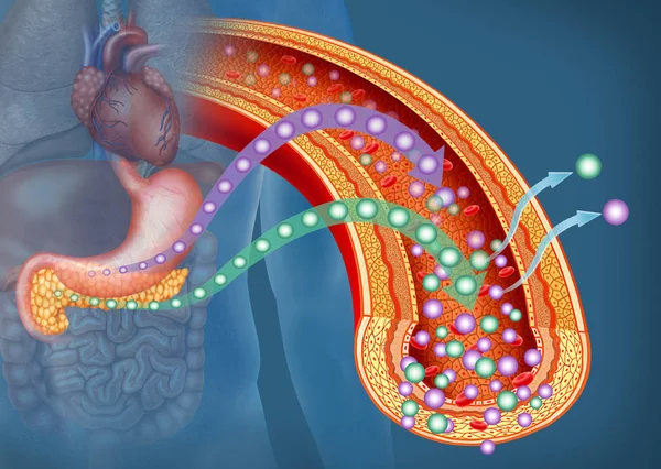
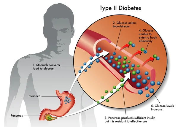
Descriptive Illustration Which Highlights The Pancreas And The Stomach In The Production Of Glucose And Insulin. Insulin Helps Glucose Enter The Cells Of The Body. If Glucose Can Not Enter The Cells, It Accumulates In The Blood.
Image, 5.41MB, 3794 × 2699 jpg

Epidermis Anatomy. Layers And Cell Structure Of The Human Skin. Close-up Of Epidermis. Vector Illustration
Vector, 2.34MB, 4444 × 4444 eps

Somatostatinoma. Malignant Tumor Of The Endocrine Part Of The Pancreas Arising From Delta Cells. Malignant Tumor. Isolated Vector On A White Background.
Vector, 0.33MB, 5653 × 2988 eps
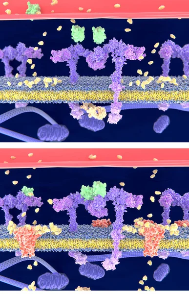
Insulin (green) Binding To The Insulin Receptor (violet) Activates The Transport Of Glucose (yellow) Into The Cell (depicted In 2 Phases) - Illustration
Image, 4.57MB, 4000 × 6200 jpg
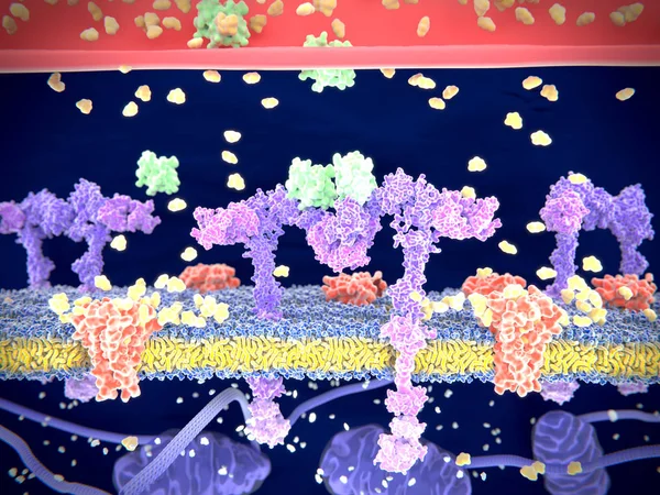
Insulin (green) Binding To The Insulin Receptor (violet) Activates The Transport Of Glucose (yellow) Into The Cell. Illustration
Image, 6.21MB, 8000 × 6000 jpg
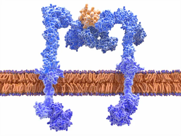
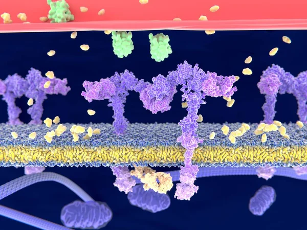
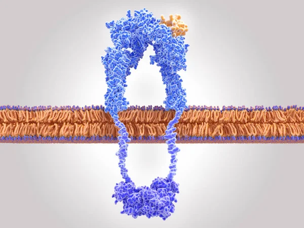
The Insulin Receptor (blue) Is A Transmembrane Protein, That Is Activated By Insulin (orange). Insulin Binding Induces Structural Changes Within The Receptor That Finally Leads To The Activation Of The Glucose Transporter Protein.
Image, 12.2MB, 8000 × 6000 jpg

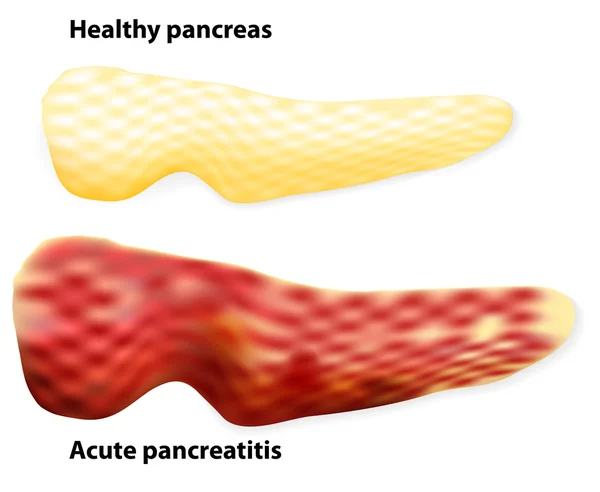
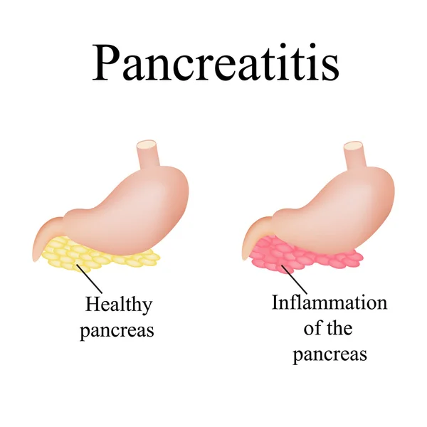
Inflammation Of The Pancreas. Pancreatitis. Vector Illustration On Isolated Background
Vector, 3.8MB, 5000 × 5000 eps
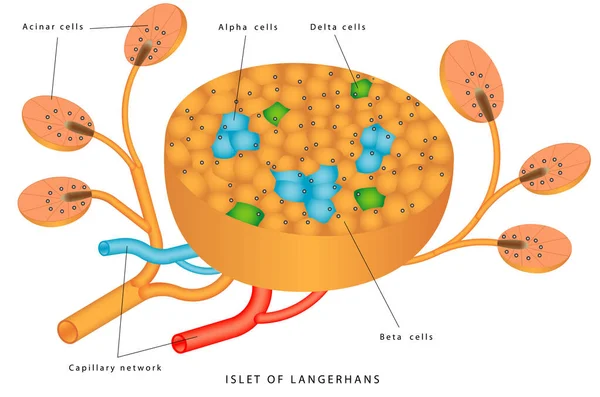
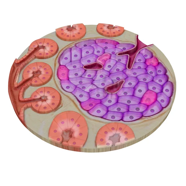
Pancreatic Islet. The Islets Of Langerhans Are Responsible For Endocrine Function Of Pancreas. Each Islet Contains Beta, Alpha, And Delta Cells That Are Responsible For The Secretion Of A Hormones
Vector, 0.62MB, 5395 × 3569 eps

Insulin (green) Binding To The Insulin Receptor (violet) Activates The Transport Of Glucose (yellow) Into The Cell. Illustration
Image, 3.71MB, 8000 × 6000 jpg


Insulin (green) Binding To The Insulin Receptor (violet) Activates The Transport Of Glucose (yellow) Into The Cell (phase 1). Illustration
Image, 3.96MB, 8000 × 6000 jpg

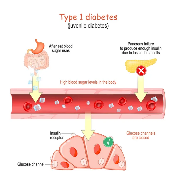
Type 1 Diabetes. Juvenile Diabetes. High Blood Sugar Levels In The Body. Glucose Channels In The Cells Are Closed. Vector Poster For Educational And Medical Use
Vector, 9.65MB, 4444 × 4444 eps

Histological Structure Of Epidermis - Skin Layers Shcematic Vector Illustration Showing Stratum Basale, Spinosum, Granulosum, Lucidum And Corneum
Vector, 7.1MB, 3090 × 3090 eps
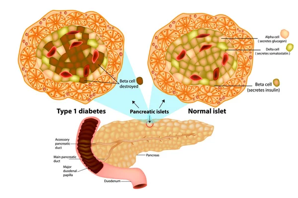
The Pancreas Has Many Islets That Contain Insulin-producing Beta Cells And Glucagon-producing. Type 1 Diabetes ( Beta Cell Destroyed).
Vector, 5.99MB, 5000 × 3333 eps


Page 1 >> Next