
Stock image Micrograph
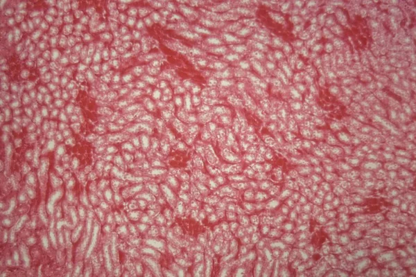


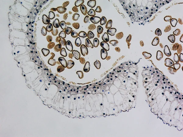
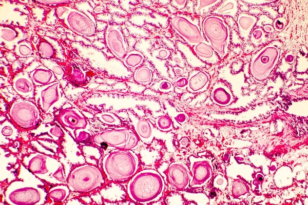
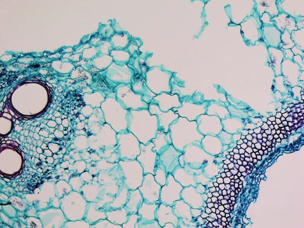
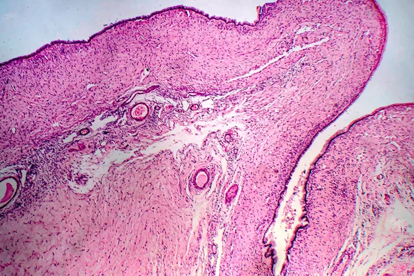
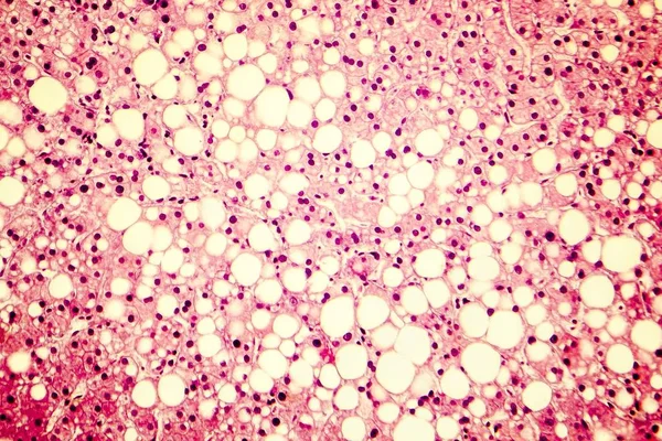
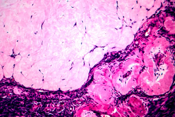
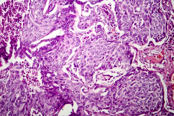
Ovarian Mucinous Cystadenoma, A Benign Tumor Of Ovary, Light Micrograph, Photo Under Microscope
Image, 10.29MB, 4436 × 2957 jpg

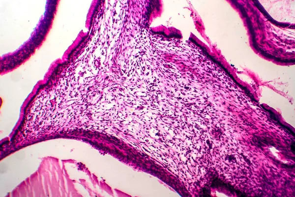
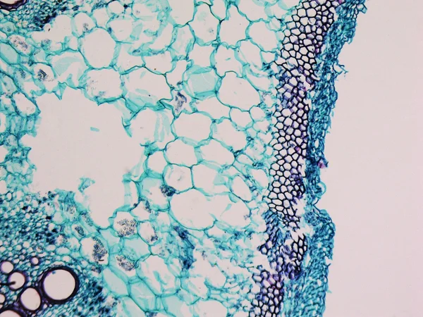
Ovarian Mucinous Cystadenoma, A Benign Tumor Of Ovary, Light Micrograph, Photo Under Microscope
Image, 11.27MB, 4436 × 2957 jpg
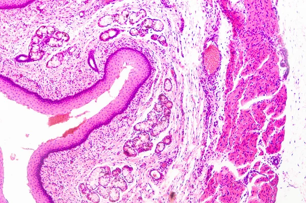

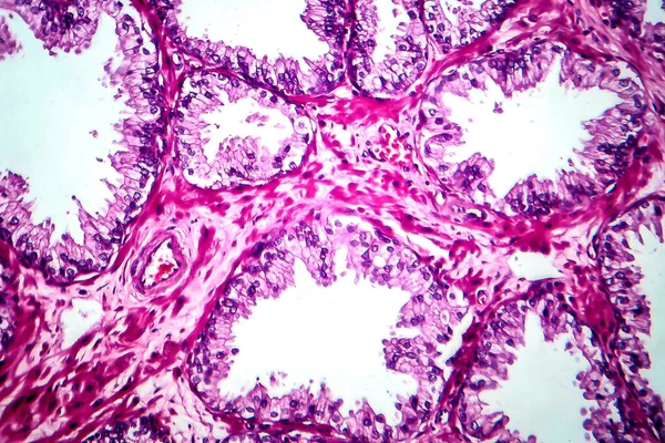
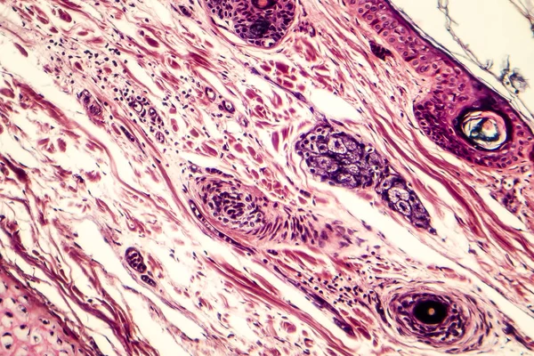
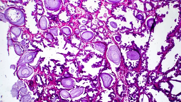
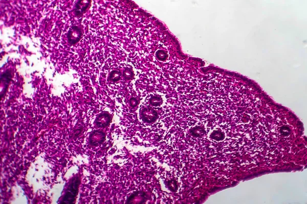
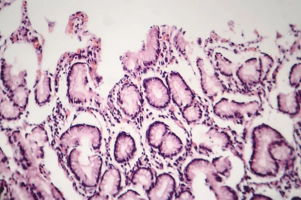
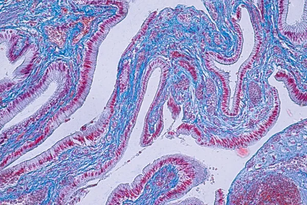
Uterus Adenofibroma, Light Micrograph, Photo Under Microscope. A Rare Benign Tumor Of The Uterus Composed Of Glandular And Fibrous Tissues
Image, 9.96MB, 4602 × 3068 jpg


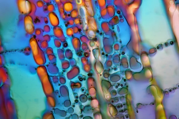
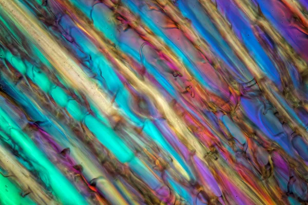
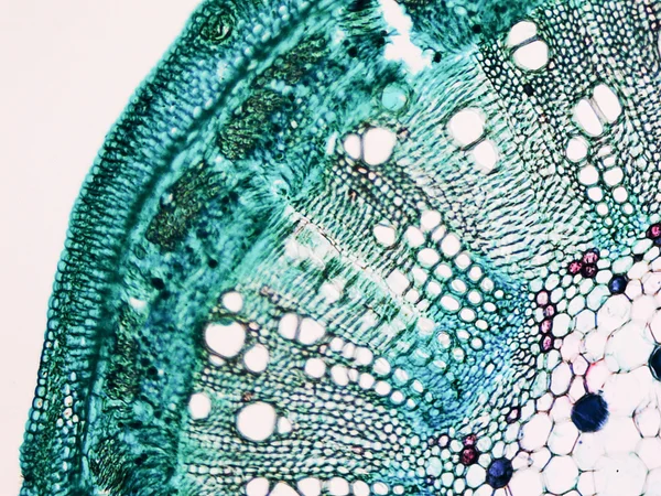
Characteristics Of Anatomy And Histological Sample Striated (Skeletal) Muscle Of Mammal Tissue Under The Microscope.
Image, 22.55MB, 8192 × 5464 jpg


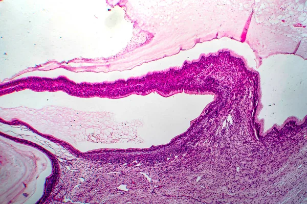
Histopathology Of Diffuse Sclerosing Glomerulonephritis, Light Micrograph, Photo Under Microscope
Image, 8.1MB, 4342 × 2895 jpg


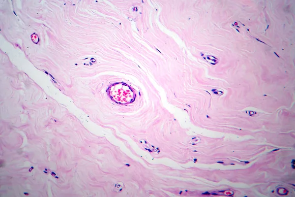
Areolar Connective Tissue Under The Microscope View. Histological For Human Physiology.
Image, 15.32MB, 6000 × 4000 jpg

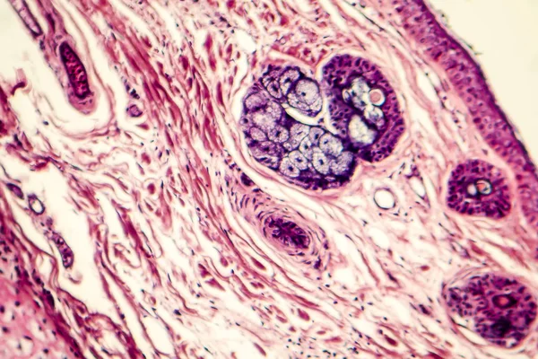
Light Microscopic Of Human Ovary Showing Primary And Secondary Follicles. Human Physiology Education.
Image, 26.67MB, 6000 × 4000 jpg
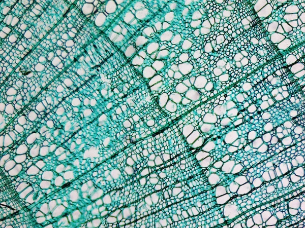
Cross Section Of Ciliated Epithelium Under The Microscope For Education Histology. Human Tissue.
Image, 14.75MB, 5168 × 3448 jpg

Page 1 >> Next