
Stock image Amyloid Plaque
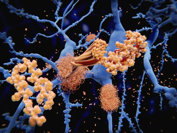
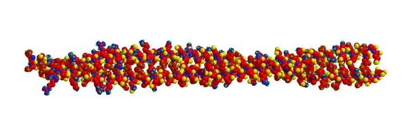
After Being Cleaved By The Gamma And Beta Secretases The Amyloid Beta Peptide, Which Has About 40 Amino Acid Residues, Leaves The Membrane, Changes Shape And Aggregates Into Long Fibrils. These Fibrils Form Dense Plaques On Nerve Cells, Which Are In
Image, 9.72MB, 8000 × 6000 jpg
A Microglia Cell. It Plays An Important Role In The Pathogenesis Of Alzheimer's Disease
Image, 13.48MB, 8000 × 6000 jpg
The Amyloid Precursor Protein (APP) Is A Complex Protein With Many Functions. It Is Found On The Surface Of Cells Throughout The Body. The Intact Protein Binds To Many Stuctural Proteins Outside Cells, Such As Heparin And Laminin And Sends Signals Th
Image, 6.49MB, 8000 × 6000 jpg
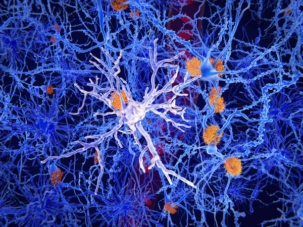
3d Computer Illustration Of The Alzheimer Disease. The Yellow Structures Are Amyloid Plaques Damaging Neurons. The Violet Cells Are Microglia Cells That Phagocyte And Degrade Sick Neurons.
Image, 5.09MB, 8000 × 6000 jpg


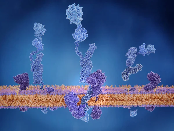
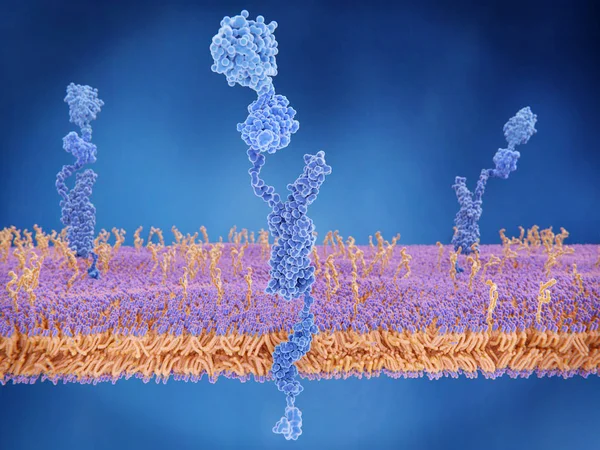
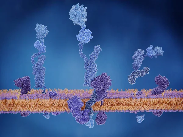
The Amyloid Precursor Protein (APP) Being Cleaved By Gamma And Beta Secretase And Setting The Beta Amyloid Peptide Free. APP Is A Complex Protein With Many Functions. It Is Found On The Surface Of Cells Throughout The Body.
Image, 5.87MB, 8000 × 6000 jpg
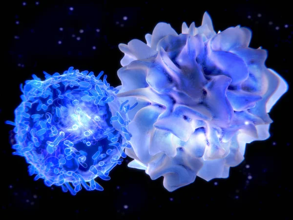
Interaction Between A Dendritic Cell And A T-lymphocyte. 3d-rendering. Dendritic Cells Are Antigen-presenting Cells Of The Immune System
Image, 6.52MB, 8000 × 6000 jpg
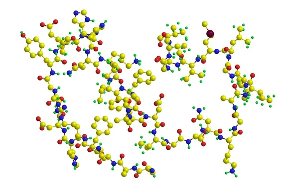
The Amyloid Precursor Protein. When Cleaved, The Membrane Domain Is Involved In The Alzheimer Disease Building Amyloid Plaques. 3d Rendering. Illustration
Image, 3.29MB, 8000 × 6000 jpg
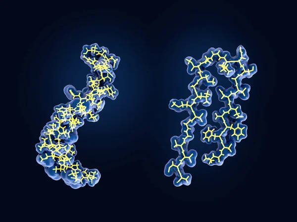
After Being Cleaved By The Gamma And Beta Secretases The Amyloid Beta Peptide, Which Has About 40 Amino Acid Residues, Leaves The Membrane, Changes Shape And Aggregates Into Long Fibrils. These Fibrils Form Dense Plaques On Nerve Cells.
Image, 2.61MB, 8000 × 6000 jpg
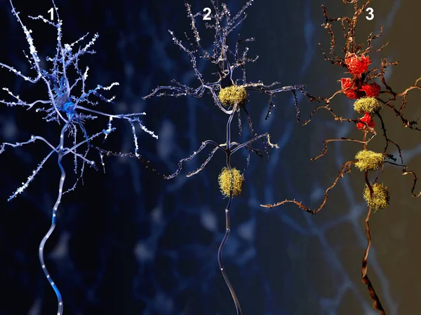
3 Phases Of The Alzheimer Disease. 1. Healthy Neuron. 2. Neuron With Amyloid Plaques (yellow). 3. Dead Neuron Being Digested By Microglia Cells (red). Illustration
Image, 6.76MB, 8000 × 6000 jpg
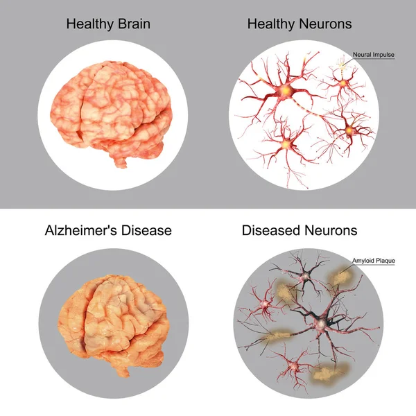
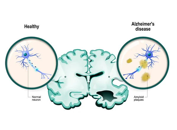

Alzheimer's Disease. Disease Associated With Amyloid Plaques, Neurofibrillary Tangles, And Loss Of Neuronal Connections In The Brain. Human Brain With Alzheimer's Disease. Close-up Of Neurons With Amyloid Plaques. Vector Illustration
Vector, 14.34MB, 4444 × 4444 eps

3 Phases Of The Alzheimer Disease. 1. Healthy Neuron. 2. Neuron With Amyloid Plaques (yellow). 3. Dead Neuron Being Digested By Microglia Cells (red). Illustration
Image, 6.76MB, 8000 × 6000 jpg
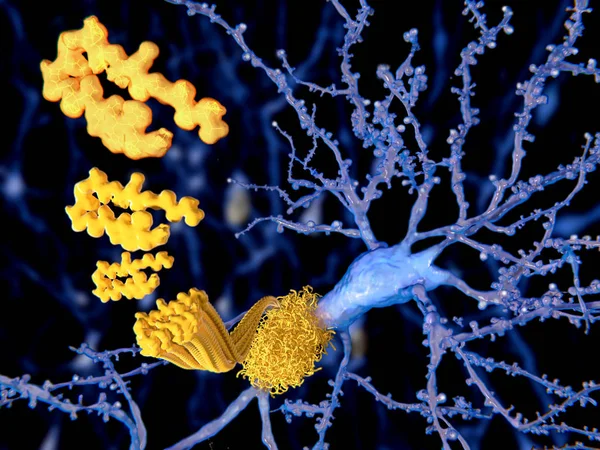
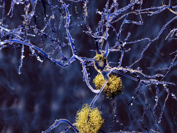
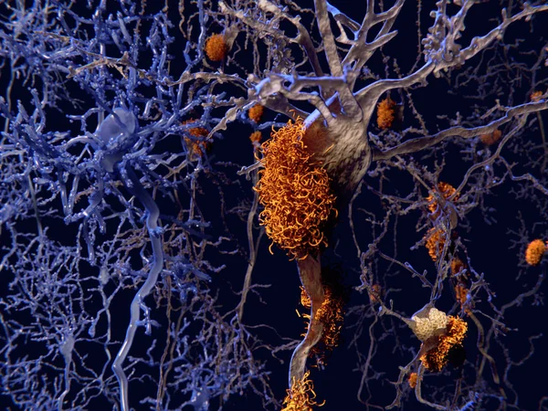
The Beta Amyloid Peptid, Amyloid Plaques Growing On A Neuron. It Consists Of About 30 Amino Acids And Aggregates To Amyloid Plaques, That May Damage And Kill Neurons. Illustration
Image, 4.49MB, 8000 × 6000 jpg
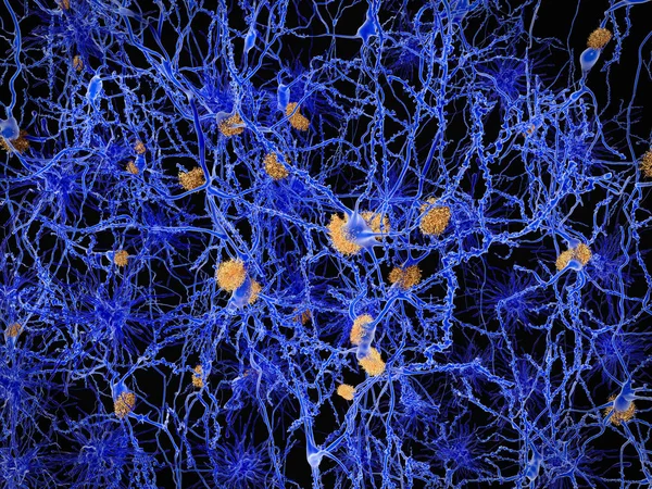
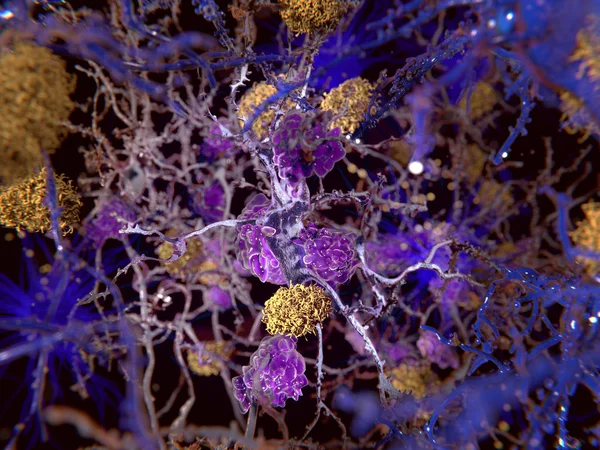
Alzheimer Disease, Neuron Network With Amyloid Plaques. Illustration
Image, 28.11MB, 8000 × 6000 jpg


Page 1 >> Next