
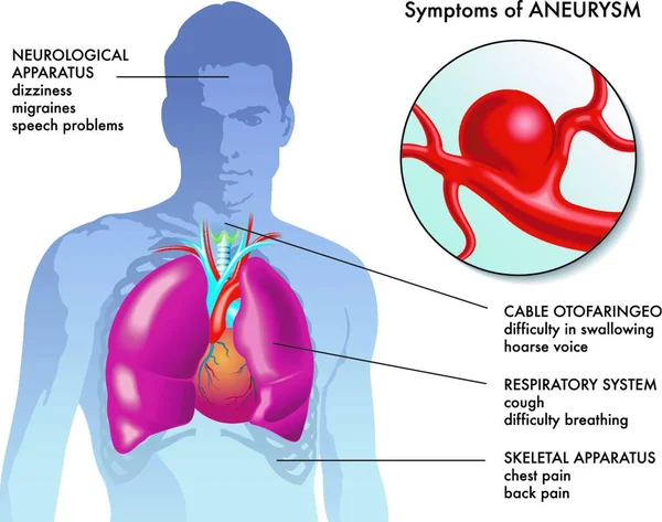
Stock image Aneurysms
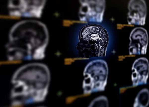
Selective Focus Of MRI Brain Sagittal Plane For Detect A Variety Of Conditions Of The Brain Such As Cysts, Tumors, Bleeding, Swelling, Developmental And Structural Abnormalities Or Infections .
Image, 7.51MB, 7584 × 5484 jpg
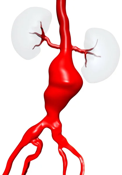
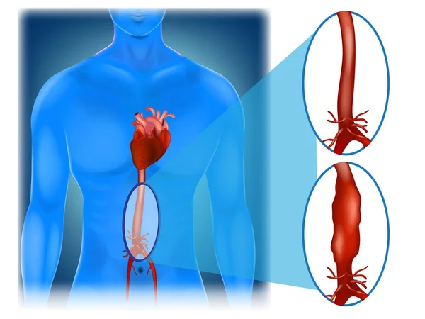
Abdominal Aortic Aneurysm (AAA) Located Below The Arteries That Supply Blood To The Kidneys. 3D Illustration.
Image, 6.94MB, 6977 × 10000 jpg

Open Surgery For Abdominal Aortic Aneurysm (AAA) Located Below The Renal Arteries That Supply Blood To The Kidneys. Aorta Are Opened, Before Insert A Graft. 3D Illustration.
Image, 9.35MB, 6977 × 10000 jpg
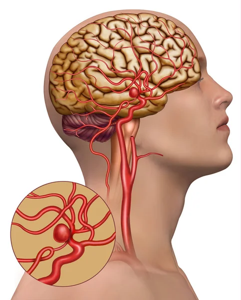
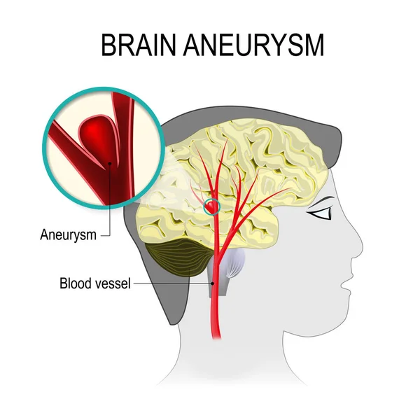
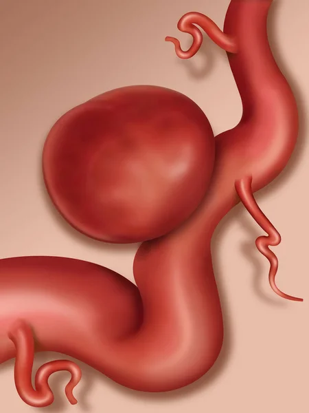
Descriptive Illustration Of A Cerebral Artery Affected By A Cerebral Aneurysm.
Image, 2.6MB, 2894 × 3593 jpg
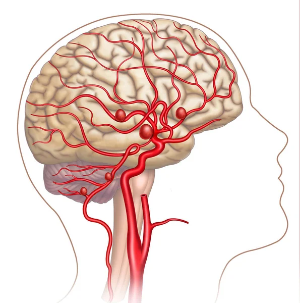
Schematic Illustration Composed By Human Head In Which We Can See The Brain And The Main Arteries, In The Podemo See The Development Of Cerebral Aneurysms.
Image, 2.16MB, 3180 × 3206 jpg


Word Writing Text Abdominal Aortic Aneurysmquestion. Business Concept For Getting To Know The Enlargement Of Aorta Keyboard Key Intention To Create Computer Message Pressing Keypad Idea.
Image, 5.95MB, 5616 × 3744 jpg
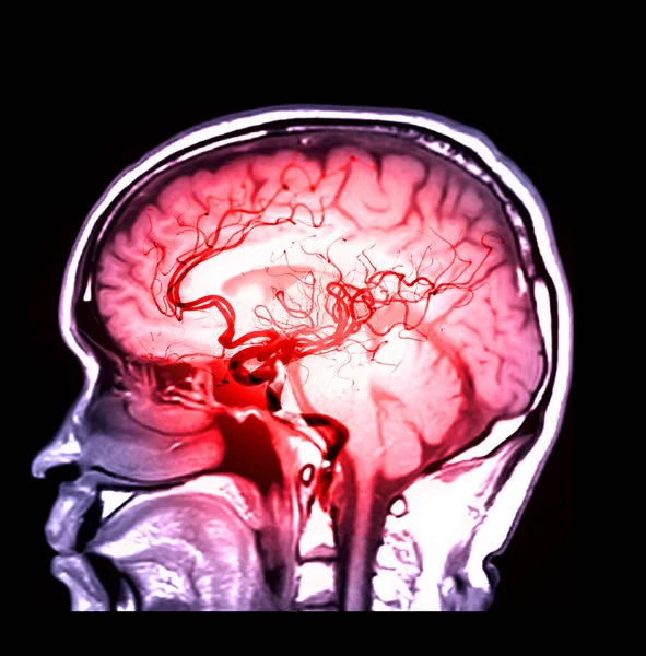
MRA Brain Or Magnetic Resonance Angiography ( MRA ) Of Cerebral Artery And Common Carotid Artery AP And Lateral View For Evaluate Them Stenosis And Stroke Disease.
Image, 3.83MB, 4980 × 2576 jpg

Abdominal Aneurysm 3d Medical Vector Illustration Isolated On White Background Eps 10 Education Vector
Vector, 33.37MB, 7500 × 5000 eps
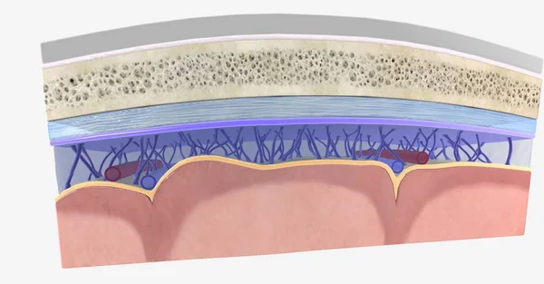
The Meninges Contain Cerebrospinal Fluid And Help Support And Protect The Central Nervous System. 3D Rendering
Image, 20.05MB, 11115 × 5802 jpg
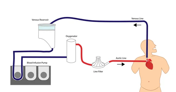
Cardiopulmonary Bypass. Extracorporeal Circulation. Support Technique For Cardiac Surgery. Machine Utilized For Heart And Lung Function
Image, 2.14MB, 11499 × 6015 jpg

Selective Focus Of MRI Brain Sagittal Plane For Detect A Variety Of Conditions Of The Brain Such As Cysts, Tumors, Bleeding, Swelling, Developmental And Structural Abnormalities Or Infections .
Image, 6.81MB, 7056 × 5208 jpg

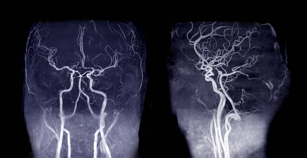
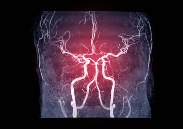
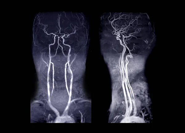
MRA Brain And Neck Or Magnetic Resonance Angiography ( MRA ) Of Cerebral Artery And Common Carotid Artery For Evaluate Them Stenosis And Stroke Disease.
Image, 2.72MB, 3199 × 3871 jpg

Diabetic Retinopathy Anatomical Poster. Human Eye Disease, Vision Loss Or Blindness. Damaged Blood Vessels In The Retina. Proliferative Or Nonproliferative Eye Condition Medical Vector Illustration
Vector, 0.65MB, 5924 × 2997 eps

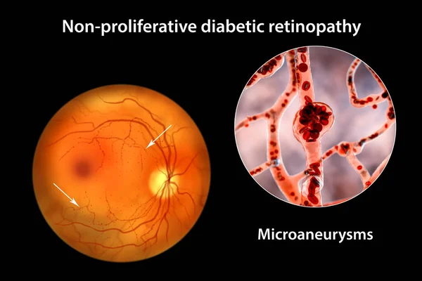
Non-proliferative Diabetic Retinopathy, 3D Illustration Showing Multiple Microaneurysms On The Eye Retina And Closeup View Of Microaneurysms, Microscopic Buldges In The Artery Walls Filled With Blood
Image, 11.11MB, 10431 × 6954 jpg
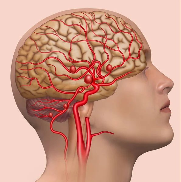
Human Head, Rendered, On A Blue Background, With An Aneurysm In The Internal Carotid Artery.
Image, 2.5MB, 3180 × 3206 jpg


Collection Transparent Image Of The Skull Blue Color With MRI Brain For Medical Background Concept.
Image, 5.96MB, 6544 × 4112 jpg

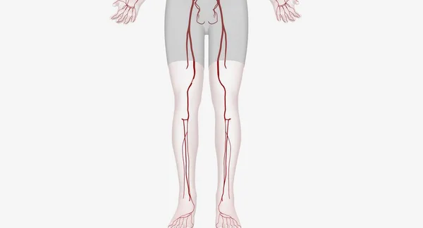
Lower Extremity Arterial Interventions Are Procedures Designed To Restore Blood Flow To Your Legs And Feet. 3D Rendering
Image, 2.3MB, 7258 × 3920 jpg

Non-proliferative Diabetic Retinopathy, Illustration Showing Normal Eye Retina And Retina With Hard Exudates, Microaneurysms, Dot Haemorrhages, Flame-shaped And Splinter Retinal Haemorrhages
Image, 6.89MB, 11738 × 6603 jpg
Non-proliferative Diabetic Retinopathy, 3D Illustration Showing Multiple Microaneurysms On The Eye Retina And Closeup View Of Microaneurysms, Microscopic Buldges In The Artery Walls Filled With Blood
Image, 10.46MB, 10431 × 6954 jpg

Diabetic Retinopathy Anatomical Poster. Human Eye Disease, Vision Loss Or Blindness. Damaged Blood Vessels In The Retina. Proliferative Or Nonproliferative Eye Condition Medical Vector Illustration
Vector, 0.46MB, 5924 × 2996 eps

Diabetic Retinopathy Anatomy. Healthy Eyeball And Damaged Organ. Human Eye Disease, Vision Loss Or Blindness. Proliferative Or Nonproliferative Eye Condition. Retina Medical Flat Vector Illustration
Vector, 1.18MB, 9182 × 2526 eps

Diabetic Retinopathy Anatomical Poster. Human Eye Disease, Vision Loss Or Blindness. Damaged Blood Vessels In The Retina. Proliferative Or Nonproliferative Eye Condition Medical Vector Illustration
Vector, 0.57MB, 5924 × 2997 eps

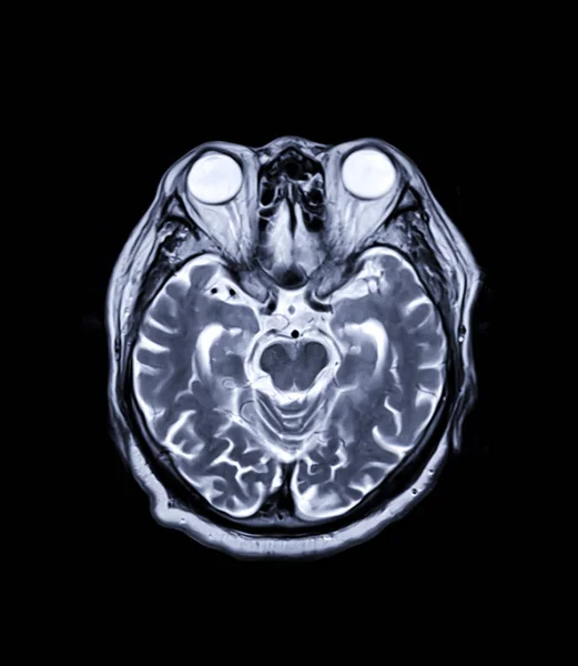
Compare MRI Brain Axial T2W Flair And T2W View For Showing Enchepalomalacia Disease.
Image, 4.43MB, 7194 × 4272 jpg

Handwriting Text Abdominal Aortic Aneurysmquestion. Concept Meaning Getting To Know The Enlargement Of Aorta Hu Analysis Hand In Suit Offering Blank Solid Color Circle For Logo Posters.
Image, 2.15MB, 7000 × 7000 jpg
MRA Brain Or Magnetic Resonance Angiography Of Cerebral Artery In The Brain Sagittal View For Evaluate Them Stenosis And Stroke Disease.
Image, 3.59MB, 3083 × 3126 jpg
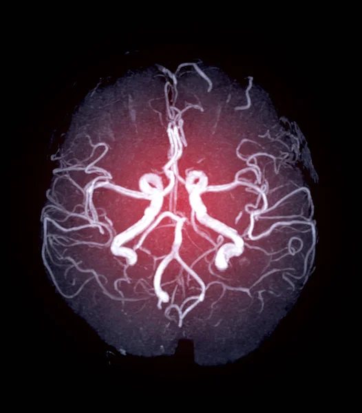
MRA Brain Or Magnetic Resonance Angiography (MRI) Of Vessel In The Brain Axial Mip View For Evaluate Them Stenosis , Occlusions, Aneurysms .
Image, 1.22MB, 2128 × 2424 jpg

Diabetic Retinopathy Anatomy. Healthy Eyeball And Damaged Organ. Human Eye Disease, Vision Loss Or Blindness. Proliferative Or Nonproliferative Eye Condition. Retina Medical Flat Vector Illustration
Vector, 0.66MB, 5924 × 2996 eps

Skull Image Fusion With MRI MRA Brain For Evaluate Them Stenosis And Stroke Disease.
Image, 3.51MB, 4491 × 2886 jpg
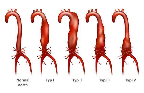
Abdominal Aortic Aneurysm (Thoracoabdominal). Crawford Classification.
Vector, 4.48MB, 5166 × 3334 eps

Non-proliferative Diabetic Retinopathy, 3D Illustration Showing Hard Exudates, Microaneurysms, Dot Haemorrhages, Flame-shaped And Splinter Retinal Haemorrhages, Ophthalmoscope View
Image, 13.11MB, 5352 × 5352 jpg

Diabetic Retinopathy Anatomy. Healthy Eyeball And Damaged Organ. Human Eye Disease, Vision Loss Or Blindness. Proliferative Or Nonproliferative Eye Condition. Retina Medical Flat Vector Illustration
Vector, 0.89MB, 6928 × 2526 eps

Diabetic Retinopathy Anatomical Poster. Human Eye Disease, Vision Loss Or Blindness. Damaged Blood Vessels In The Retina. Proliferative Or Nonproliferative Eye Condition Medical Vector Illustration
Vector, 0.64MB, 5924 × 2997 eps
MRA Brain Or Magnetic Resonance Angiography ( MRA ) Of Cerebral Artery In The Brain For Evaluate Them Stenosis And Stroke Disease
Image, 1.97MB, 3392 × 2400 jpg

Magnetic Resonance (MR) Spectroscopy Of The Brain For Identifies The Anatomical Location Of A Tumor.
Image, 3.43MB, 5120 × 3840 jpg

MRI Of The Brain Sagittal Plane For Detect A Variety Of Conditions Of The Brain Such As Cysts, Tumors, Bleeding, Swelling, Developmental And Structural Abnormalities, Infections.
Image, 4.62MB, 3104 × 4000 jpg
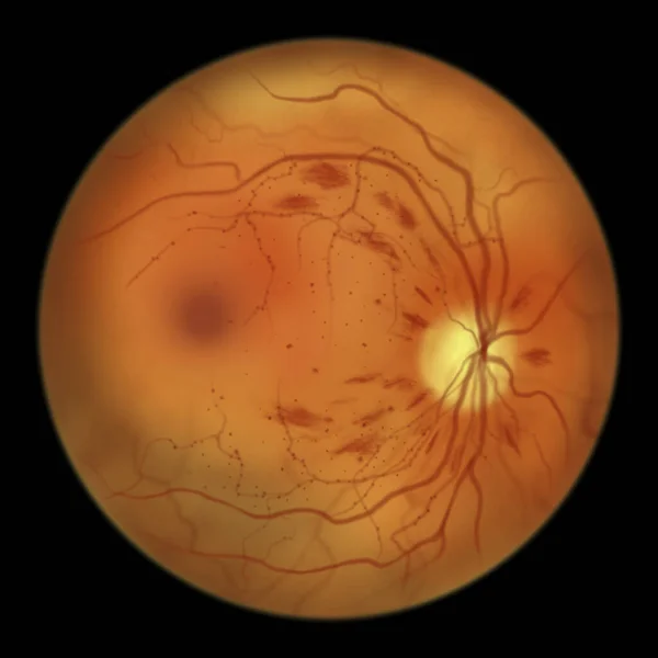
Non-proliferative Diabetic Retinopathy, Illustration Showing Microaneurysms, Dot Haemorrhages, Flame-shaped And Splinter Retinal Haemorrhages, Ophthalmoscope View
Image, 2.86MB, 5000 × 5000 jpg
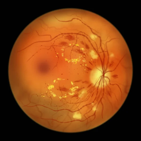
Diabetic Retinopathy Non-proliferative, Illustration Showing Hard Exudates, Cotton Wool Spots, Microaneurysms, Dot Haemorrhages, Flame-shaped And Splinter Retinal Haemorrhages, IRMAs, Venous Beading
Image, 3.07MB, 5000 × 5000 jpg

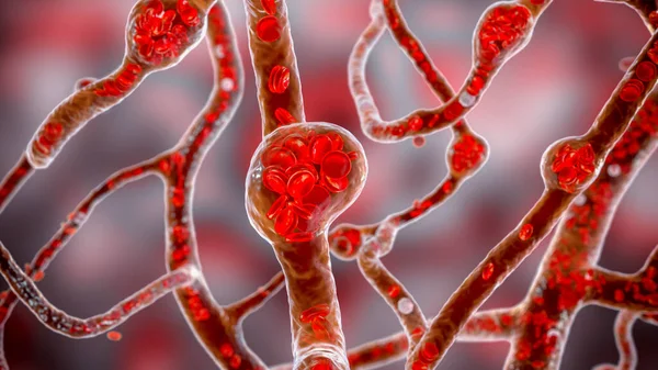
Microaneurysms, Microscopic Buldges In The Artery Walls Filled With Blood, 3D Illustration. Found In The Eye Retina In Diabetic Retinopathy, And Also In Brain (Charcot-Bouchard Aneurysms)
Image, 7.58MB, 7200 × 4050 jpg
MRI Brain Axial T2 Technique For Detect Variety Of Conditions Of The Brain Such As Cysts, Tumors, Bleeding, Swelling, Developmental And Structural Abnormalities Infections.
Image, 1.83MB, 2904 × 3339 jpg
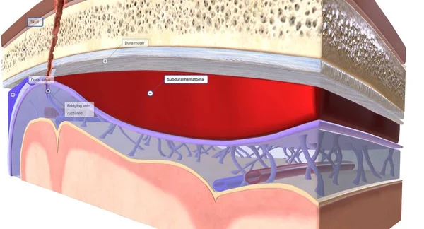
The Blood That Collects Between The Dura Mater And The Arachnoid Mater Is Called A Subdural Hematoma. 3D Rendering
Image, 12.41MB, 7258 × 3920 jpg
MRA Brain And Neck Or Magnetic Resonance Angiography ( MRA ) Of Cerebral Artery And Common Carotid Artery AP And Lateral View For Evaluate Them Stenosis And Stroke Disease.
Image, 4.76MB, 5696 × 4112 jpg
Page 1 >> Next