
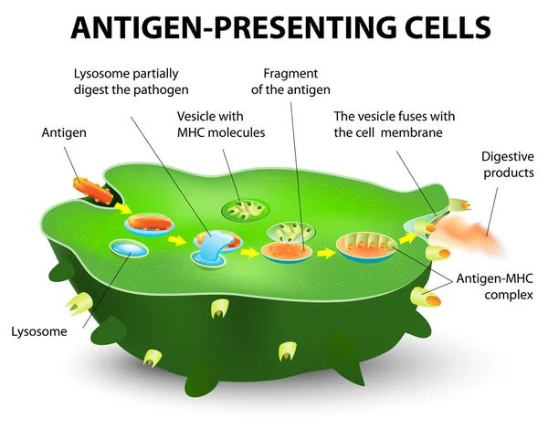
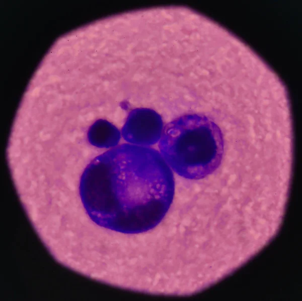
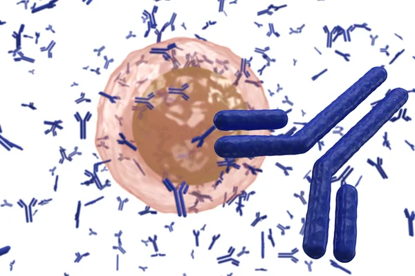
Stock image Antigen Presenting Cell
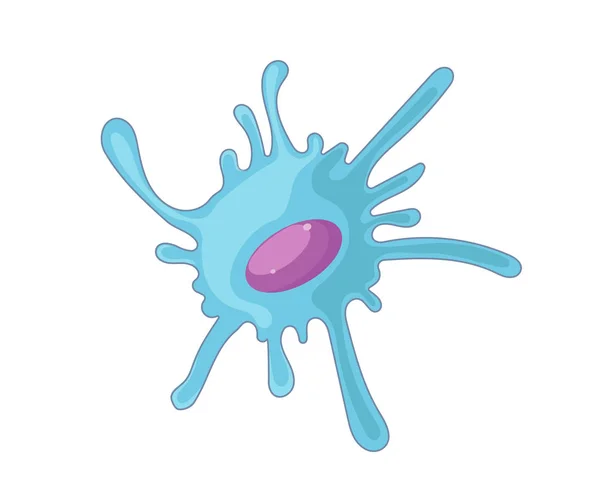

Dendritic Cells Vector Illustration. Anatomical Labeled Closeup Scheme With Progenitor, Immature, Nucleus And Membrane Extensions. Antigen, Receptor And T Cell Diagram.
Vector, 4.58MB, 4800 × 3386 eps


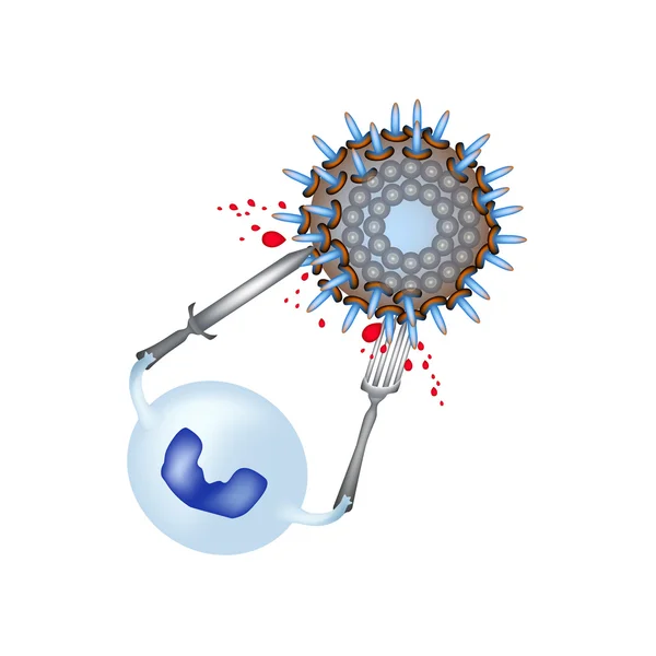
Vector Illustration Of Mechanisms Of Microbial Cell Resistance To Antibiotics
Vector, 3.53MB, 6688 × 3296 eps

Vector Illustration Of Mechanisms Of Microbial Cell Resistance To Antibiotics
Vector, 3.53MB, 6688 × 3296 eps

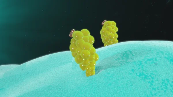
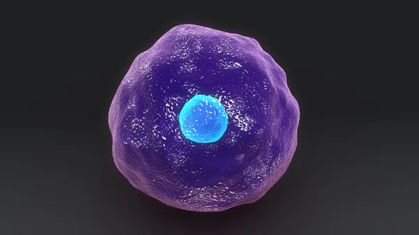
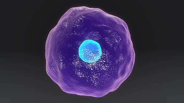
Dendritic Cell. Dendritic Cells Are Antigen-presenting Cells Of The Immune System. They Process Antigen Material And Present It On The Cell Surface To The T Cells Of The Immune System. 3D Rendering. Illustration
Image, 2.94MB, 8000 × 6000 jpg
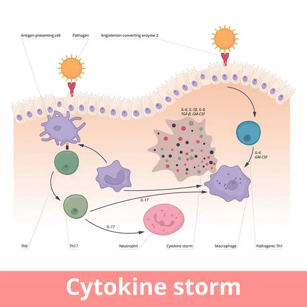
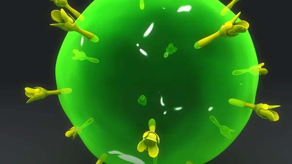
Cytokine Storm. Hypercytokinemia During Which The Immune System Causes A Release Of Cytokines With Help Of Macrophages, T Helper Cells, Neutrophils.
Vector, 6.7MB, 6250 × 6250 eps
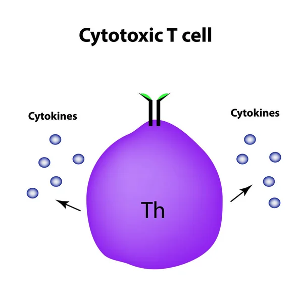
Cytotoxic Cells. Cytokines. Cell Immunity. Infographics. Vector Illustration
Vector, 1.39MB, 5000 × 5000 eps
3d Computer Illustration Of A Dendritic Cell. They Areantigen-presenting Cells Of The Immune System. Their Main Function Is To Process Antigen Material And Present It On The Cell Surface To The T Cells Of The Immune System. They Are Messengers Betwe
Image, 5.2MB, 8000 × 6000 jpg
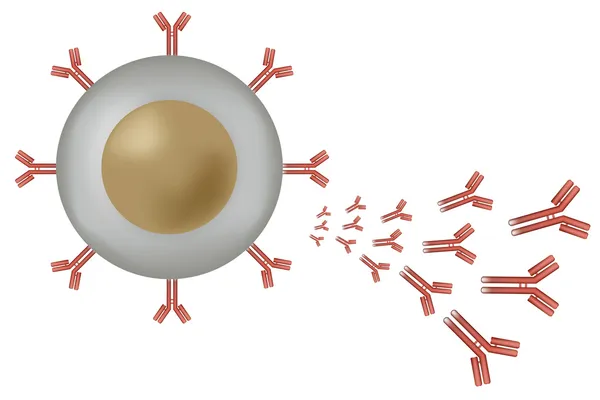
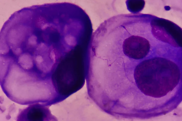
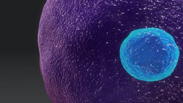
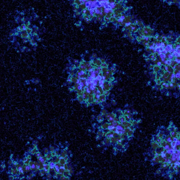
Dendritic Cells Present Antigens (green) To Lymphocytes Through Their Membran Bound MHC-molecules (violet). CD4 Molecules (light Blue) Bind To Other Portions Of The MHC, Strengthening The Interaction.
Image, 10.24MB, 8000 × 6000 jpg
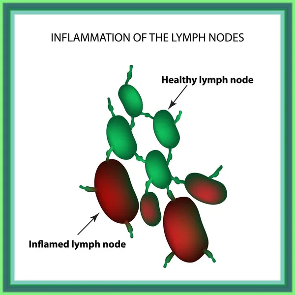
Inflammation Of The Lymph Nodes. Infographics. Vector Illustration On Isolated Background
Vector, 1.13MB, 5000 × 5000 eps
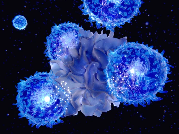
Interaction Between A Dendritic Cell And A T-lymphocyte. 3d-rendering. Dendritic Cells Are Antigen-presenting Cells Of The Immune System
Image, 6.52MB, 8000 × 6000 jpg


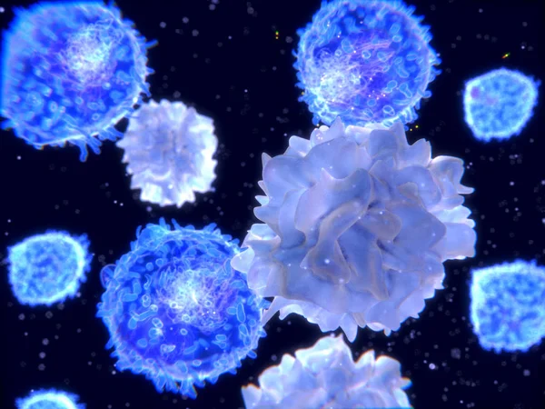
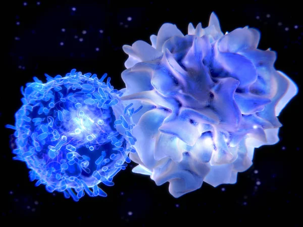
T-lymphocytes And Dendritic Cells, 3D-rendering; Dendritic Cells Are Antigen-presenting Cells Of The Immune System. They Process Antigen Material And Present It On The Cell Surface To The T-cells. Illustration
Image, 2.23MB, 4000 × 3000 jpg

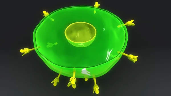
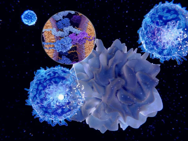
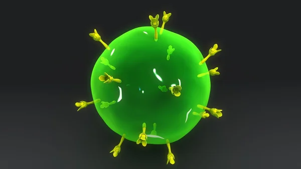
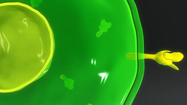
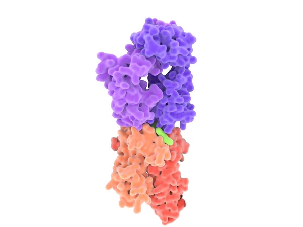
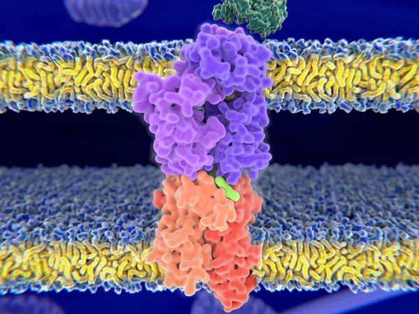
T-cell Receptor In Complex With The MHC Class II-peptide Complex. The Antigen (light Green) Is A Peptide From A Tumor Cell, Bacteria Or Virus. Different Stages Of The Interaction. 3D-Rendering. Illustration
Image, 7.31MB, 8000 × 6000 jpg
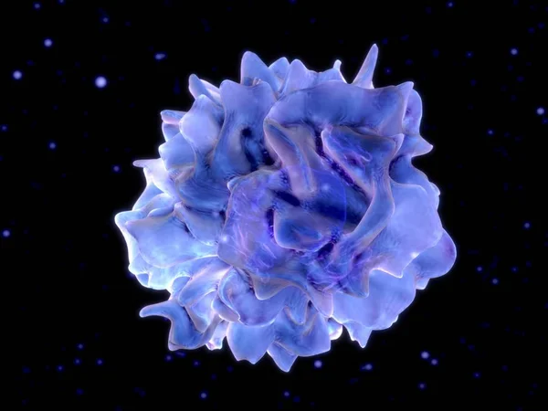
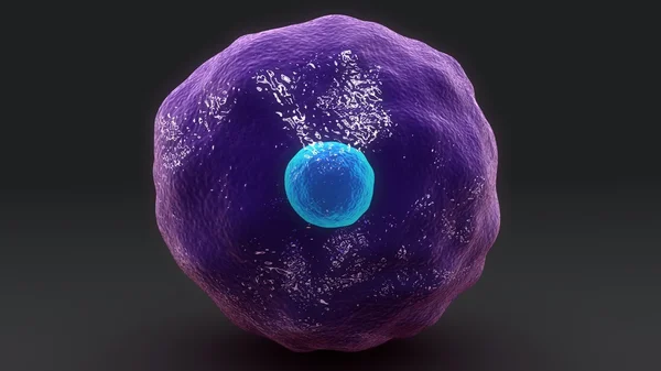
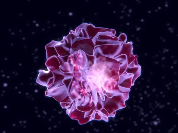
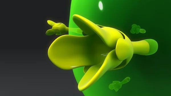
Dendritic Cell. 3D-rendering. Dendritic Cells Are Antigen-presenting Cells Of The Immune System. Illustration
Image, 5.07MB, 8000 × 6000 jpg
T-cell Receptor In Complex With The MHC Class II-peptide Complex. The Antigen (light Green) Is A Peptide From A Tumor Cell, Bacteria Or Virus. Different Stages Of The Interaction. 3D-Rendering. Illustration
Image, 2.17MB, 8000 × 6000 jpg
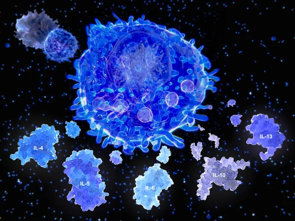
After Activation By An Antigen Presenting Cell, A T Helper Cell Segregates Several Cytokines.
Image, 13.65MB, 8000 × 6000 jpg
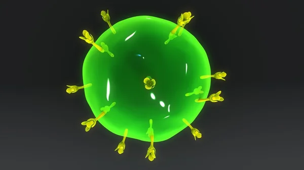
T-cell Receptor In Complex With The MHC Class II-peptide Complex. The Antigen (light Green) Is A Peptide From A Tumor Cell, Bacteria Or Virus. Different Stages Of The Interaction. 3D-Rendering. Illustration
Image, 9.21MB, 8000 × 6000 jpg
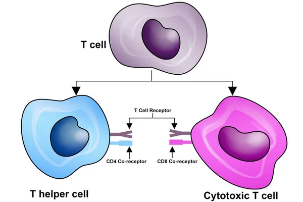
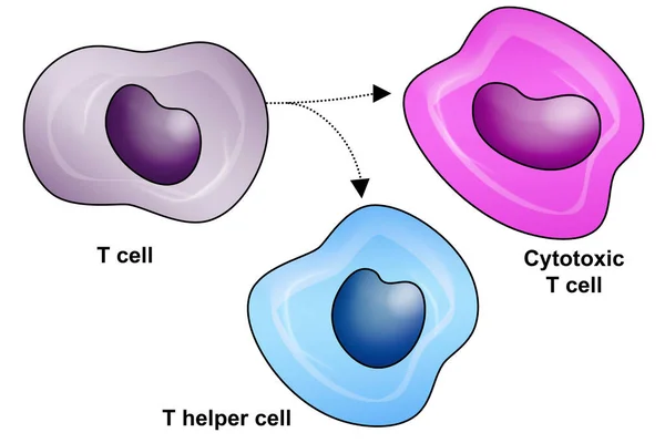
T Cell, Helper T Cell And Cytotoxic T Cell, CD Antigen Types, CD4 And CD8, 3d Rendering
Image, 3.44MB, 6000 × 4000 jpg
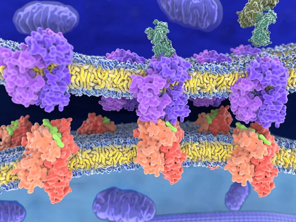
T-cell Receptor In Complex With The MHC Class II-peptide Complex. The Antigen (light Green) Is A Peptide From A Tumor Cell, Bacteria Or Virus. Complex Embedded In The Membranes. 3D-Rendering. Illustration
Image, 7.59MB, 8000 × 6000 jpg
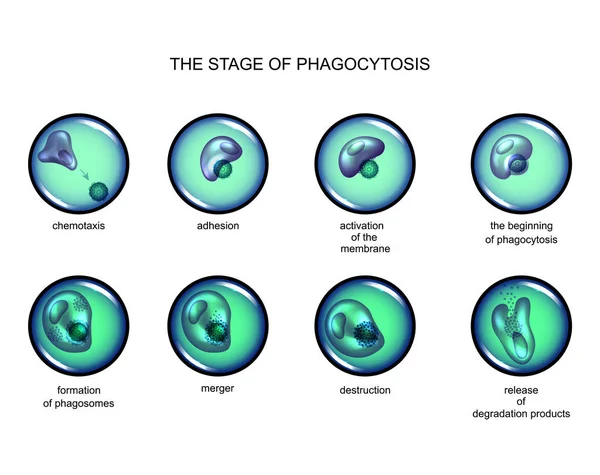
Phagocytosis. Leukocyte Absorbs The Virus. Infographics. Leukocyte Eats Bacteria. Vector Illustrations In Cartoon Style On Isolated Background
Vector, 6.91MB, 5000 × 5000 eps
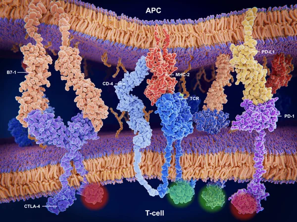
Interactions Of MHC-II With The T-cell Receptor And CD4 And B7-1 With CD-28 Activates T-cells While The Interactions Of P7-1 With CTLA-4 And PD-L1 With PD-1 Deactivates T-cells.
Image, 10.7MB, 8000 × 6000 jpg
Page 1 >> Next