
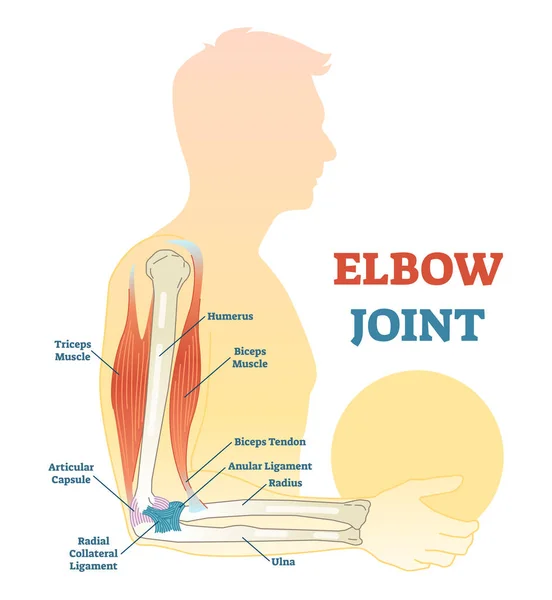
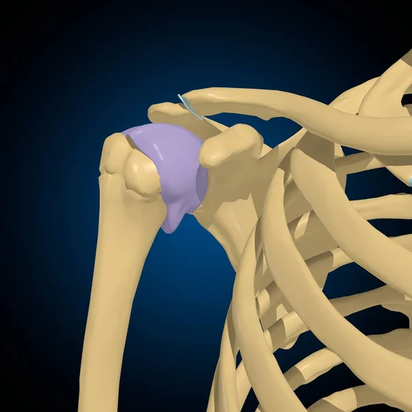
Stock image Articular Capsule
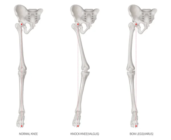
Alignment Types Disease Of Lower Half Limbs Or Leg Bone Problem- Normal- Knock Knee And Bowlegs Or Valgus And Varus Knee- 3D Medical Illustration-human Anatomy And Educational Concept White Background
Image, 6.62MB, 12000 × 9830 jpg
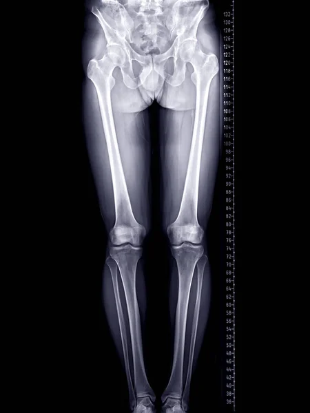
Scanogram Of Lower Limb Or X-ray Image Of Total Lower Extremity With Scale.
Image, 3.15MB, 3280 × 4375 jpg
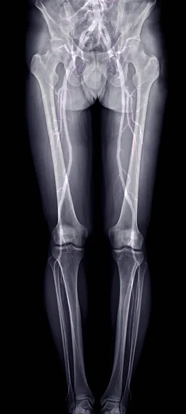
Scanogram Of Lower Limb Or X-ray Image With Merge CTA Femoral Run Off Showing Bone And Vessel Of Lower Limb.
Image, 2.19MB, 1936 × 4303 jpg
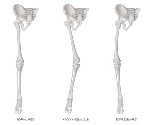
Types Disease Of Lower Half Limbs Or Leg Bone Problem- Normal- Knock Knee And Bowlegs Or Valgus And Varus Knee- 3D Medical Illustration- Human Anatomy And Educational Concept-Isolated White Background
Image, 6.31MB, 12000 × 9830 jpg
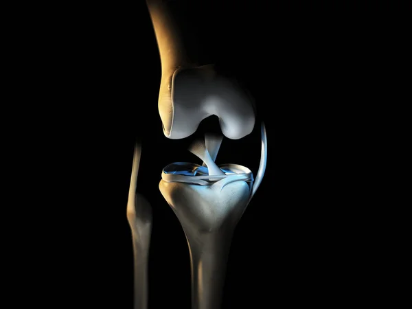
3D Illustration Showing Knee Joint With Ligaments, Meniscus, Articular Cartilage, Fibula And Tibia.
Image, 3.82MB, 6666 × 5000 jpg
Synovitis Icon. Trendy Modern Flat Linear Vector Synovitis Icon On White Background From Thin Line Diseases Collection, Editable Outline Stroke Vector Illustration
Vector, 0.6MB, 6944 × 6944 eps

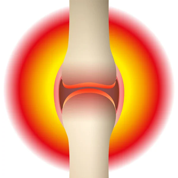
Joint Pain - Schematic Anatomical Graphic Of A Synovial Joint With Arthritis, Rheumatism, Gout, Osteoarthritis Or Inflammation. Isolated Vector Illustration On White Background.
Vector, 2.07MB, 8055 × 8055 eps

Synovitis Icon. Trendy Flat Vector Synovitis Icon On White Background From Diseases Collection, Vector Illustration Can Be Use For Web And Mobile, Eps10
Vector, 0.57MB, 6944 × 6944 eps

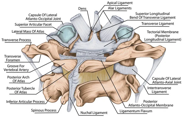
Ligaments And Joints Of The Cervical Vertebrae And The Occipital Bone. Back View. Vector Illustration
Vector, 2.91MB, 6010 × 4167 eps

3D Illustration Showing Knee Joint With Ligaments, Meniscus, Articular Cartilage, Fibula And Tibia.
Image, 5.22MB, 6666 × 5000 jpg
The Ligaments Of The Median Atlantoaxial Joint. Atlas And Axis Ligaments. Cervical Spine, Vertebral Morphology, First And Second Cervical Vertebra, Cervical Vertebrae, Atlas, Axis, Atlantoaxial Joint, Posterosuperior View
Image, 5.91MB, 5906 × 3830 jpg
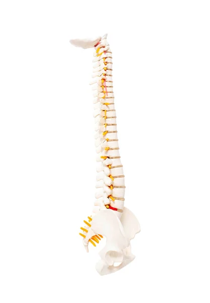
Mock Up Human Spine On A White Background. The Concept Of Segments And Divisions Of The Spine, The Structure And Anatomy Of The Bone Marrow, Nerves And Vertebrae
Image, 0.82MB, 2553 × 3674 jpg
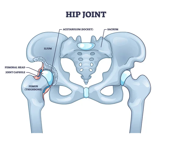
Hip Joint Structure With Anatomical Bone Parts Description Outline Concept
Vector, 8.5MB, 4500 × 3825 eps

Knee And Meniscus Anatomy Medical Vector Illustration Isolated On White Background Eps 10
Vector, 26.81MB, 5000 × 5000 eps


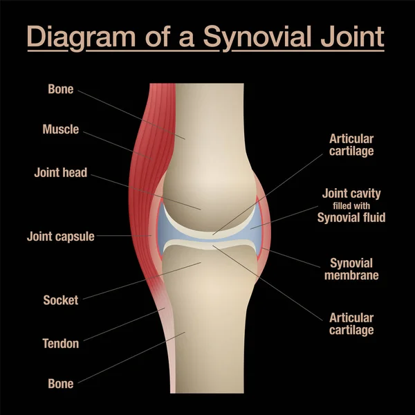
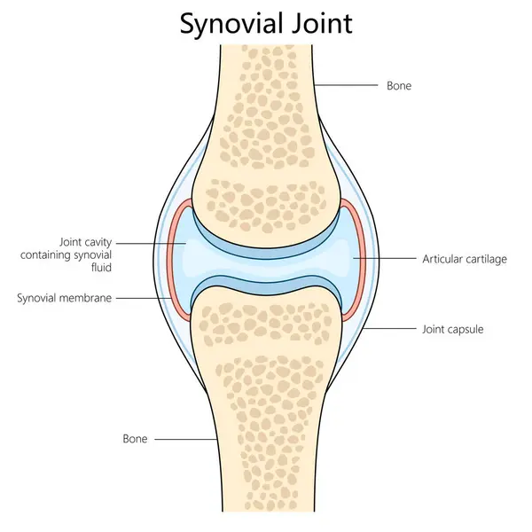
Synovial Joint Diagram. Labeled Anatomy Chart With Two Bones, Articular Cartilage, Joint Cavity, Synovial Fluid, Muscle And Tendon. Isolated Vector Illustration On Black.
Vector, 6.9MB, 8055 × 8055 eps
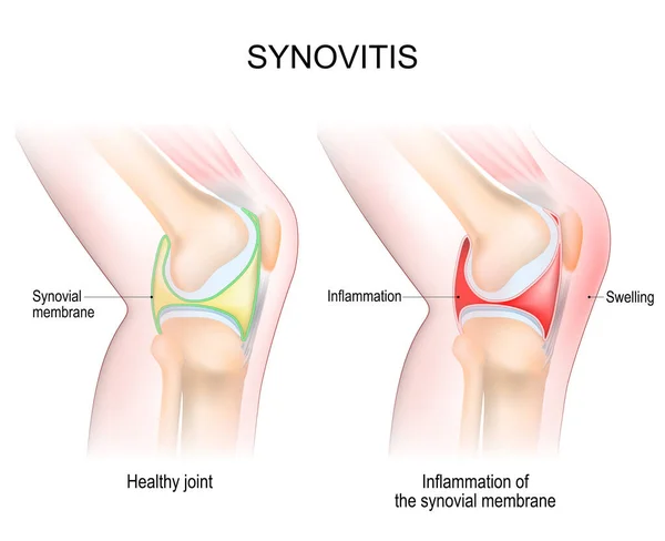
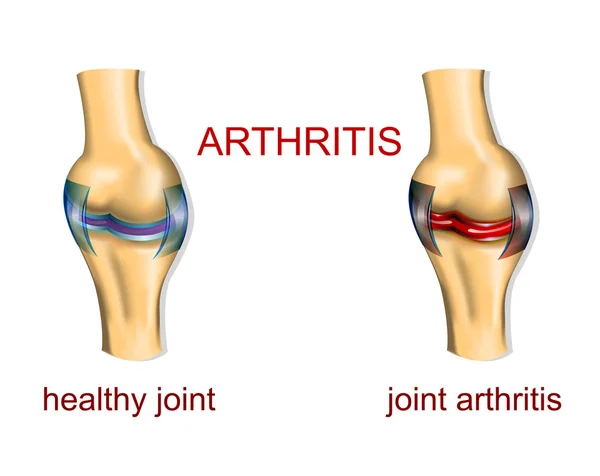
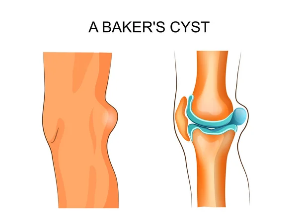
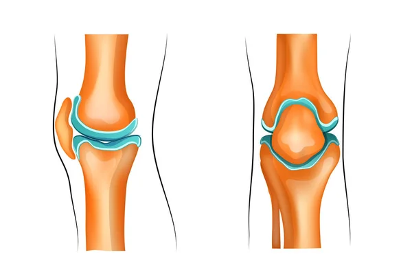
Synovitis Of A Knee. Close-up Of Normal Joint, And Knee With Inflammation Of The Synovial Membrane. Signs And Symptoms Of The Disease. Side View Of Human Knee Joint. Vector Illustration
Vector, 4.54MB, 5000 × 4070 eps
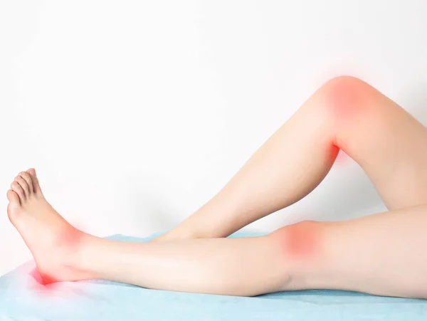
Female Legs On A White Background With Sore Reddened Knee Joints And Ankle Joints With Heels. Concept Of Disease And Treatment Of Arthritis And Joint Inflammation.
Image, 1.64MB, 4854 × 3648 jpg

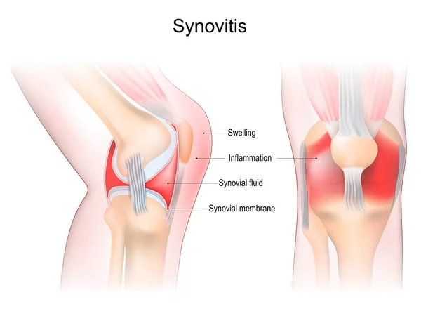
Synovitis Of A Knee. Close-up Of Joint With Inflammation Of The Synovial Membrane. Signs And Symptoms Of The Disease. Synovial Joint Anatomy. Frontal And Side View Of Human Knee Joint. Vector Illustration
Vector, 8.37MB, 5241 × 3930 eps
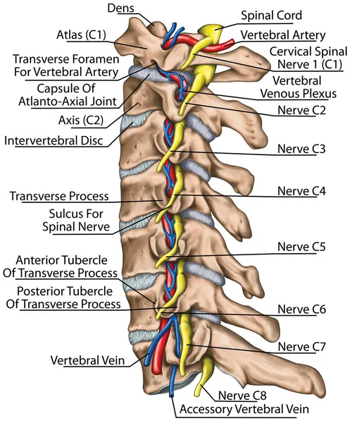
Cervical Spine With Both Vertebral Arteries In Transverse Foramen And The Emerging Spinal Nerves. Topographic Relationship Of The Spinal Nerve And Vertebral Artery. Lateral View.
Image, 6.51MB, 4926 × 5906 jpg

Synovial Joint Chart. Labeled Anatomy Infographic With Two Bones, Articular Cartilage, Joint Cavity, Synovial Fluid, Muscle And Tendon. Isolated Vector Illustration On White.
Vector, 8.73MB, 8055 × 8055 eps

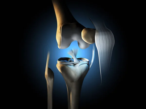
3D Illustration Showing Knee Joint With Ligaments, Meniscus, Articular Cartilage, Fibula And Tibia.
Image, 4.95MB, 6666 × 5000 jpg
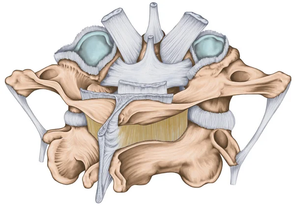
The Ligaments Of The Median Atlantoaxial Joint. Atlas And Axis Ligaments. Cervical Spine, Vertebral Morphology, First And Second Cervical Vertebra, Cervical Vertebrae, Atlas, Axis, Atlantoaxial Joint, Posterosuperior View
Image, 5.87MB, 5906 × 4183 jpg

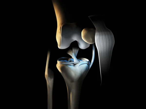
Accurate Medically Illustration Showing Knee Joint With Ligaments, Meniscus, Articular Cartilage, Femur And Tibia.
Image, 5.34MB, 6666 × 5000 jpg
3D Illustration Showing Knee Joint With Ligaments, Meniscus, Articular Cartilage, Fibula And Tibia.
Image, 4.39MB, 6666 × 5000 jpg
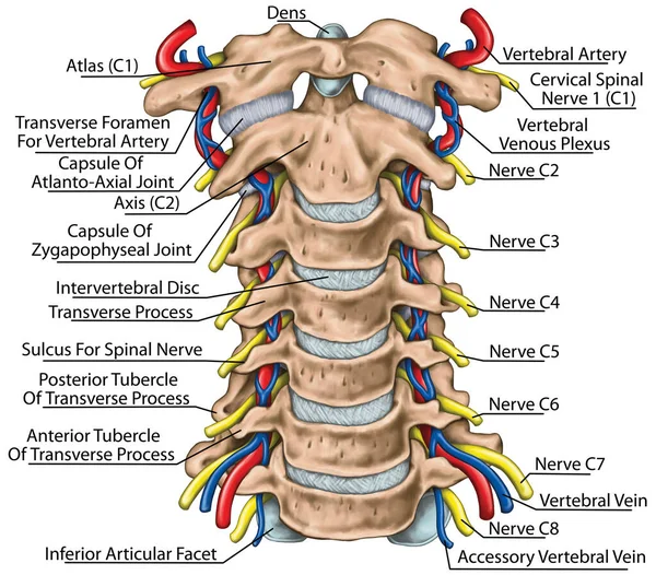
Cervical Spine With Both Vertebral Arteries In Transverse Foramen And The Emerging Spinal Nerves. Topographic Relationship Of The Spinal Nerve And Vertebral Artery. Anterior View.
Image, 7.41MB, 5906 × 5180 jpg

Accurate Medically Illustration Showing Knee Joint With Ligaments, Meniscus, Articular Cartilage, Femur And Tibia.
Image, 3.5MB, 6828 × 5000 jpg
Human Synovial Joint Structure Diagram Hand Drawn Schematic Raster Illustration. Medical Science Educational Illustration
Image, 3.67MB, 6000 × 6000 jpg
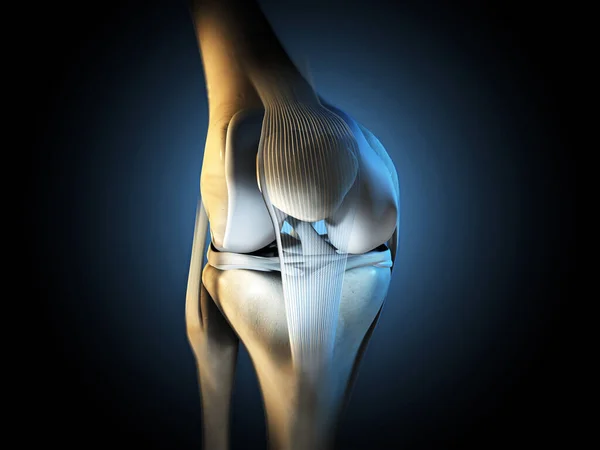
3D Illustration Showing Knee Joint With Ligaments, Meniscus, Articular Cartilage, Fibula And Tibia.
Image, 5.83MB, 6666 × 5000 jpg

Arthroscopy Of The Knee Joint. Surgery Or Diagnosis Using A Puncture With An Arthroscope. Medical Vector Illustration.
Vector, 9.29MB, 2000 × 2000 eps


Knee Anatomy. Side And Front View. Cross Section Of The Joint Showing The Main Parts: Femur, Fibula, Articular Capsule, Menisci, Muscles And Ligaments. Vector Illustration
Vector, 3.91MB, 4444 × 3973 eps
Page 1 >> Next