
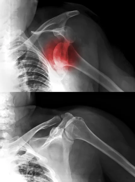
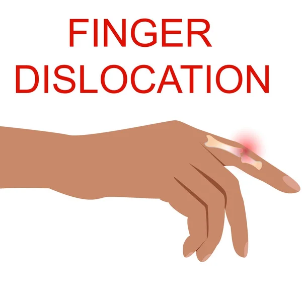
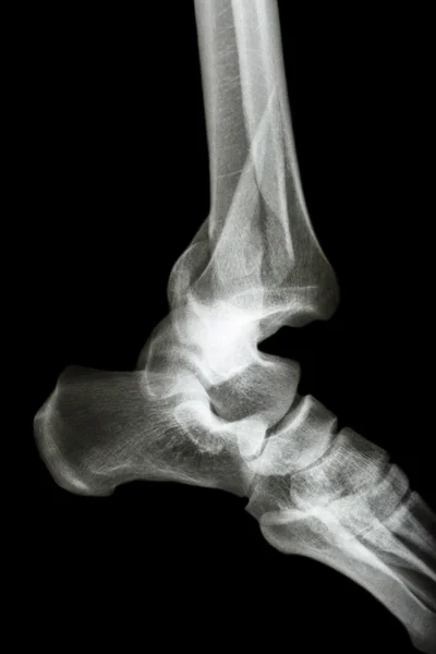
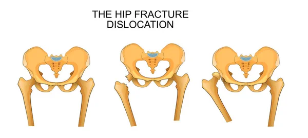
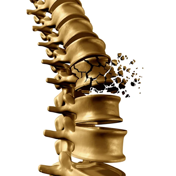
Stock image Bone Dislocation


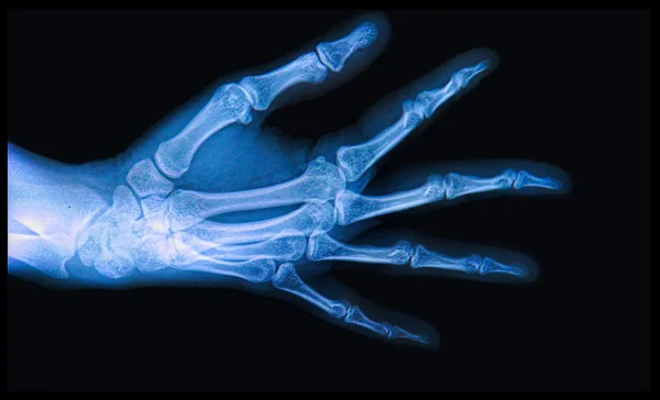
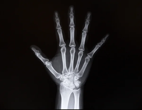
A Mans Wrist Hurts. Traumatologist Examines Hand. Wrist Pain As A Sign Of Tunnel Syndrome Or Sprain, Tendon Degeneration, Arthritis
Image, 6.96MB, 5115 × 3332 jpg

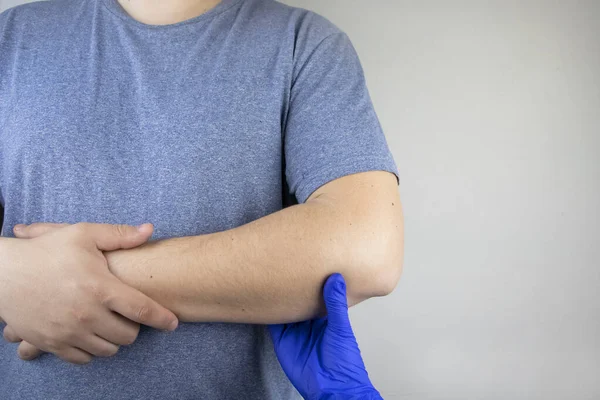
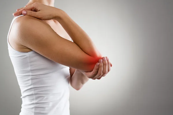
A Man Suffers From Elbow Pain. Damaged Elbow Joint, Bone Fracture, Or Sprain. Hand Injury And Flexion Pain Concept
Image, 8.78MB, 6000 × 4000 jpg
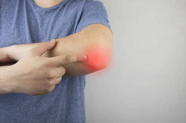
A Man Suffers From Elbow Pain. Damaged Elbow Joint, Bone Fracture, Or Sprain. Hand Injury And Flexion Pain Concept
Image, 12.44MB, 5864 × 3909 jpg
A Man Suffers From Elbow Pain. Damaged Elbow Joint, Bone Fracture, Or Sprain. Hand Injury And Flexion Pain Concept
Image, 8.36MB, 5640 × 3736 jpg

Types Of Shoulder Dislocation, Normal, Anterior, Posterior, Inferior Dislocations, Positions Of Humeral Head Diagram Hand Drawn Schematic Vector Illustration. Medical Science Educational Illustration
Vector, 0.65MB, 4000 × 4000 eps

Types Of Shoulder Dislocation, Normal, Anterior, Posterior, Inferior Dislocations, Positions Of Humeral Head Diagram Hand Drawn Schematic Raster Illustration. Medical Science Educational Illustration
Image, 2.24MB, 6000 × 6000 jpg
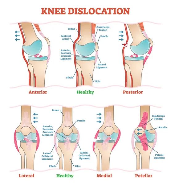
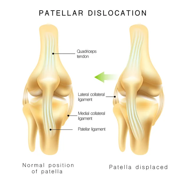
Knee Dislocations - Medical Vector Illustration Diagrams. Anatomical Knee Injury Types Scheme.
Vector, 7.05MB, 4528 × 4891 eps

Shoulder Dislocation And Humerus Bone Trauma Explanation Outline Diagram
Vector, 5.87MB, 5000 × 3750 eps

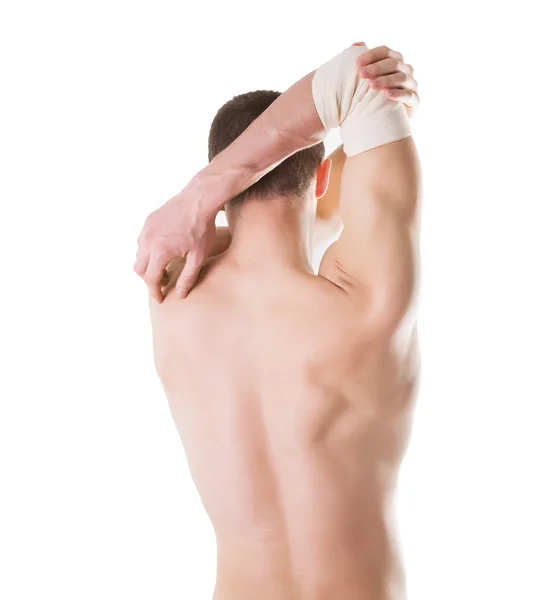
Elastic Bandage On A Hand Of The Man With Ideal Skin. Isolated On A White Background
Image, 2.24MB, 2404 × 2603 jpg
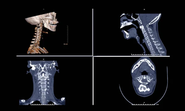
Comparison Of CT C-Spine Or Cervical Spine 3D Rendering Image , Sagittal ,Corona And Axiall View In Patient Trauma Head Injury.
Image, 2.06MB, 5008 × 3008 jpg


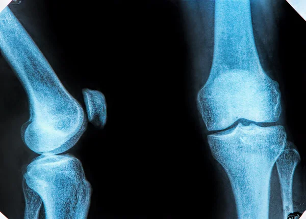
Human Skeleton And Damajed Joints . Xray View. Medically Accurate 3D Illustration
Image, 3.66MB, 3128 × 4000 jpg
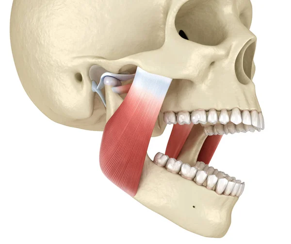
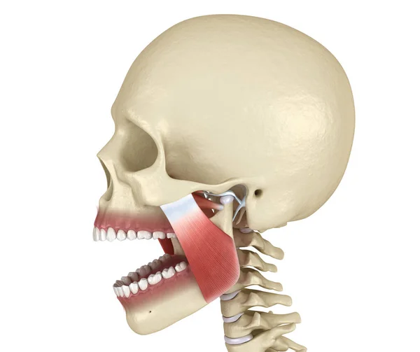
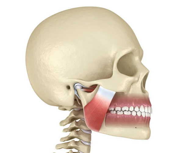
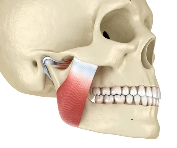
TMJ: The Temporomandibular Joints And Muscles. Medically Accurate 3D Illustration.
Image, 7.3MB, 5000 × 4200 jpg
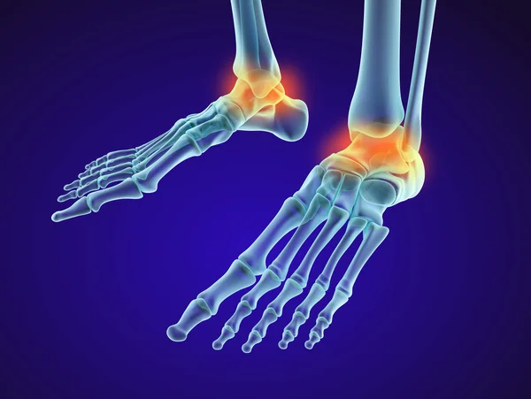
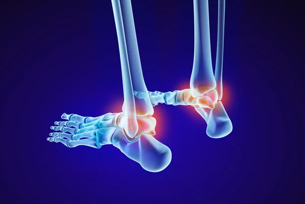
Skeletal Foot - Injuryd Talus Bone. Xray View. Medically Accurate 3D Illustration
Image, 4.43MB, 5322 × 4000 jpg

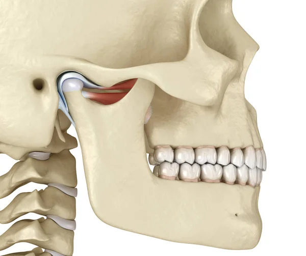
TMJ: The Temporomandibular Joints. Healthy Occlusion Anatomy. Medically Accurate 3D Illustration Of Human Teeth And Dentures Concept
Image, 8.07MB, 5000 × 4200 jpg


TMJ: The Temporomandibular Joints And Muscles. Medically Accurate 3D Illustration.
Image, 8.22MB, 6000 × 5040 jpg
TMJ: The Temporomandibular Joints. Healthy Occlusion Anatomy. Medically Accurate 3D Illustration Of Human Teeth And Dentures Concept
Image, 8.49MB, 5000 × 4200 jpg
TMJ: The Temporomandibular Joints And Muscles. Medically Accurate 3D Illustration.
Image, 8.95MB, 6000 × 5040 jpg
TMJ: The Temporomandibular Joints And Muscles. Medically Accurate 3D Illustration.
Image, 8.15MB, 5000 × 4200 jpg
Skeletal Foot - Injuryd Talus Bone. Xray View. Medically Accurate 3D Illustration
Image, 3.69MB, 5322 × 3568 jpg
Page 1 >> Next