
Stock image Brain Plaques
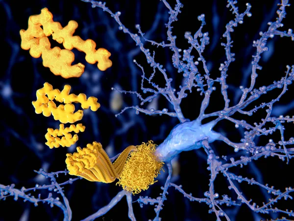
The Beta Amyloid Peptid, Amyloid Plaques Growing On A Neuron. It Consists Of About 30 Amino Acids And Aggregates To Amyloid Plaques, That May Damage And Kill Neurons. Illustration
Image, 4.49MB, 8000 × 6000 jpg


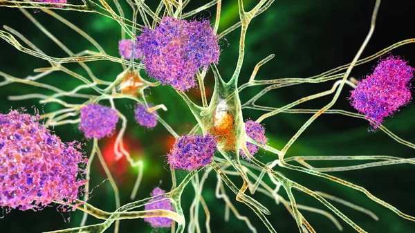
Neurons In Dementia. Alzheimer's Disease, Huntington's Disease. 3D Illustration Showing Amyloid Plaques In Brain Tissue, Neurofibrillary Tangles And Distruction Of Neuronal Networks
Image, 10.46MB, 6000 × 4000 jpg
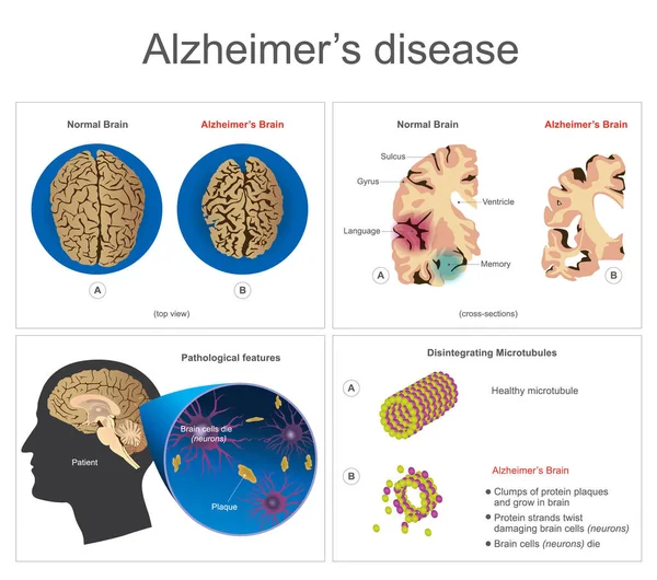
Alzheimers Disease. Brain Cells Die, Neuron Diseased, Certain Areas Of Brain Shrink Memory Loss Or Changes In Memory For People At Risk Could Affect Younger People. Info Graphic Vector.
Vector, 4.02MB, 6000 × 5306 eps

Brain In Severe Brain Disease, Dementia, Alzheimer, Chorea Huntington -- 3D Rendering
Image, 19MB, 8000 × 4500 jpg

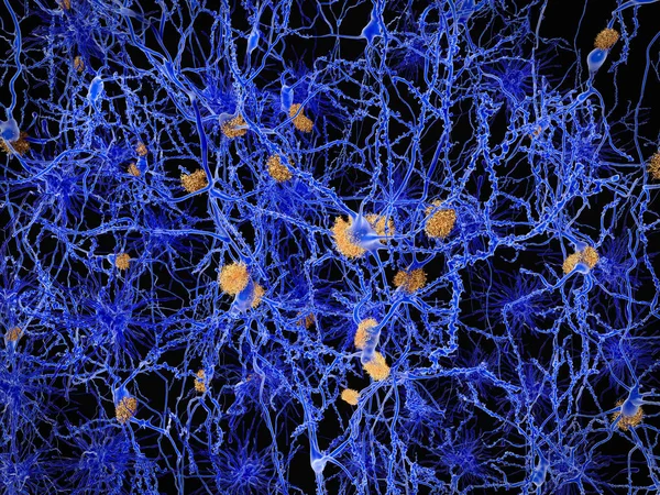
Neurons In Dementia. Alzheimer's Disease, Huntington's Disease, Other Types Of Dementia. Degradation Of Neuronal Networks, Formation Of Amyloid Plaques, 3D Illustration
Image, 10.38MB, 6000 × 4000 jpg
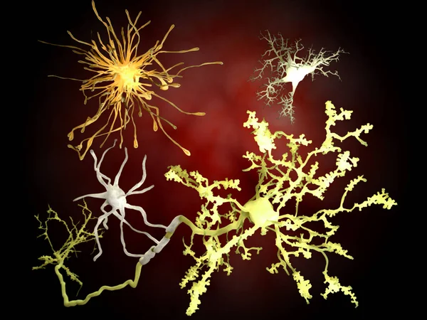
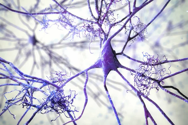
The Main Cells Of The Brain: Yellow: Neurons; Orange: Astrocytes; Grey: Oligodendrocytes; White: Microglia, Brain Cells. Illustration
Image, 4.91MB, 4000 × 3000 jpg

Neurons In Alzheimer's Disease. 3D Illustration Showing Amyloid Plaques In Brain Tissue, Neurofibrillary Tangles And Distruction Of Neuronal Networks
Image, 12.01MB, 7200 × 4050 jpg


Alzheimer Disease, Neuron Network With Amyloid Plaques. Illustration
Image, 28.11MB, 8000 × 6000 jpg
Neurons In Alzheimer's Disease. 3D Illustration Showing Amyloid Plaques In Brain Tissue, Neurofibrillary Tangles And Distruction Of Neuronal Networks
Image, 8.51MB, 7200 × 4050 jpg

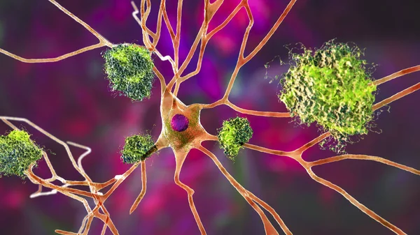
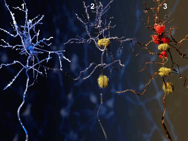
3 Phases Of The Alzheimer Disease. 1. Healthy Neuron. 2. Neuron With Amyloid Plaques (yellow). 3. Dead Neuron Being Digested By Microglia Cells (red). Illustration
Image, 6.76MB, 8000 × 6000 jpg
3 Phases Of The Alzheimer Disease. 1. Healthy Neuron. 2. Neuron With Amyloid Plaques (yellow). 3. Dead Neuron Being Digested By Microglia Cells (red). Illustration
Image, 6.76MB, 8000 × 6000 jpg

Neurons In Alzheimer's Disease. 3D Illustration Showing Amyloid Plaques In Brain Tissue, Neurofibrillary Tangles And Distruction Of Neuronal Networks
Image, 9.65MB, 7200 × 4050 jpg
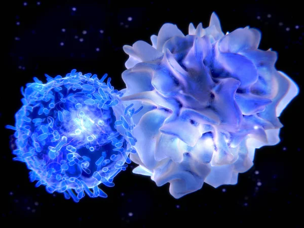
Interaction Between A Dendritic Cell And A T-lymphocyte. 3d-rendering. Dendritic Cells Are Antigen-presenting Cells Of The Immune System
Image, 6.52MB, 8000 × 6000 jpg

Neurons In Alzheimer's Disease. 3D Illustration Showing Amyloid Plaques In Brain Tissue, Neurofibrillary Tangles And Distruction Of Neuronal Networks
Image, 8.23MB, 7200 × 4050 jpg

Alzheimer's Disease. Disease Associated With Amyloid Plaques, Neurofibrillary Tangles, And Loss Of Neuronal Connections In The Brain. Human Brain With Alzheimer's Disease. Close-up Of Neurons With Amyloid Plaques. Vector Illustration
Vector, 14.34MB, 4444 × 4444 eps
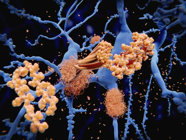
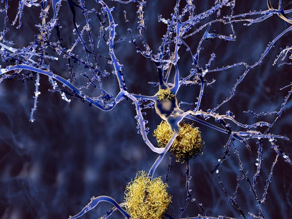
After Being Cleaved By The Gamma And Beta Secretases The Amyloid Beta Peptide, Which Has About 40 Amino Acid Residues, Leaves The Membrane, Changes Shape And Aggregates Into Long Fibrils. These Fibrils Form Dense Plaques On Nerve Cells, Which Are In
Image, 9.72MB, 8000 × 6000 jpg
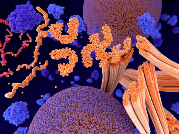
Pathological Phosphorylation (yellow) Of Tau Proteins (red-orange) Leads To Disintegration Of Microtubuli In The Neuron Axon An Aggregation Of The Tau Proteins. The Transport Of Synaptic Vesicles (orange-violet Spheres) Is Interrupted.
Image, 9.48MB, 8000 × 6000 jpg

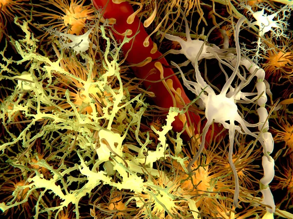
The Main Cells Of The Brain: Yellow: Neurons; Orange: Astrocytes; Grey: Oligodendrocytes; White: Microglia, Brain Cells. Illustration
Image, 9.17MB, 4000 × 3000 jpg
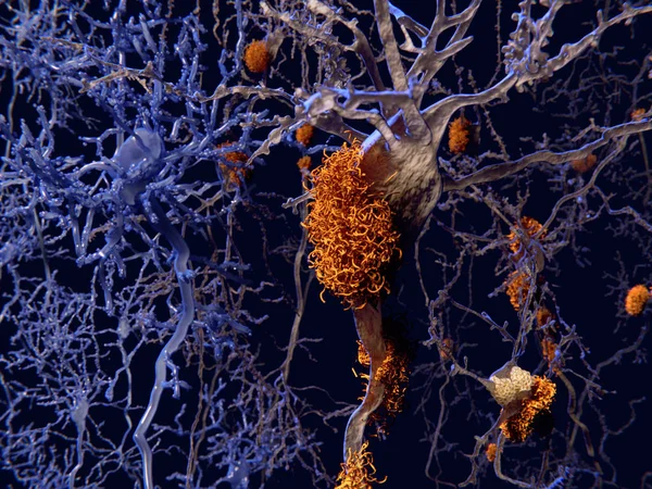
Neurons In Dementia. Alzheimer's Disease, Huntington's Disease, Other Types Of Dementia. Degradation Of Neuronal Networks, Formation Of Amyloid Plaques, 3D Illustration
Image, 6.8MB, 6000 × 4000 jpg
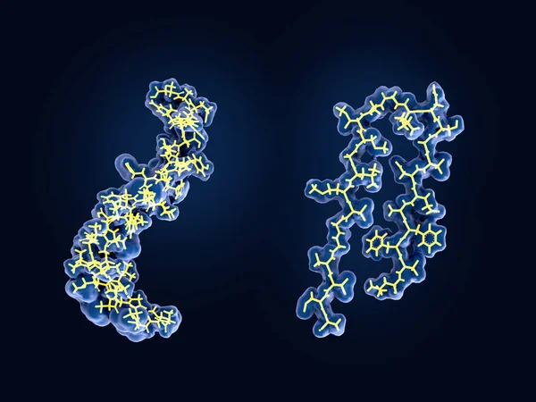
After Being Cleaved By The Gamma And Beta Secretases The Amyloid Beta Peptide, Which Has About 40 Amino Acid Residues, Leaves The Membrane, Changes Shape And Aggregates Into Long Fibrils. These Fibrils Form Dense Plaques On Nerve Cells.
Image, 2.61MB, 8000 × 6000 jpg

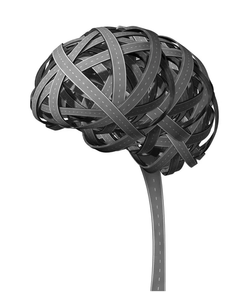
Alzheimer Disease As A Cognitive Decline As A Degenerative Dementia Brain Illness Resulting In Memory Loss As A Neurology Symbol For Aging Of The Mind.
Image, 9.61MB, 6277 × 4421 jpg

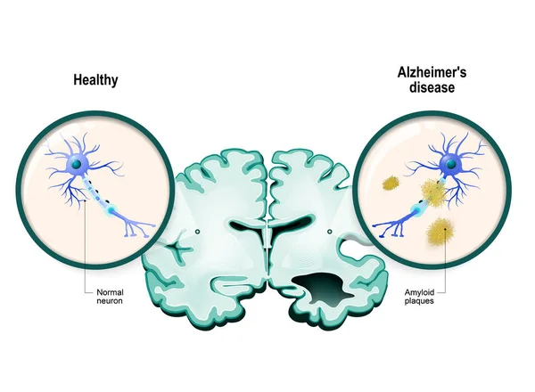
Alzheimer's Disease. Comparison Of Neurons In A Healthy Brain And Nerve Cells In Neurodegenerative Disease. Dementia. Close-up Of Neurons With Neurofibrillary Tangles And Amyloid Plaques. Vector Illustration
Vector, 11.61MB, 5000 × 3472 eps

Alzheimer's Disease. Neurodegeneration. Cross Section Of Normal And Alzheimer Brain, With Atrophy Of The Cerebral Cortex, Enlarged Ventricles And Hippocampus. Close-up Of Neurons With Neurofibrillary Tangles And Amyloid Plaques. Vector Illustration
Vector, 13.59MB, 5000 × 3572 eps
Neurons In Alzheimer's Disease. 3D Illustration Showing Amyloid Plaques In Brain Tissue, Neurofibrillary Tangles And Distruction Of Neuronal Networks
Image, 10.2MB, 7200 × 4050 jpg
Page 1 >> Next