
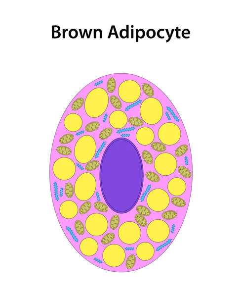
Stock image Brown Adipocyte
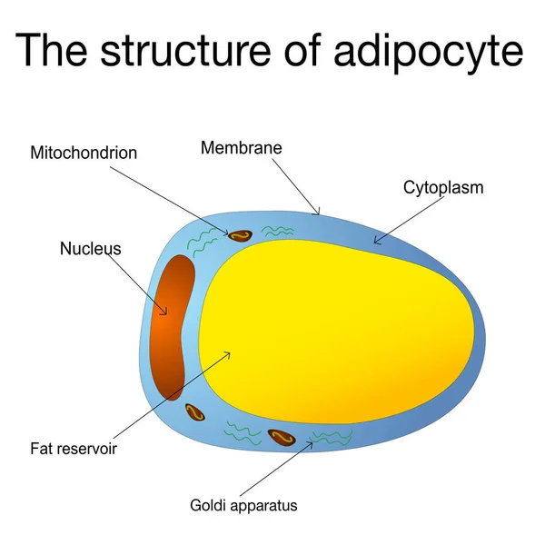
Adipocytes, Lipocytes And Fat Cells. Illustration Depicting Structure White Adipose Cells
Vector, 0.14MB, 3000 × 3000 eps
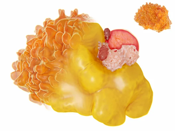
Structure Of A Fat Cell. 3D Rendering. Fat Cells Contain A Large Lipid Droplet (yellow), A Nucleus (red) And Cell Organelles Located In The Periphery. Illustration
Image, 2.77MB, 8000 × 6000 jpg

Adipocytes, Lipocytes And Fat Cells. Fat Cell Structure Vector Illustration.
Vector, 5.45MB, 4909 × 4909 eps
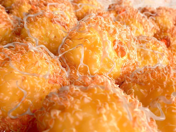
Fat Cells, 3d Rendering. White Fat Cells Contain A Large Lipid Droplet (yellow) And A Nucleus (red) Located In The Periphery. Illustration
Image, 6.78MB, 8000 × 6000 jpg

Vector Set Of Brown, Beige And White Fat Cells. Illustration Of Adipose Tissue
Vector, 22.69MB, 9539 × 4494 eps

Hormones Produced By Adipocytes. Brown Adipose Tissue (BAT) And White Adipose Tissue (WAT). Cytokines, Resistin, Leptin, And Estrogen. Human Endocrine System. Vector Illustration For Medical, Education And Science Use.
Vector, 1.58MB, 4444 × 4444 eps


Fat Cell Types As Adipocyte Division In Brown, Beige Or White Outline Diagram
Vector, 5.97MB, 5300 × 3347 eps

Low Magnification Light Micrograph Showing Interscapular Brown Adipose Tissue. Brown Fat Is Very Developed In Rodents And Hibernating Animals. Brown Adipocytes Show A Spongy Eosinophilic Aspect Because They Stored Fat As Small Lipid Droplets.
Image, 12.57MB, 3840 × 3072 jpg

Very Low Magnification Light Micrograph Showing Interscapular Brown Adipose Tissue. Brown Fat Is Very Developed In Rodents And Hibernating Animals. Brown Adipocytes Show A Spongy Eosinophilic Aspect Because They Stored Fat As Small Lipid Droplets.
Image, 12.95MB, 3840 × 3072 jpg
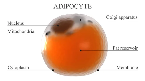
Adipocyte Structure. 3d Rendered Illustration Showing The Names Of The Main Elements Of A White Fat Cell
Image, 1.04MB, 3840 × 2160 jpg
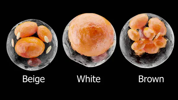
Brown, Beige And White Fat Cells, Adipocyte And Lipocyte, Cholesterol In A Cells, Adipose Tissue, Lipid Droplet, Fat In Body, Obesity, Types Of Lipocytes Dermis And Hypodermis, Nucleus, 3d Render
Image, 6.65MB, 8000 × 4500 jpg

Very Low Magnification Light Micrograph Showing Interscapular Brown Adipose Tissue. Brown Fat Is Very Developed In Rodents And Hibernating Animals. Brown Adipocytes Show A Spongy Eosinophilic Aspect Because They Stored Fat As Small Lipid Droplets.
Image, 13.28MB, 3840 × 3072 jpg
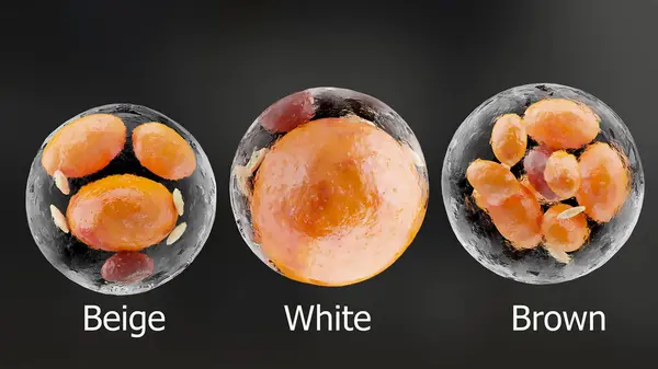
Brown, Beige And White Fat Cells, Adipocyte And Lipocyte, Cholesterol In A Cells, Adipose Tissue, Lipid Droplet, Fat In Body, Obesity, Types Of Lipocytes Dermis And Hypodermis, Nucleus, 3d Render
Image, 6.25MB, 8000 × 4500 jpg

3D Isometric Flat Vector Conceptual Illustration Of Adipoce Tissue, Pathology Of Obesity
Vector, 1.87MB, 6000 × 4000 eps

Page 1 >> Next