
Stock image Cajal

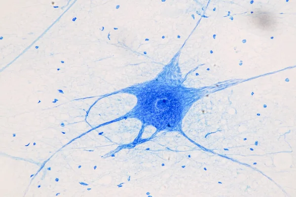

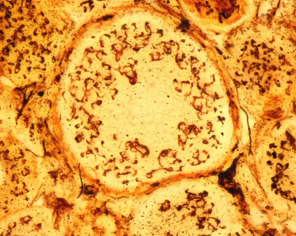
High Magnification Micrograph Of Pseudounipolar Neurons Of A Dorsal Root Ganglion Stained With The Cajal's Formol-uranium Silver Method That Demonstrates The Golgi Apparatus. It Appears As A Brown Network Located In The Neuron Cell Body Around The Nu
Image, 5.13MB, 3840 × 3072 jpg

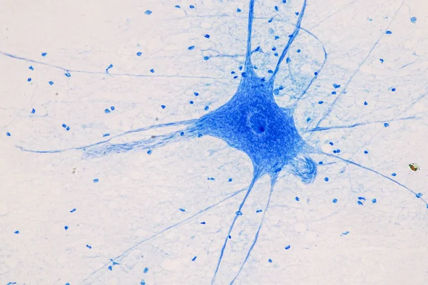


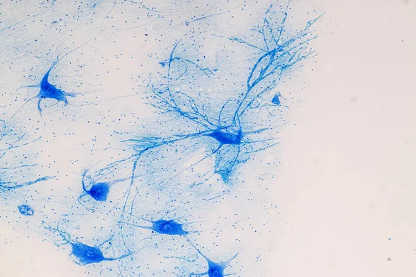
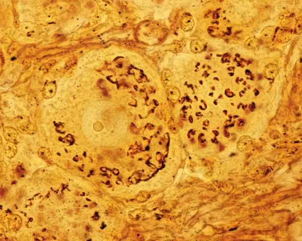
Light Micrograph Showing The Golgi Apparatus In Neurons Of Dorsal Root Ganglion. Cajal's Formol-uranium Silver Method. The Golgi Apparatus Is Distributed Throughout The Cell Body Cytoplasm Around The Nucleus.
Image, 15.85MB, 3840 × 3072 jpg

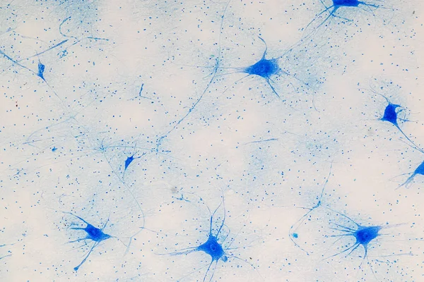
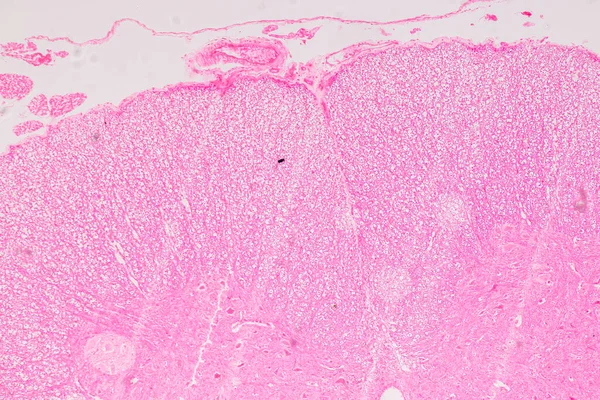
Low Magnification Light Microscope Micrograph Of A Dorsal Root Ganglion Stained With The Cajal's Formol-uranium Silver Method That Demonstrates The Golgi Apparatus In The Dorsal Root Ganglion Neurons. The Golgi Apparatus Is Distributed Throughout The
Image, 17.72MB, 3840 × 3072 jpg

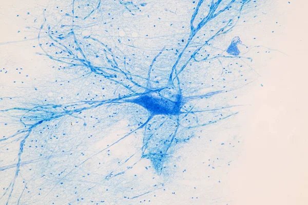
High Magnification Micrograph Of Pseudounipolar Neurons Of A Dorsal Root Ganglion Stained With The Cajal's Formol-uranium Silver Method That Demonstrates The Golgi Apparatus. It Appears As A Brown Network Located In The Neuron Cell Body Around The Nu
Image, 9.6MB, 3840 × 3072 jpg

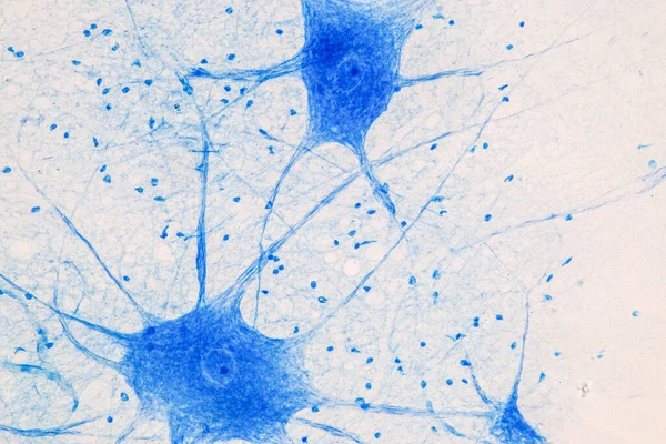
High Magnification Micrograph Of Pseudounipolar Neurons Of A Dorsal Root Ganglion Stained With The Cajal's Formol-uranium Silver Method That Demonstrates The Golgi Apparatus. It Appears As A Brown Network Located In The Neuron Cell Body Around The Nu
Image, 9.14MB, 3840 × 3072 jpg


High Magnification Micrograph Of Pseudounipolar Neurons Of A Dorsal Root Ganglion Stained With The Cajal's Formol-uranium Silver Method That Demonstrates The Golgi Apparatus. It Appears As A Brown Network Located In The Neuron Cell Body Around The Nu
Image, 10.8MB, 3840 × 3072 jpg
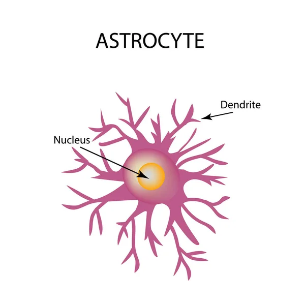
Astrocyte Structure. Nerve Cell. Infographics. Vector Illustration On Isolated Background.
Vector, 0.84MB, 5000 × 5000 eps


High Magnification Micrograph Of Pseudounipolar Neurons Of A Dorsal Root Ganglion Stained With The Cajal's Formol-uranium Silver Method That Demonstrates The Golgi Apparatus. It Appears As A Brown Network Located In The Neuron Cell Body Around The Nu
Image, 9.79MB, 3840 × 3072 jpg

High Magnification Micrograph Of Pseudounipolar Neurons Of A Dorsal Root Ganglion Stained With The Cajal's Formol-uranium Silver Method That Demonstrates The Golgi Apparatus. It Appears As A Brown Network Located In The Neuron Cell Body Around The Nu
Image, 9.43MB, 3840 × 3072 jpg


Madrid, Spain. Monument To Santiago Ramon Y Cajal, Spanish Neuroscientist, Specialized In Neuroanatomy And Central Nervous System, In Parque El Retiro
Image, 18.54MB, 5794 × 3863 jpg


High Magnification Micrograph Of Pseudounipolar Neurons Of A Dorsal Root Ganglion Stained With The Cajal's Formol-uranium Silver Method That Demonstrates The Golgi Apparatus. It Appears As A Brown Network Located In The Neuron Cell Body Around The Nu
Image, 10.98MB, 3840 × 3072 jpg
High Magnification Micrograph Of Pseudounipolar Neurons Of A Dorsal Root Ganglion Stained With The Cajal's Formol-uranium Silver Method That Demonstrates The Golgi Apparatus. It Appears As A Brown Network Located In The Neuron Cell Body Around The Nu
Image, 8.79MB, 3840 × 3072 jpg
High Magnification Micrograph Of Pseudounipolar Neurons Of A Dorsal Root Ganglion Stained With The Cajal's Formol-uranium Silver Method That Demonstrates The Golgi Apparatus. It Appears As A Brown Network Located In The Neuron Cell Body Around The Nu
Image, 9.35MB, 3840 × 3072 jpg

High Magnification Micrograph Of Pseudounipolar Neurons Of A Dorsal Root Ganglion Stained With The Cajal's Formol-uranium Silver Method That Demonstrates The Golgi Apparatus. It Appears As A Brown Network Located In The Neuron Cell Body Around The Nu
Image, 5.39MB, 3840 × 3072 jpg


Light Micrograph Showing The Golgi Apparatus In Neurons Of Dorsal Root Ganglion. Cajal's Formol-uranium Silver Method. The Golgi Apparatus Is Distributed Throughout The Cell Body Cytoplasm Around The Nucleus.
Image, 9.83MB, 3840 × 3072 jpg

Neuron Network, Pyramidal Neurons Of The Cortex In The Style Of Ramon Y Cajals Drawings. Illustration
Image, 16.89MB, 8000 × 6000 jpg

High Magnification Micrograph Of Pseudounipolar Neurons Of A Dorsal Root Ganglion Stained With The Cajal's Formol-uranium Silver Method That Demonstrates The Golgi Apparatus. It Appears As A Brown Network Located In The Neuron Cell Body Around The Nu
Image, 8.53MB, 3840 × 3072 jpg
Page 1 >> Next