
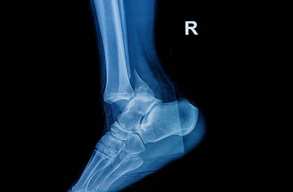
Stock image Calcaneus Fracture
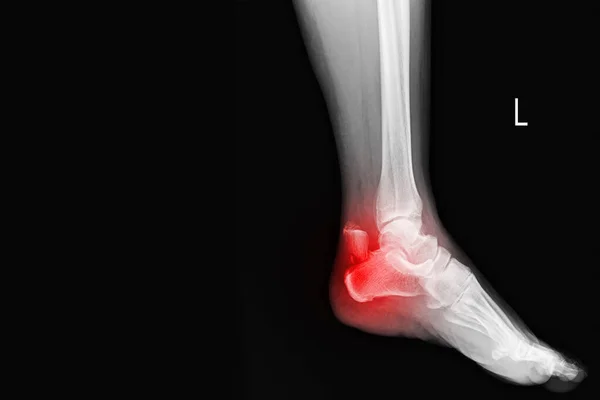
Film Ankle X-ray Radiograph Showing Heel Bone Broken On Red Mark(close Fracture Calcaneus) . Medical Technology And Healthcare Concept
Image, 4.41MB, 6000 × 4000 jpg


Heel Bone Fracture. Violation Of Integrity As A Result Of Injury. A Hand-drawn Diagram Of This Injury. Black And White Illustration For Different Uses.
Vector, 0.28MB, 5780 × 3214 eps

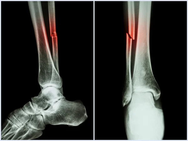
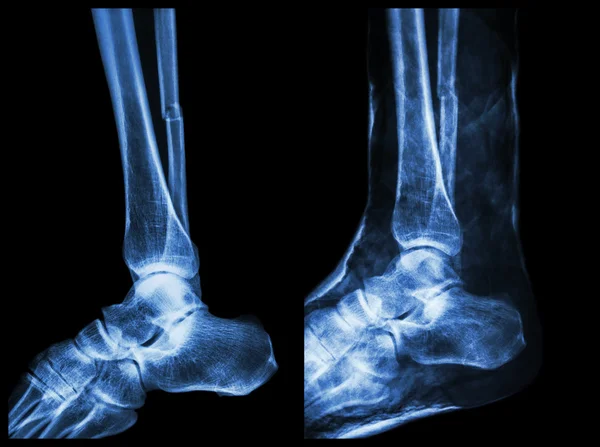
Fracture Shaft Of Fibula Bone ( Leg Bone ) . X-ray Of Leg ( 2 Position : Side And Front View )
Image, 11.03MB, 7032 × 5264 jpg

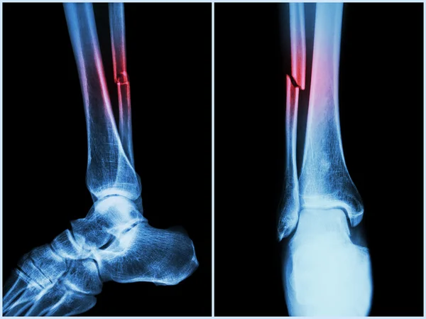
Fracture Shaft Of Fibula Bone ( Leg Bone ) . X-ray Of Leg ( 2 Position : Side And Front View )
Image, 10.87MB, 7032 × 5264 jpg
Fracture Shaft Of Fibula Bone ( Leg Bone ) . X-ray Of Leg ( 2 Position : Side And Front View )
Image, 8.72MB, 7032 × 5264 jpg
Fracture Shaft Of Fibula Bone ( Leg Bone ) . X-ray Of Leg ( 2 Position : Side And Front View )
Image, 9.02MB, 7032 × 5264 jpg

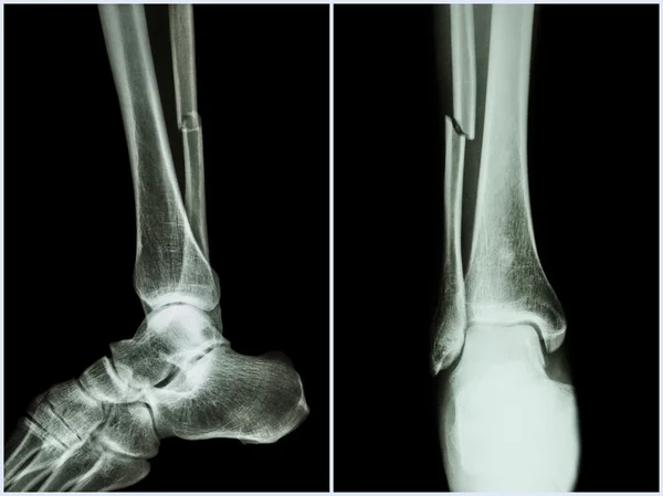
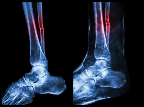
Left Image : Fracture Shaft Of Fibula (calf Bone) , Right Image : It Was Splinted With Plaster Cast
Image, 10.77MB, 7142 × 5331 jpg

Left Image : Fracture Shaft Of Fibula (calf Bone) , Right Image : It Was Splinted With Plaster Cast
Image, 11.19MB, 7142 × 5331 jpg
Left Image : Fracture Shaft Of Fibula (calf Bone) , Right Image : It Was Splinted With Plaster Cast
Image, 13.06MB, 7142 × 5331 jpg
Left Image : Fracture Shaft Of Fibula (calf Bone) , Right Image : It Was Splinted With Plaster Cast
Image, 13.26MB, 7142 × 5331 jpg
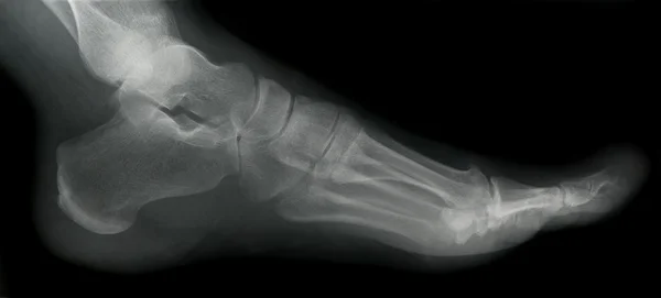

Fracture Of The Metatarsal Bones In The Foot. Anatomical Structure Of The Foot. Skeleton. Broken Bones. Vector Illustration On Isolated Background
Vector, 2.68MB, 5000 × 5000 eps
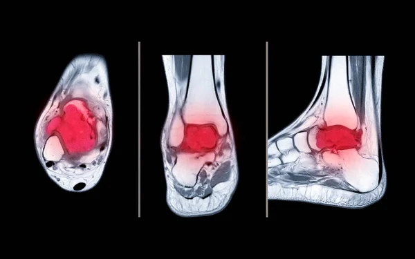
Compare Of MRI Ankle Axial, Coronal And Sagittal PDW View Showing Bone Metastasis To The Talus.
Image, 6.73MB, 7680 × 4800 jpg

Fracture Of The Metatarsal Bones In The Foot. Anatomical Structure Of The Foot. Skeleton. Broken Bones. Vector Illustration On Isolated Background
Vector, 2.74MB, 5000 × 5000 eps

Ankle Joint Fracture Type C Medical Vector Illustration On White Background
Vector, 13.49MB, 5000 × 5000 eps

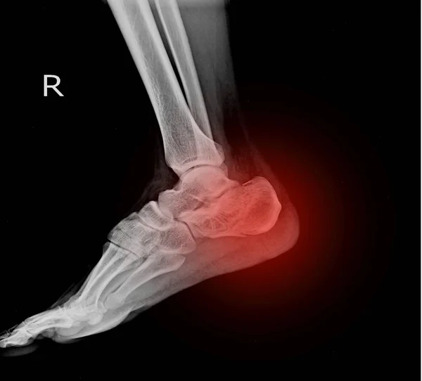
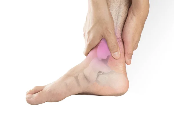
Young Man Doctor Explain To The Woman Patient The Result Of X Ray That Show Fracture Of Her Calcaneus
Image, 1.38MB, 6016 × 4016 jpg

Ankle Joint Fracture Type B Medical Vector Illustration On White Background
Vector, 12.76MB, 5000 × 5000 eps


Plantar Fasciitis As Fascia Muscle Inflammation And Tearing Outline Diagram. Labeled Educational Scheme With Painful Foot Condition And Medical Xray Explanation Vector Illustration. Feet Skeleton.
Vector, 6.84MB, 4000 × 4000 eps

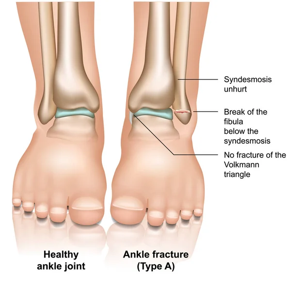
Ankle Joint Fracture Type A Medical Vector Illustration On White Background
Vector, 11.29MB, 5000 × 5000 eps
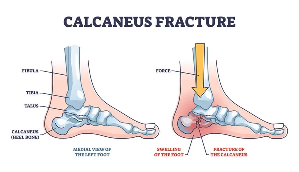
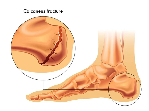
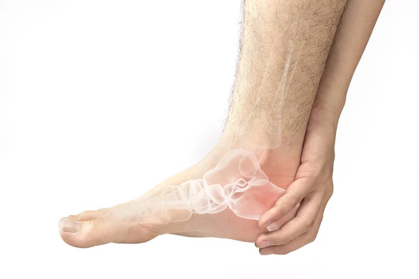
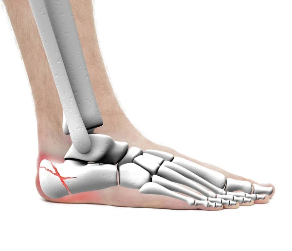
Calcaneus Fracture Anatomy With Broken Heel Bone Structure Outline Diagram. Labeled Educational Scheme With Physical Force Direction To Broke Leg Skeletal Vector Illustration. Medial View Of Left Foot
Vector, 6.35MB, 5500 × 3163 eps
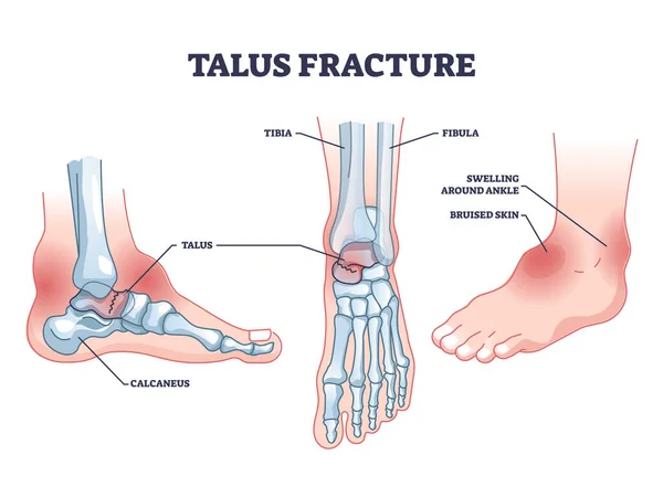
Talus Fracture As Broken Leg With Swelling Ankle Symptom Outline Diagram. Labeled Educational Scheme With Medical Bone Trauma Vector Illustration. Human Leg Foot Anatomical Structure With Painful Part
Vector, 7.1MB, 4800 × 3600 eps
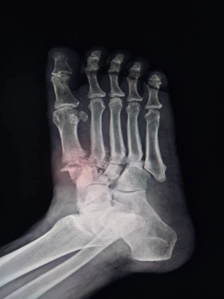
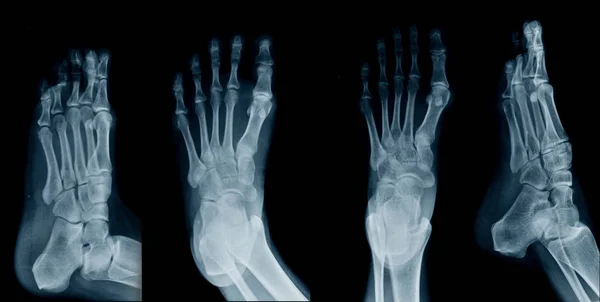
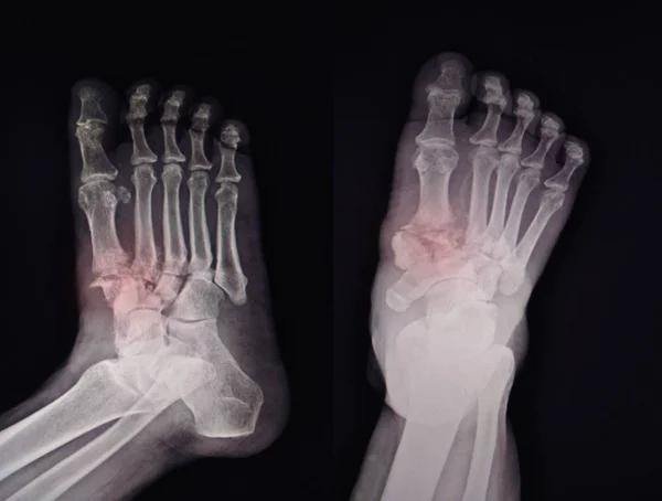
X-ray Foot Finidngs Fracture Base Of 1st-2nd Metatarsal Bone.color Mark,too Soft And Blurry Image.
Image, 5.2MB, 3072 × 4096 jpg

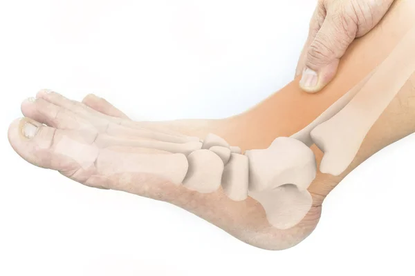
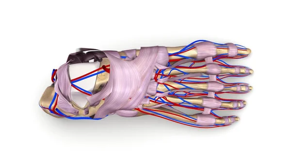
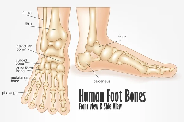
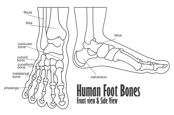
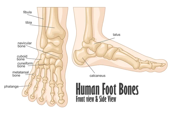
Foot Anatomy. Human Foot With The Name And Description Of All Bones And Sites. Top View And Side View. Arches Of The Feet. Skeleton Anatomy. Vector Illustration
Vector, 3.99MB, 5000 × 3663 eps


Page 1 >> Next