photo free
Home Images Vectors Videos

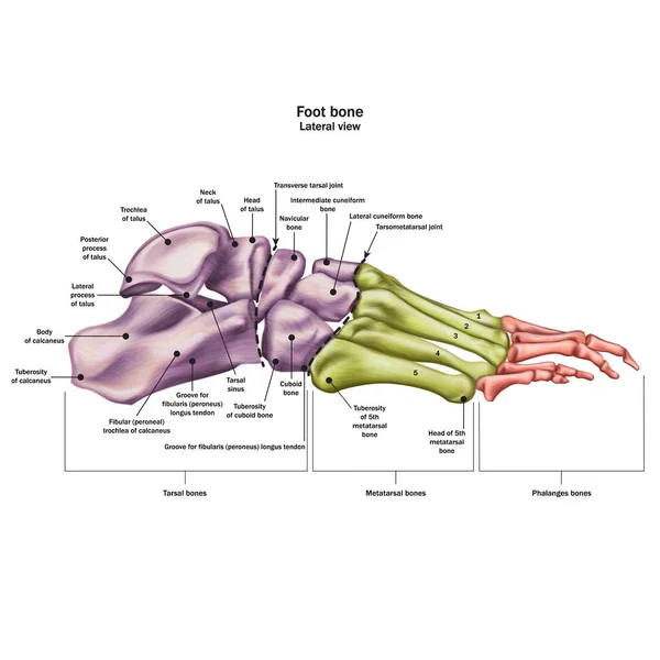
Stock image Calcaneus page 2

stock vector Foot

Foot

Vector, 2.79MB, 4807 × 4576 eps
stock vector Heel Spur.

Heel Spur.

Vector, 0.52MB, 3494 × 4881 eps
stock image Section of bone with osteoporosis

Section Of Bone With Osteoporosis

Image, 4.66MB, 2567 × 4409 jpg
stock vector Medical illustrations of symptoms of peroneal tendonitis, with enlargement of the affected area, with annotations.

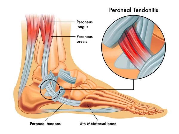
Medical Illustrations Of Symptoms Of Peroneal Tendonitis, With Enlargement Of The Affected Area, With Annotations.

Vector, 8.82MB, 2668 × 2001 eps
stock image Human Foot Muscles Anatomy Model for study medicine.

Human Foot Muscles Anatomy Model For Study Medicine.

Image, 3.97MB, 5282 × 3264 jpg
stock vector Peroneus tertius leg muscle with longus and brevis location outline diagram

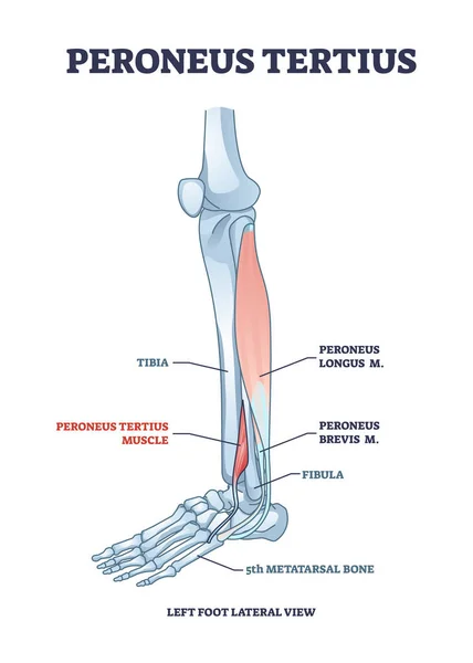
Peroneus Tertius Leg Muscle With Longus And Brevis Location Outline Diagram

Vector, 6.04MB, 3500 × 4900 eps
stock vector Extensor digitorum longus muscle with foot skeletal system outline diagram

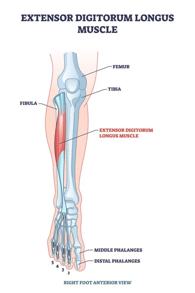
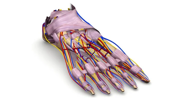
Extensor Digitorum Longus Muscle With Foot Skeletal System Outline Diagram

Vector, 6.73MB, 3500 × 5250 eps
stock vector Flexor hallucis longus muscle with human leg and foot bones outline diagram

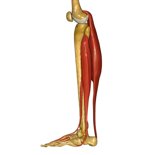
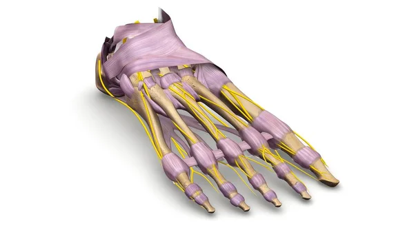
Flexor Hallucis Longus Muscle With Human Leg And Foot Bones Outline Diagram

Vector, 7.14MB, 4000 × 6667 eps
stock vector Bones the of foot

Bones The Of Foot

Vector, 2.23MB, 4208 × 4208 eps
stock vector Light blue visualization of bones of foot.

Light Blue Visualization Of Bones Of Foot.

Vector, 7.05MB, 6250 × 6250 eps
stock vector Foot Bone Anatomy Vector Illustration

Foot Bone Anatomy Vector Illustration

Vector, 5.22MB, 5000 × 5000 eps
stock vector Achilles tendon rupture as painful injury and leg trauma outline diagram. Labeled educational anatomical scheme with orthopedic problem explanation vector illustration. Medical body muscle condition.

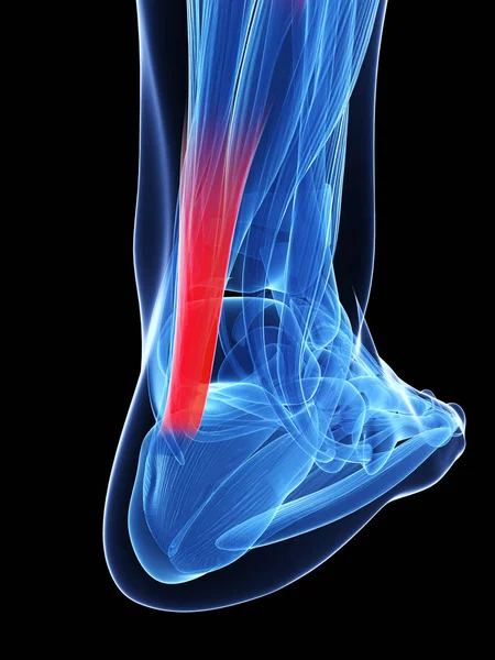
Achilles Tendon Rupture As Painful Injury And Leg Trauma Outline Diagram. Labeled Educational Anatomical Scheme With Orthopedic Problem Explanation Vector Illustration. Medical Body Muscle Condition.

Vector, 10.37MB, 4706 × 4000 eps
stock image Fibula and Tibia Muscles

Fibula And Tibia Muscles

Image, 2.24MB, 4096 × 4096 jpg
stock image Fibula and Tibia Muscles

Fibula And Tibia Muscles

Image, 1.59MB, 4096 × 4096 jpg
stock vector Bursitis inflammation concept

Bursitis Inflammation Concept

Vector, 0.42MB, 4168 × 4168 eps
stock image Foot bones 3d illustration

Foot Bones 3d Illustration

Image, 2.32MB, 3840 × 2160 jpg
stock vector Bones of foot

Bones Of Foot

Vector, 1.25MB, 4188 × 5488 eps
stock image X-ray normal human foot . Lateral view . Invert color style .

X-ray Normal Human Foot . Lateral View . Invert Color Style .

Image, 4.62MB, 5184 × 3456 jpg
stock image Foot bones 3d illustration

Foot Bones 3d Illustration

Image, 2.25MB, 3840 × 2160 jpg
stock image Foot bones 3d illustration

Foot Bones 3d Illustration

Image, 1.92MB, 3840 × 2160 jpg
stock image Osteomyelitis on the diagnosis list, medical concept

Osteomyelitis On The Diagnosis List, Medical Concept

Image, 4.36MB, 3800 × 2533 jpg
stock image 3d rendered illustration of the achilles tendon

3d Rendered Illustration Of The Achilles Tendon

Image, 0MB, 3750 × 5000 jpg
stock image Left image : Fracture shaft of fibula (calf bone)  ,  Right image : It was splinted with plaster cast

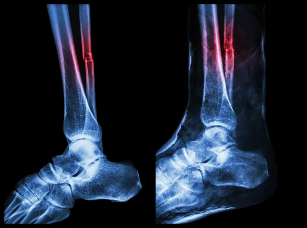
Left Image : Fracture Shaft Of Fibula (calf Bone) , Right Image : It Was Splinted With Plaster Cast

Image, 13.06MB, 7142 × 5331 jpg
stock image 3D rendering  of the foot bones for diagnosis bone fracture and rheumatoid arthritis from CT scannner.

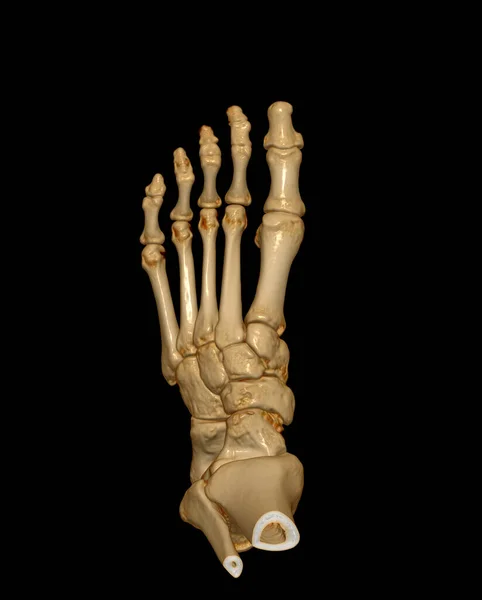
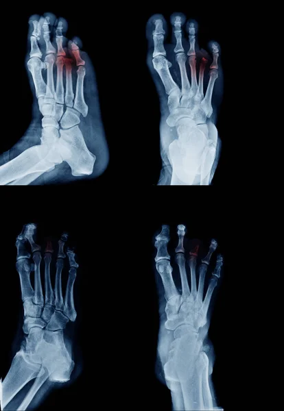
3D Rendering Of The Foot Bones For Diagnosis Bone Fracture And Rheumatoid Arthritis From CT Scannner.

Image, 1.63MB, 3220 × 4008 jpg
stock vector Bones of the foot. Vector drawing

Bones Of The Foot. Vector Drawing

Vector, 0.5MB, 5554 × 4182 eps
stock vector Illustration of the heel spur, which is a calcium deposit that promotes the appearance of a bone protrusion on the heel

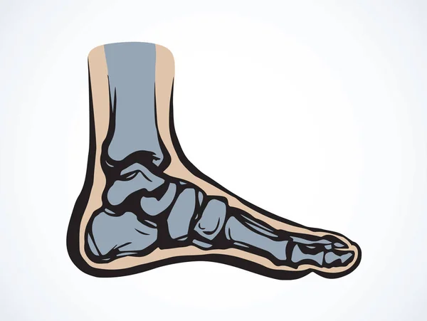
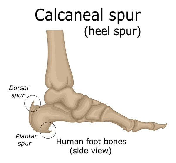
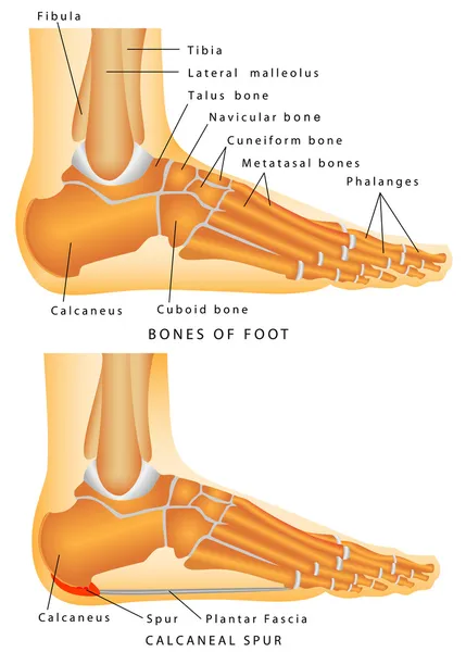
Illustration Of The Heel Spur, Which Is A Calcium Deposit That Promotes The Appearance Of A Bone Protrusion On The Heel

Vector, 2.29MB, 4500 × 4167 eps
stock image X-ray both foot showing Calcaneal spur on red color.

X-ray Both Foot Showing Calcaneal Spur On Red Color.

Image, 4.25MB, 4292 × 3380 jpg
stock image X-ray of the foot broken calcaneal/Heel  on red color.

X-ray Of The Foot Broken Calcaneal/Heel On Red Color.

Image, 2.72MB, 6334 × 5729 jpg
stock vector X ray of bones the of foot. Medial view

X Ray Of Bones The Of Foot. Medial View

Vector, 8.39MB, 5500 × 4417 eps
stock vector Vector illustration set of human cubit skeletal anatomy

Vector Illustration Set Of Human Cubit Skeletal Anatomy

Vector, 0.84MB, 6854 × 3021 eps
stock vector vector illustration of the ankle bones of the foot

Vector Illustration Of The Ankle Bones Of The Foot

Vector, 3.95MB, 8802 × 3518 eps
stock vector Extensor hallucis longus muscle with foot skeletal system outline diagram

Extensor Hallucis Longus Muscle With Foot Skeletal System Outline Diagram

Vector, 6.92MB, 3000 × 5600 eps
stock vector Soleus muscle with anatomical leg bones skeletal structure outline diagram

Soleus Muscle With Anatomical Leg Bones Skeletal Structure Outline Diagram

Vector, 7.36MB, 3500 × 5091 eps
stock image Woman suffering from heel pain. Inflammation or sprain of the tendon in the foot, heel spur, bursitis. The concept of diseases and pains in the leg

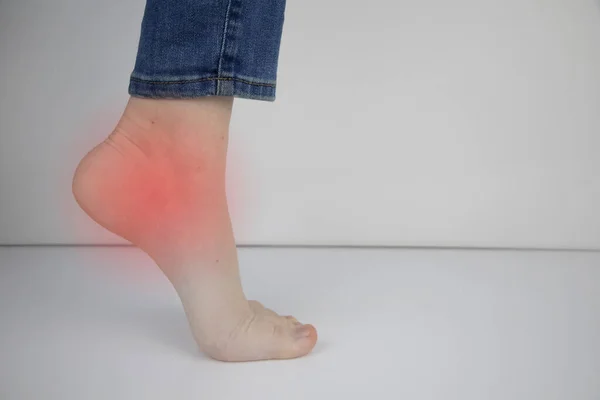
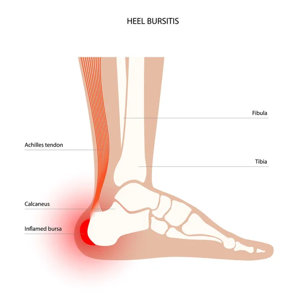
Woman Suffering From Heel Pain. Inflammation Or Sprain Of The Tendon In The Foot, Heel Spur, Bursitis. The Concept Of Diseases And Pains In The Leg

Image, 6.61MB, 6000 × 4000 jpg
stock image Human anatomy. Nerves of the sole of the right foot on a white background. 3D illustration

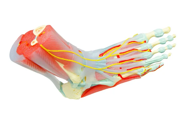
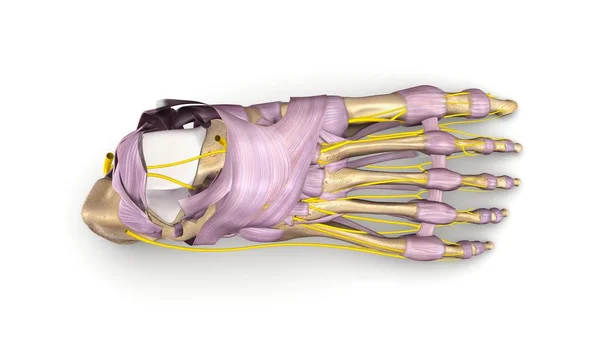
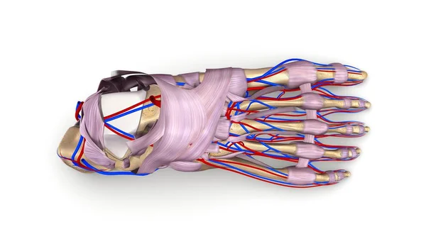
Human Anatomy. Nerves Of The Sole Of The Right Foot On A White Background. 3D Illustration

Image, 2.74MB, 4167 × 6471 jpg
stock vector Bones of the human foot with the name and description of all sit

Bones Of The Human Foot With The Name And Description Of All Sit

Vector, 2.66MB, 5556 × 5556 eps
stock vector Foot Bone Anatomy Vector Illustration

Foot Bone Anatomy Vector Illustration

Vector, 5.51MB, 5000 × 5000 eps
stock image Woman with plantar fasciitis uses a spiky ball to massage her ac

Woman With Plantar Fasciitis Uses A Spiky Ball To Massage Her Ac

Image, 13.63MB, 5338 × 3559 jpg
stock vector Sprain. A soft tissue injury in the human foot. Bruising and swelling of the leg skin. Close-up of a Achilles tendon, foot bones and ligaments. Torn of Calcaneofibular ligament. Vector illustration

Sprain. A Soft Tissue Injury In The Human Foot. Bruising And Swelling Of The Leg Skin. Close-up Of A Achilles Tendon, Foot Bones And Ligaments. Torn Of Calcaneofibular Ligament. Vector Illustration

Vector, 7.84MB, 5000 × 3345 eps
stock image Medial view of accurate human left foot bones with body contours on blue background 3D rendering illustration. Anatomy, osteology, orthopedics concept.

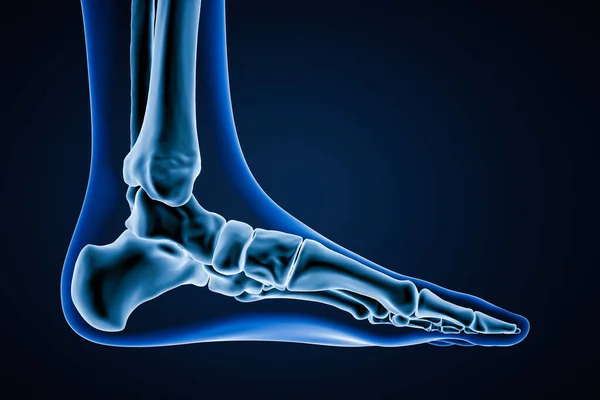
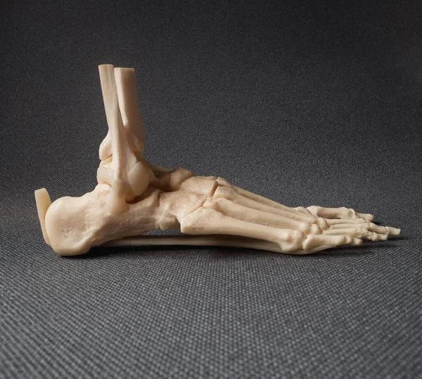
Medial View Of Accurate Human Left Foot Bones With Body Contours On Blue Background 3D Rendering Illustration. Anatomy, Osteology, Orthopedics Concept.

Image, 1.7MB, 3000 × 2000 jpg
stock image Foot anatomy. Medically accurate 3D illustration of the foot bones

Foot Anatomy. Medically Accurate 3D Illustration Of The Foot Bones

Image, 4.47MB, 6000 × 4000 jpg
stock vector Bones of the human foot with the name and description of all sit

Bones Of The Human Foot With The Name And Description Of All Sit

Vector, 2.59MB, 5556 × 5556 eps
stock vector Illustration of the joints of the foot. The bones of the foot are depicted and the joints between them are schematically shown.

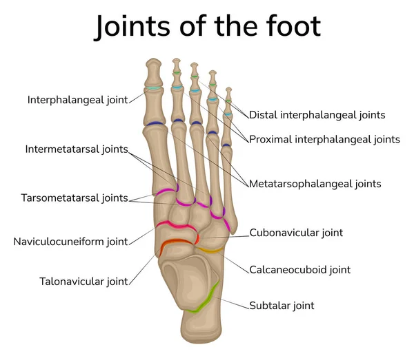
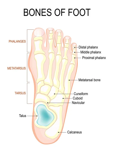
Illustration Of The Joints Of The Foot. The Bones Of The Foot Are Depicted And The Joints Between Them Are Schematically Shown.

Vector, 5.83MB, 8858 × 7677 eps
stock vector Heel bone fracture. Violation of integrity as a result of injury. A hand-drawn diagram of this injury. Black and white illustration for different uses.

Heel Bone Fracture. Violation Of Integrity As A Result Of Injury. A Hand-drawn Diagram Of This Injury. Black And White Illustration For Different Uses.

Vector, 0.28MB, 5780 × 3214 eps
stock image Doctor examining X-ray scan of Calcaneus or heel bone destruction. Bone of the tarsus of the foot. Medical treatment concept. Diagnosis of the disease humans bones.

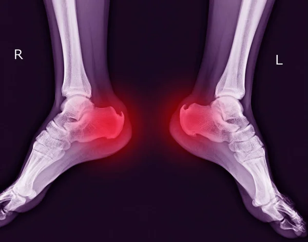
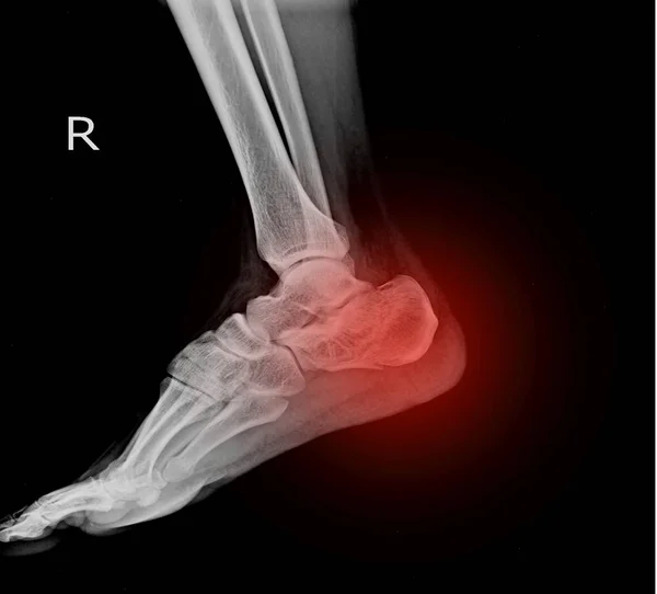
Doctor Examining X-ray Scan Of Calcaneus Or Heel Bone Destruction. Bone Of The Tarsus Of The Foot. Medical Treatment Concept. Diagnosis Of The Disease Humans Bones.

Image, 5.26MB, 3334 × 5000 jpg
stock image Foot bones 3d illustration

Foot Bones 3d Illustration

Image, 2.5MB, 3840 × 2160 jpg
stock image amputee foot x-ray image in many view and many case

Amputee Foot X-ray Image In Many View And Many Case

Image, 2.11MB, 2269 × 3284 jpg
stock image Feet bones model recreation over a grey background.

Feet Bones Model Recreation Over A Grey Background.

Image, 5.75MB, 3499 × 3141 jpg
stock image 3d rendering illustration of calcaneus bone

3d Rendering Illustration Of Calcaneus Bone

Image, 0.66MB, 2048 × 2048 jpg
stock image 3d render medical illustration of the calcaneus bone

3d Render Medical Illustration Of The Calcaneus Bone

Image, 3.33MB, 4637 × 3072 jpg
Previous << Page 2 >> Next