
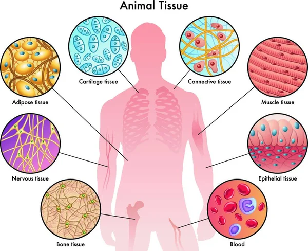
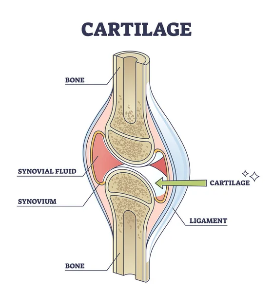
Stock image Cartilage Tissue

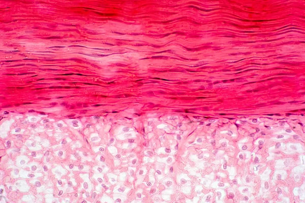
Knee Muscle, Tendon And Cartilage Tissue Anatomy For Physiology Education.
Image, 16.85MB, 6000 × 4000 jpg
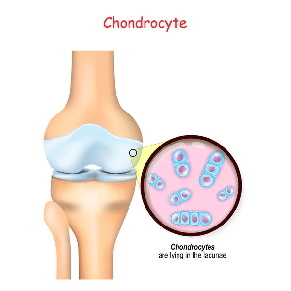
Knee And Close Up Of Cells Of A Cartilage. Chondrocytes Are Lying In The Lacunae And Produce And Maintain The Cartilaginous Matrix, And Collagen.
Vector, 9.79MB, 4444 × 4444 eps
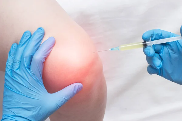
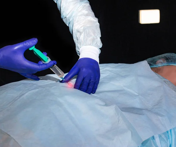
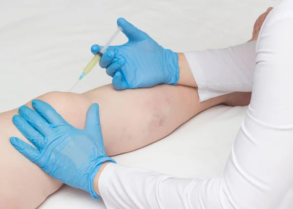
The Doctor Injects Plasma Into A Sore, Inflamed Female Knee For Arthrosis And Arthritis, Plasma-lifting, Close-up, Medical, Professional
Image, 9.68MB, 5184 × 3456 jpg
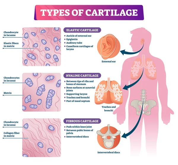
Types Of Cartilage Vector Illustration. Labeled Educational Tissue Scheme.
Vector, 9.14MB, 4320 × 4000 eps
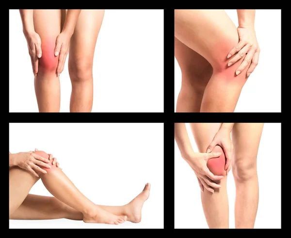
Collection OA Knee Or Pain Or Sprain, Set Of Woman Pain At Her Knee Joint Because Of Osteoarthritis Or Ligament Sprain Or Muscle Around Joint Injury, Hand Palpation Around Knee Joint
Image, 3.45MB, 4386 × 3585 jpg
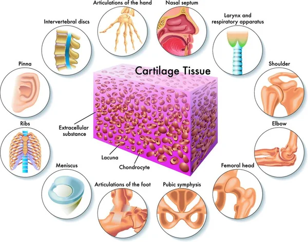
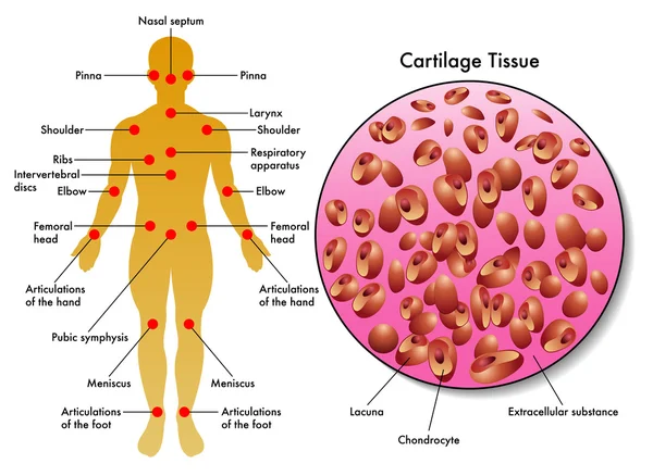
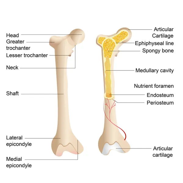
Medical Illustration Of Cartilage Tissue And Its Position In The Human Body
Vector, 0MB, 5144 × 4048 zip

Knee Joint Mockup On A Blue Background And Pills With A Syringe. The Concept Of Treatment Of The Knee Joint With Chondroprotectors And Anti-inflammatory Drugs, Glucosteroids
Image, 1.26MB, 4968 × 3486 jpg
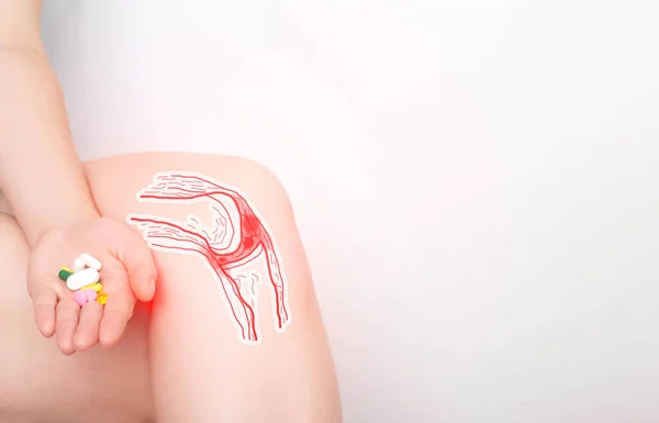
The Girl Holds A Handful Of Pills In Her Hand For Pain In The Knee Joint. Concept Of Drugs For The Treatment And Restoration Of Cartilage Tissue In The Knee Joint, Inflammatory
Image, 1.04MB, 5472 × 3514 jpg
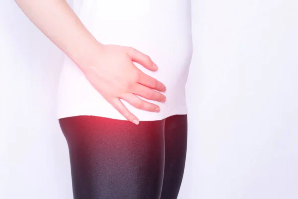
The Girl Holds On To The Hip Joint In Which Pain And Inflammation, Arthrosis And Arthritis In The Thigh, Ankylosing Spondylitis, Copy Space, Medical
Image, 3.5MB, 5184 × 3456 jpg
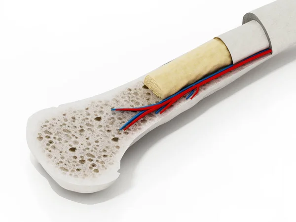
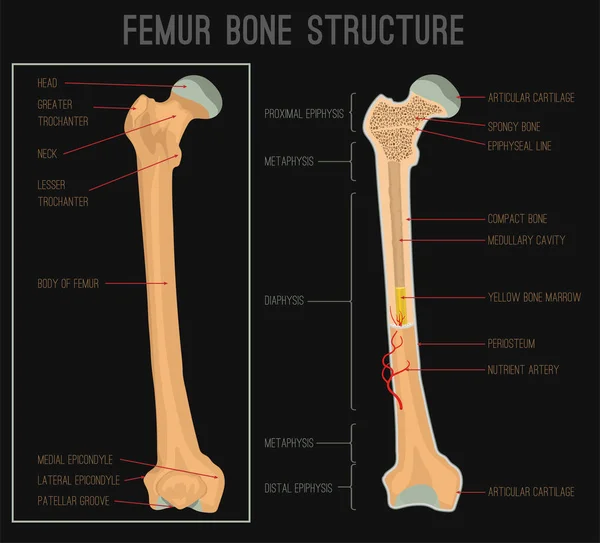
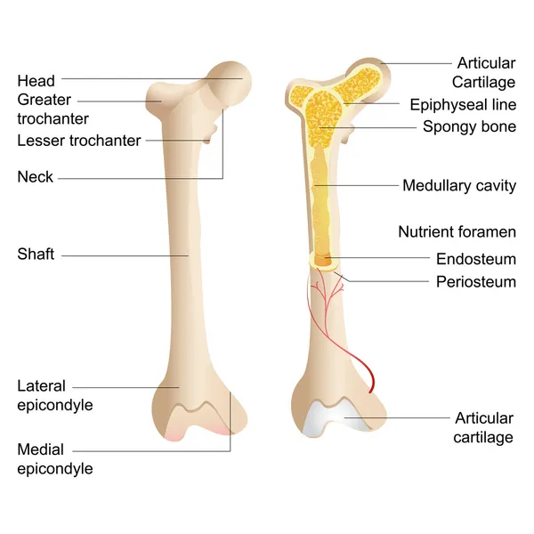
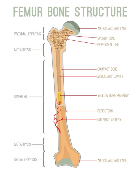
Cross Section Of A Human Bone Showing Bone Marrow, Spongy Bone And Blood Vessels. 3D Illustration.
Image, 5.61MB, 5500 × 4125 jpg
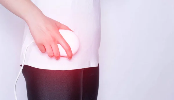
Treatment Of The Hip Joint With The Help Of Physiotherapy Magnetic And Laser Therapy, The Elimination Of Pain And Inflammation In The Joint, Copy Space, Ankylosing Spondylitis
Image, 4.28MB, 5184 × 2980 jpg
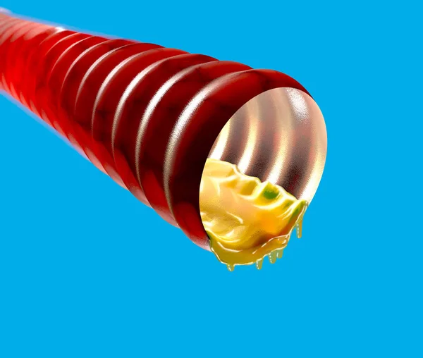
Trachea With Mucus, Catarrh, 3d Section. Duct That Serves To Transfer The Air From The Outside To The Lungs. 3d Rendering
Image, 5.13MB, 4724 × 4000 jpg
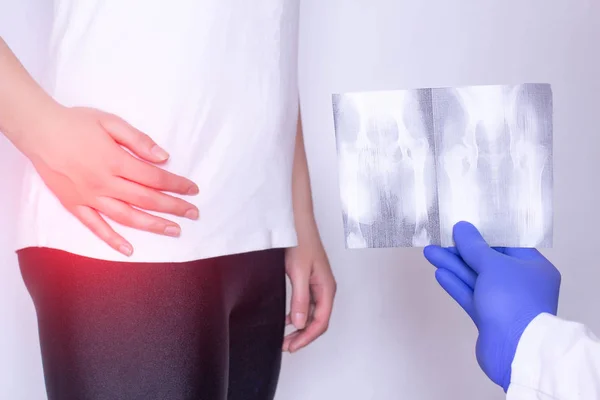
Doctor Holds X-ray Picture On The Background Of A Girl With A Sore Hip Joint And Intervertebral Hernia, Fibromyalgia, Close-up
Image, 4.57MB, 5184 × 3456 jpg

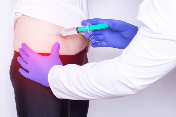
Modern Method Of Eliminating Pain And Inflammation Of The Injection Of Blood Plasma Into The Hip Joint, Plasma Therapy, Intervertebral Hernia
Image, 4.21MB, 5184 × 3456 jpg
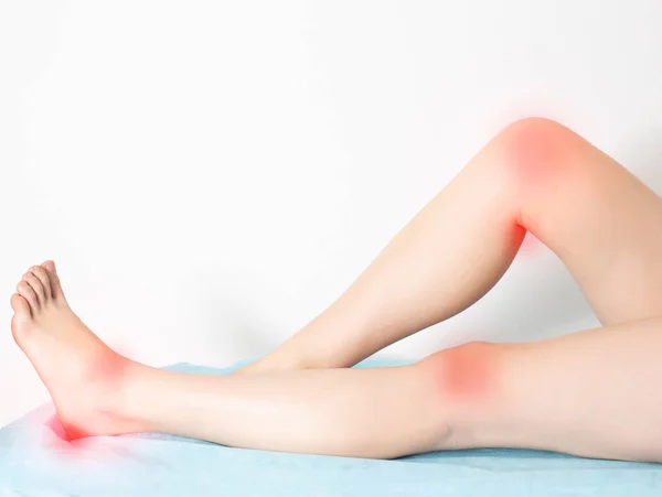
Female Legs On A White Background With Sore Reddened Knee Joints And Ankle Joints With Heels. Concept Of Disease And Treatment Of Arthritis And Joint Inflammation.
Image, 1.64MB, 4854 × 3648 jpg
Doctor Neurologist Makes A Therapeutic Injection Of Medicine Into The Patient's Hip Joint. Treatment Of Arthrosis And Joint Inflammation, Close-up
Image, 1.57MB, 3339 × 2802 jpg
The Doctor Makes An Injection Of Hyaluronic Acid And Chondroprotector With Anesthesia In The Hip Joint Of The Girl, The Treatment Of Arthritis, Medical, Osteoarthritis
Image, 3.96MB, 5184 × 3456 jpg
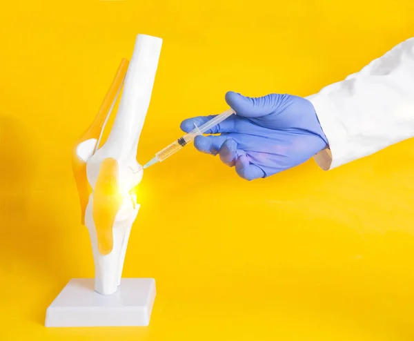
The Doctor Performs The Procedure Of Introducing A Blockade With A Chondroprotector And Hilauric Acid Into A Knee Joint Model. Knee Joint Treatment Concept With Injection Blockade, Copy Space For Text, Collagen, Stem Cells
Image, 0.6MB, 4080 × 3363 jpg
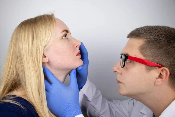
The Otolaryngologist Examines The Girl's Nasal Passages. Painful Sensations In The Nose, Polyps, Adenoids And Shortness Of Breath. Fractures Of The Cartilage Tissue Of The Nose. Closure Of The Nasopharyngeal Canals
Image, 9.48MB, 6000 × 4000 jpg
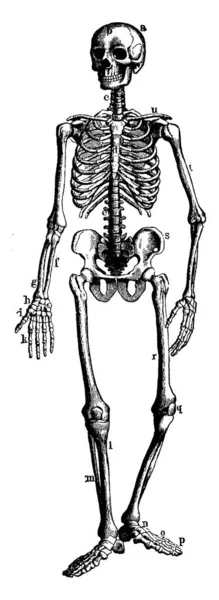
This Illustration Represents Human Skeleton Showing Bony And Cartilage Tissue, Vintage Line Drawing Or Engraving Illustration.
Vector, 2.81MB, 3474 × 9487 eps
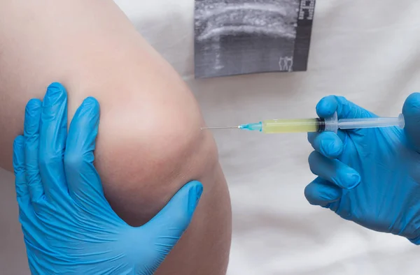
The Doctor Injects Plasma Into The Knee Joint Of A Woman To Eliminate Pain And Symptoms Of Arthritis And Arthrosis, Relieve Inflammation With A Plasma-lifting Method
Image, 7.32MB, 4841 × 3456 jpg
Doctor Introduces Blood Plasma To A Woman's Knee Joint To Treat Knee Sutsawa, Atrita, Autoplasmotherapy, Close-up
Image, 8.89MB, 4973 × 3274 jpg
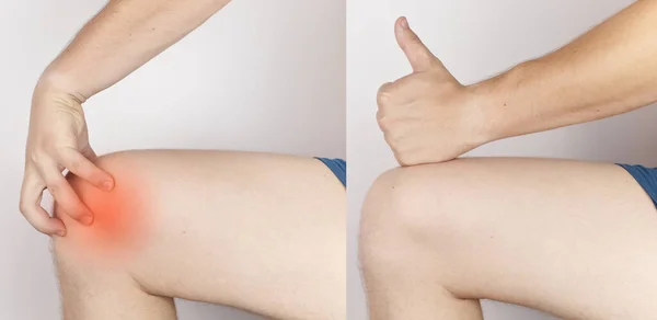
Before And After. On The Left, A Man Is Holding Onto An Injured Knee, And On The Right, Doctors Have Already Cured A Patient. Ruptured Knee Tendons, Muscles, Meniscus Injury, Bone Fracture Or Fissure
Image, 10.34MB, 8191 × 4000 jpg

Close-up Hand Inflammation. Left Hand Was Very Swollen In Region Of The Cartilages Of Index Finger. Inflammation Of Joints. Arthritis And Arthrosis. Swelling Of Adjacent Tissues. Brush Comparison
Image, 7.99MB, 5431 × 2854 jpg
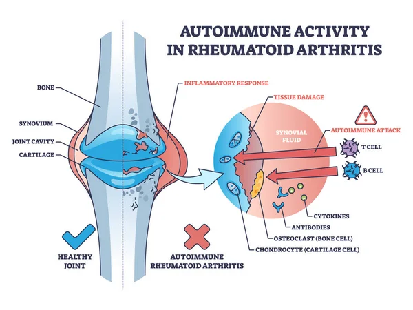
Autoimmune Activity In Rheumatoid Arthritis Skeletal Disease Outline Diagram. Labeled Educational Scheme With Body Immune System Attack To Tissues With Cells Vector Illustration. Bone Inflammation.
Vector, 6.01MB, 5000 × 3750 eps
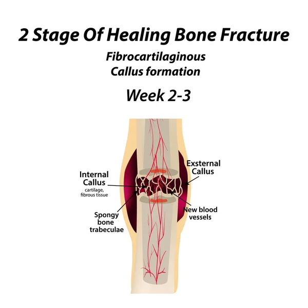
2 Stage Of Healing Bone Fracture. Formation Of Callus. The Bone Fracture. Infographics. Vector Illustration On Isolated Background.
Vector, 1.33MB, 5000 × 5000 eps

Cartilage Elastic Tissue Location In Body And Leg Structure Outline Diagram
Vector, 5.7MB, 4000 × 4286 eps

1 Stage Of Healing Bone Fracture. Formation Of Callus. The Bone Fracture. Infographics. Vector Illustration On Isolated Background.
Vector, 1.22MB, 5000 × 5000 eps

2 Stage Of Healing Bone Fracture. Formation Of Callus. The Bone Fracture. Infographics. Vector Illustration On Isolated Background.
Vector, 1.37MB, 5000 × 5000 eps

Sprain Vs Strain Anatomical Comparison As Medical Foot Injury Outline Diagram
Vector, 6.66MB, 5000 × 3250 eps

Chondromalacia Patella Knee Breakdown Compared With Healthy Outline Diagram. Labeled Educational Kneecap Tissue Damage With Cartilage Problem And Anatomical Leg Joint Structure Vector Illustration.
Vector, 7.11MB, 4200 × 3990 eps
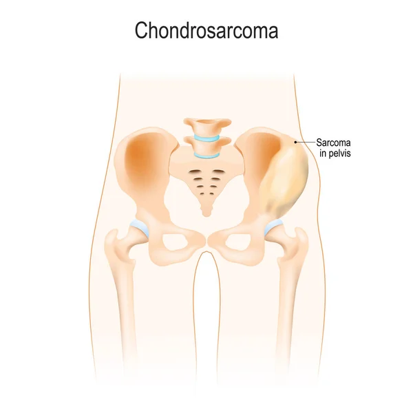
Chondrosarcoma Is A Cancer From Cartilage Cells. Malignant Neoplasm. Lumbar Region. Anatomy Of The Hip, Vertebra, And Pelvis. Vector Illustration For Biological, Science, Medical Use.
Vector, 5.22MB, 5400 × 5399 eps
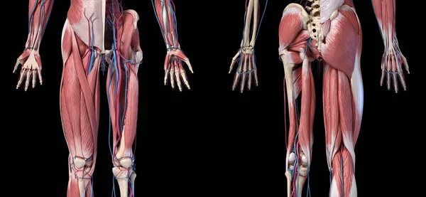
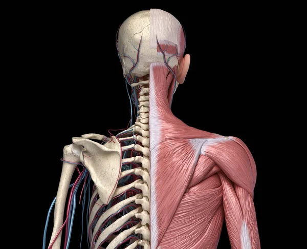
Human Anatomy, Limbs And Hip Skeletal, Muscular And Cardiovascular Systems.
Image, 23.68MB, 11905 × 5500 jpg
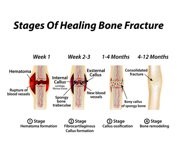
Stages Of Healing Bone Fracture. Formation Of Callus. The Bone Fracture. Infographics. Vector Illustration On Isolated Background
Vector, 2.08MB, 5000 × 4630 eps


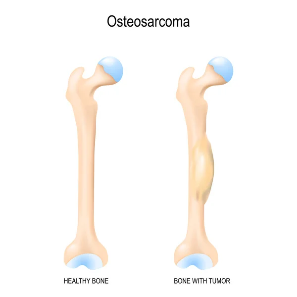
Osteosarcoma. Osteogenic Sarcoma Is A Cancerous Tumor In A Bone. Malignant Neoplasm. Two Human Bones: Healthy Femur And Bone With Osteosarcoma. Vector Illustration For Biological, Science, Medical Use.
Vector, 14.3MB, 5706 × 5707 eps
Page 1 >> Next