photo free
Home Images Vectors Videos

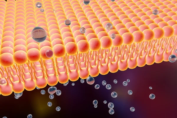
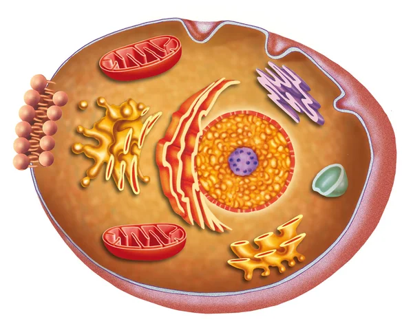
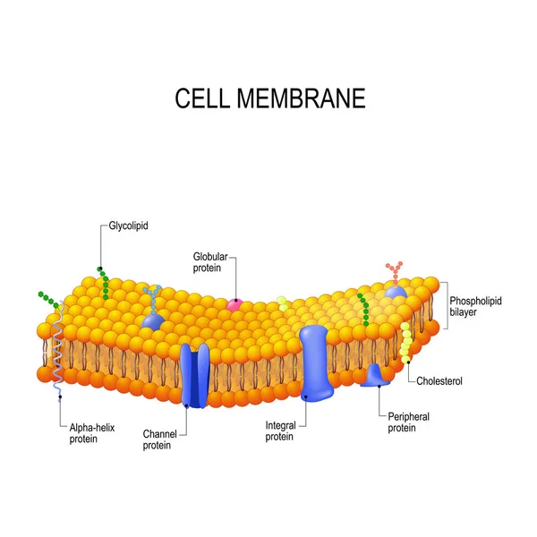
Stock image Cell Membrane

stock vector Plasma membrane

Plasma Membrane

Vector, 4.59MB, 5333 × 4000 eps
stock vector Human cell structure

Human Cell Structure

Vector, 4.22MB, 5333 × 4000 eps
stock image Macro of a blue cell with a nucleus. Abstract blurred cells background. Concept of medicine, science, research and DNA studies. 3d rendering mock up

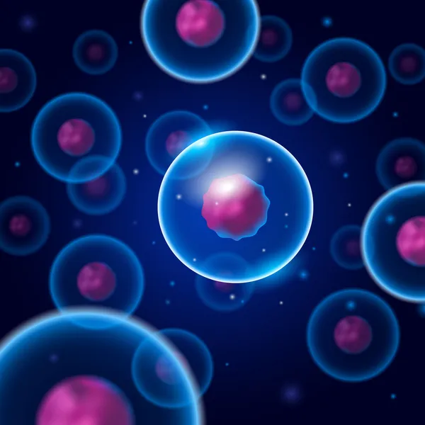
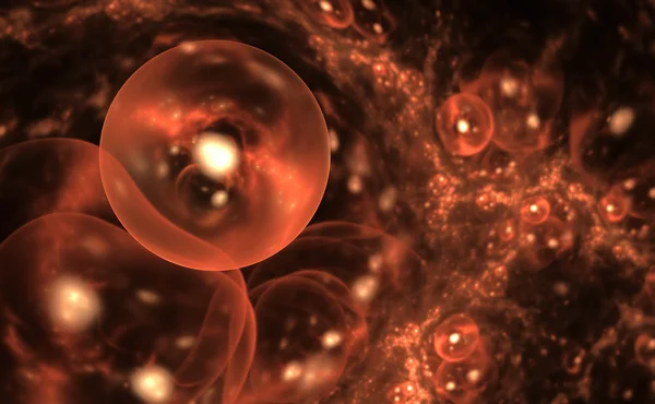
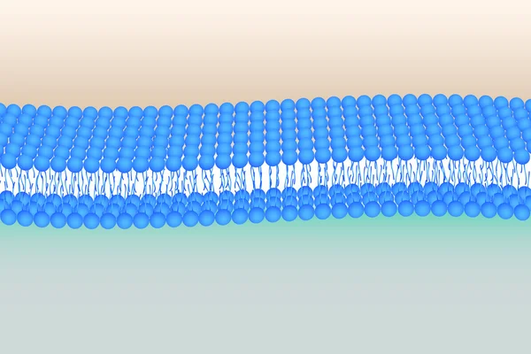
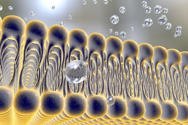
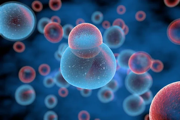
Macro Of A Blue Cell With A Nucleus. Abstract Blurred Cells Background. Concept Of Medicine, Science, Research And DNA Studies. 3d Rendering Mock Up

Image, 3.27MB, 4500 × 3000 jpg
stock vector Anatomy of an animal cell

Anatomy Of An Animal Cell

Vector, 3.87MB, 5000 × 4086 eps
stock image Cell membrane

Cell Membrane

Image, 2.21MB, 3000 × 2000 jpg
stock vector Detailed vector illustration of round membrane cells

Detailed Vector Illustration Of Round Membrane Cells

Vector, 13.66MB, 4000 × 4000 eps
stock vector Cell membrane with labeled educational structure scheme vector illustration

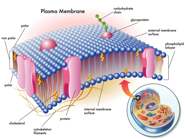
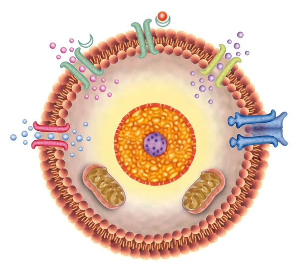
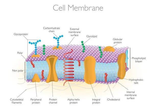
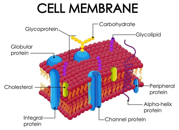
Cell Membrane With Labeled Educational Structure Scheme Vector Illustration

Vector, 7.46MB, 4500 × 3750 eps
stock image Human cell, anatomy image.

Human Cell, Anatomy Image.

Image, 3.73MB, 4200 × 3189 jpg
stock image Cell membrane

Cell Membrane

Image, 1.88MB, 3000 × 2000 jpg
stock image Synthetic cells

Synthetic Cells

Image, 8.24MB, 5000 × 3090 jpg
stock image Cell structure

Cell Structure

Image, 4.15MB, 3000 × 2250 jpg
stock vector Cell membrane transport systems illustration

Cell Membrane Transport Systems Illustration

Vector, 3.14MB, 6000 × 4000 eps
stock image Structure of plasma membrane of cell

Structure Of Plasma Membrane Of Cell

Image, 7.66MB, 4724 × 3622 jpg
stock vector Endocytosis Vesicle Transport Cell Membrane

Endocytosis Vesicle Transport Cell Membrane

Vector, 1.57MB, 6250 × 5000 eps
stock vector Animal and plant cells

Animal And Plant Cells

Vector, 7.9MB, 6629 × 3319 eps
stock image Receiving cell

Receiving Cell

Image, 3.32MB, 3336 × 3077 jpg
stock image Cells view under a microscope.

Cells View Under A Microscope.

Image, 3.68MB, 3200 × 2250 jpg
stock image Cell with nucleus

Cell With Nucleus

Image, 3.2MB, 3600 × 2400 jpg
stock vector Active and Passive transport

Active And Passive Transport

Vector, 0.76MB, 5676 × 2000 eps
stock vector Omega 3 phospholipid

Omega 3 Phospholipid

Vector, 1.45MB, 5000 × 3750 eps
stock image Receiving cell

Receiving Cell

Image, 3.19MB, 2799 × 2871 jpg
stock vector Membrane proteins labeled vector illustration. Detailed structure scheme.

Membrane Proteins Labeled Vector Illustration. Detailed Structure Scheme.

Vector, 8.91MB, 4100 × 3972 eps
stock vector Cell membrane. Vector

Cell Membrane. Vector

Vector, 2.51MB, 4037 × 4037 eps
stock vector Structure of a animal cell. Animal cell organelles.

Structure Of A Animal Cell. Animal Cell Organelles.

Vector, 5.38MB, 4500 × 4500 eps
stock vector Flexible Phosholipid bilayer

Flexible Phosholipid Bilayer

Vector, 0.92MB, 6250 × 4167 ai
stock vector Detailed Diagram Models of a Cell Membrane

Detailed Diagram Models Of A Cell Membrane

Vector, 11.14MB, 7000 × 5113 eps
stock vector Cells under a microscope. Research of stem cells. Cellular Therapy. Cell division. Vector illustration on a light background

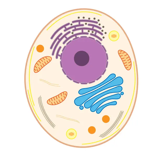
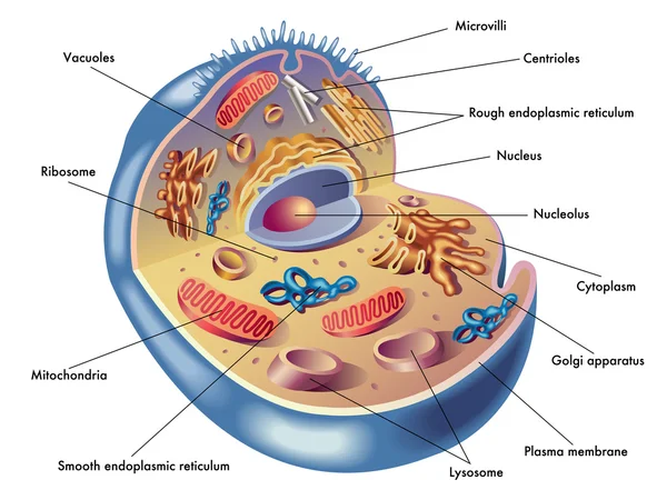
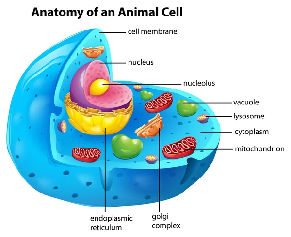
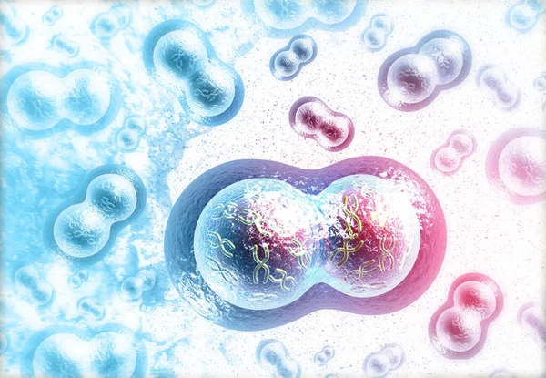
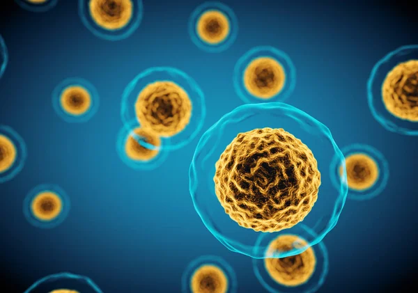
Cells Under A Microscope. Research Of Stem Cells. Cellular Therapy. Cell Division. Vector Illustration On A Light Background

Vector, 11.7MB, 7500 × 4313 eps
stock image Human cells division. medical background  3d illustration

Human Cells Division. Medical Background 3d Illustration

Image, 7.16MB, 3900 × 2700 jpg
stock image Cell Division Stages of Mitosis

Cell Division Stages Of Mitosis

Image, 4.58MB, 4500 × 3000 jpg
stock image Depolarization: phospholipid membrane with NA + and K + ion channels.

Depolarization: Phospholipid Membrane With NA + And K + Ion Channels.

Image, 2.27MB, 6600 × 5452 jpg
stock image Cell membrane

Cell Membrane

Image, 2.44MB, 3000 × 2000 jpg
stock image Depolarization: phospholipid membrane with NA + and K + ion channels.

Depolarization: Phospholipid Membrane With NA + And K + Ion Channels.

Image, 2.35MB, 6600 × 5452 jpg
stock image Ligand-dependent ion channel: attachment of a particular molecule causes the channel to open.

Ligand-dependent Ion Channel: Attachment Of A Particular Molecule Causes The Channel To Open.

Image, 1.14MB, 3020 × 4229 jpg
stock image Ligand-dependent ion channel: attachment of a particular molecule causes the channel to open.

Ligand-dependent Ion Channel: Attachment Of A Particular Molecule Causes The Channel To Open.

Image, 1.12MB, 3020 × 4229 jpg
stock vector Human cell membrane structure  illustration

Human Cell Membrane Structure Illustration

Vector, 2.06MB, 5810 × 4237 eps
stock vector Human cell membrane structure  illustration

Human Cell Membrane Structure Illustration

Vector, 2.34MB, 5812 × 4237 eps
stock vector Amphipatic property of cell membrane

Amphipatic Property Of Cell Membrane

Vector, 0.49MB, 4444 × 4444 eps
stock vector A crying tetrad cartoon character drawing concept

A Crying Tetrad Cartoon Character Drawing Concept

Vector, 5.75MB, 5000 × 5000 eps
stock image Cells under a microscope. Research of stem cells. Cellular Therapy. Cell division. 3d illustration on a light background

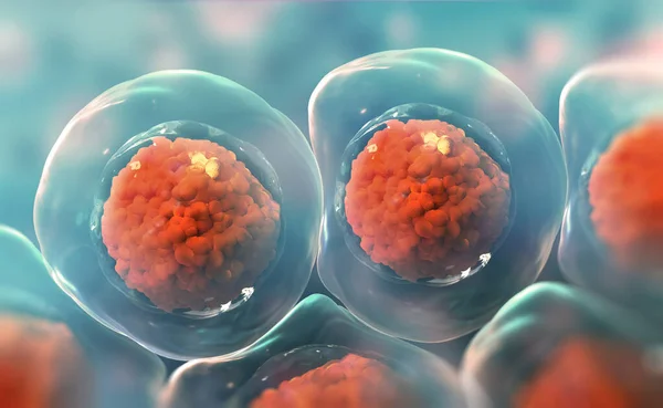
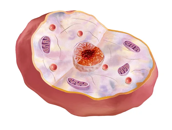
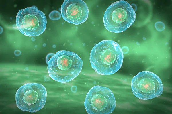
Cells Under A Microscope. Research Of Stem Cells. Cellular Therapy. Cell Division. 3d Illustration On A Light Background

Image, 6.62MB, 6500 × 4000 jpg
stock vector Membrane proteins. integral, and Peripheral membrane proteins, Single-pass, and Multi-pass transmembrane helix, Lipid-anchored protein. Vector illustration for biological, science and educational use

Membrane Proteins. Integral, And Peripheral Membrane Proteins, Single-pass, And Multi-pass Transmembrane Helix, Lipid-anchored Protein. Vector Illustration For Biological, Science And Educational Use

Vector, 4.52MB, 3921 × 3921 eps
stock image T Cells attacking Cancer Cells 3D illustration

T Cells Attacking Cancer Cells 3D Illustration

Image, 3.14MB, 6000 × 4000 jpg
stock image Anatomy and characteristics of the human cell

Anatomy And Characteristics Of The Human Cell

Image, 3.48MB, 3035 × 2400 jpg
stock image Cells view under a microscope.

Cells View Under A Microscope.

Image, 4.74MB, 3200 × 2250 jpg
stock image Human cell

Human Cell

Image, 6.24MB, 5000 × 2800 jpg
stock vector Cell membrane proteins

Cell Membrane Proteins

Vector, 10.18MB, 4444 × 4443 eps
stock vector Cell membrane. A detailed diagram models of membrane Structure

Cell Membrane. A Detailed Diagram Models Of Membrane Structure

Vector, 8.71MB, 5205 × 5205 eps
stock image Cell membrane

Cell Membrane

Image, 2.21MB, 3000 × 2000 jpg
stock image The gastric proton pump H+,K+ -ATPase with a proton pump inhibitor (PPI) bound

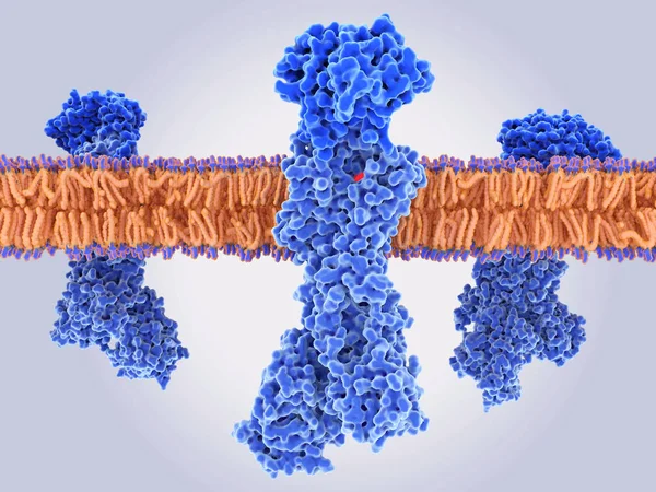
The Gastric Proton Pump H+,K+ -ATPase With A Proton Pump Inhibitor (PPI) Bound

Image, 7.06MB, 8000 × 6000 jpg
stock vector Fluid mosaic model vector illustration. Cell membrane structure infographic

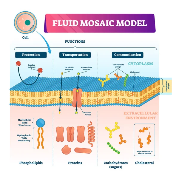
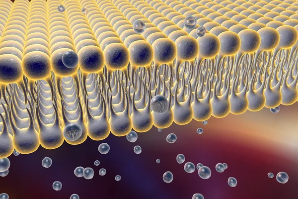
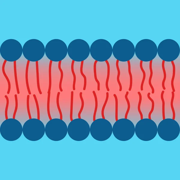
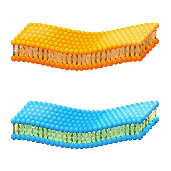
Fluid Mosaic Model Vector Illustration. Cell Membrane Structure Infographic

Vector, 9.21MB, 4000 × 4000 eps
stock vector Exocytosis Vesicle Transport Cell Membrane

Exocytosis Vesicle Transport Cell Membrane

Vector, 1.55MB, 6250 × 5417 eps
Page 1 >> Next