
Stock image Cell Nucleus page 2

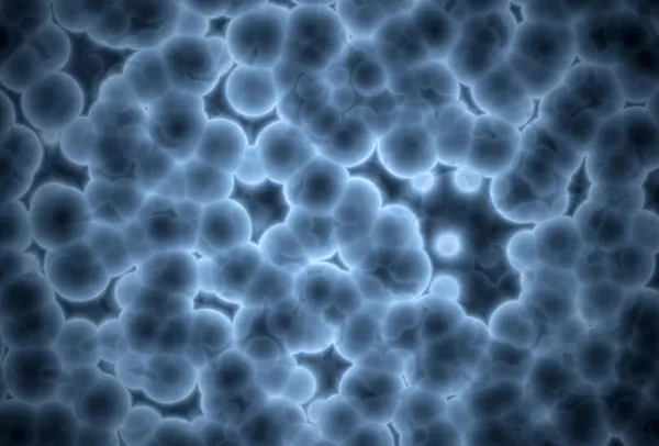
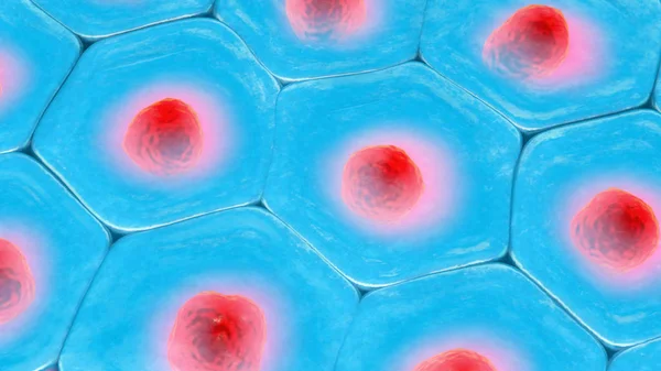
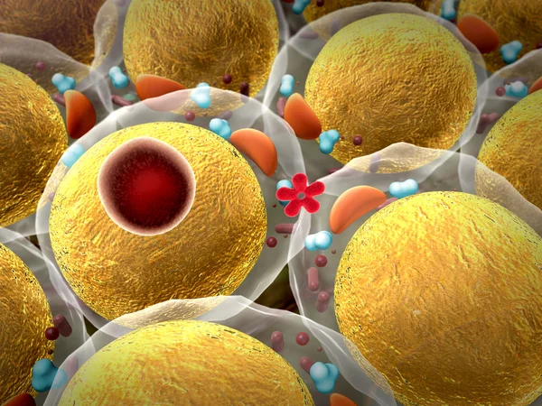
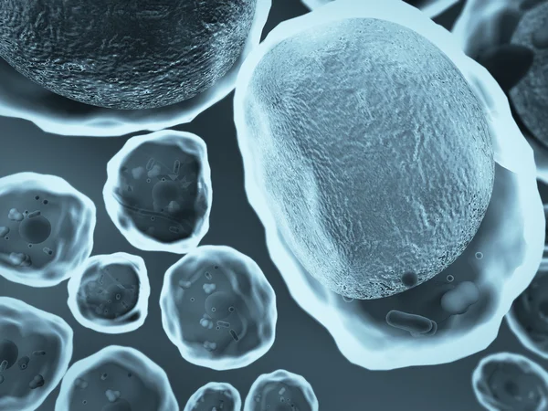
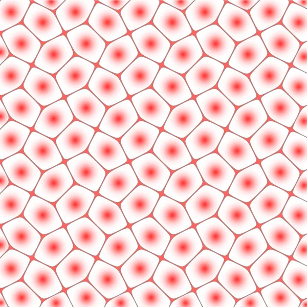
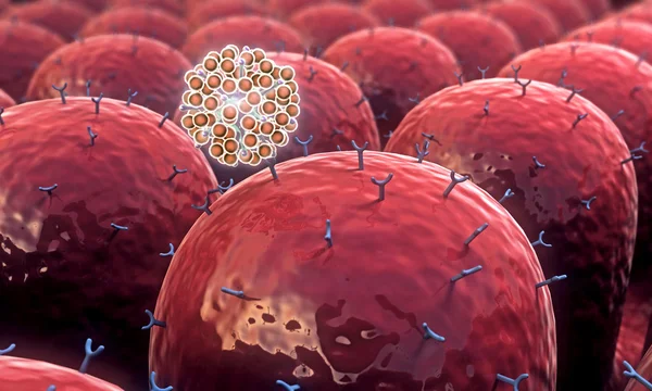
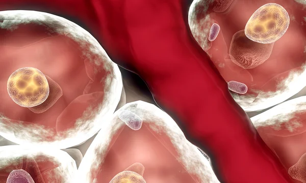
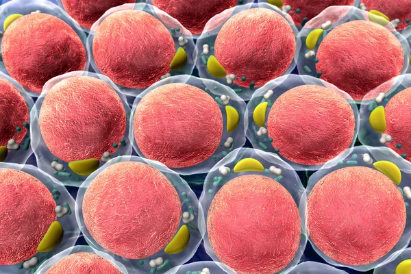
3d Illustration Of A Top View On Blue Cell Pattern With Red Cell Nucleus
Image, 32.49MB, 8400 × 4725 jpg

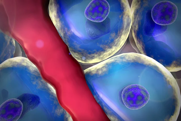
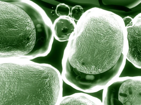
3d Illustration Of A Top View On Blue Cell Pattern With Red Cell Nucleus
Image, 25.63MB, 9200 × 6900 jpg
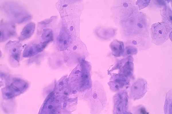
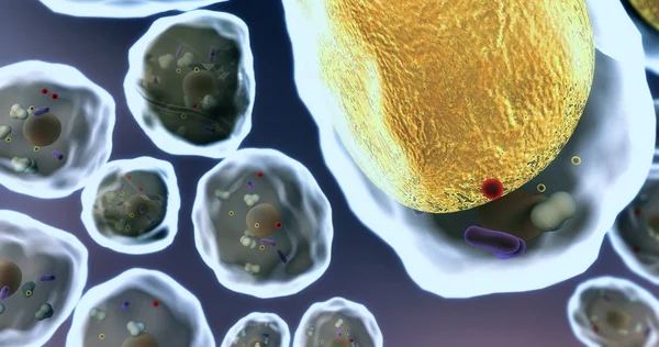
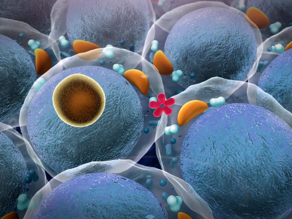
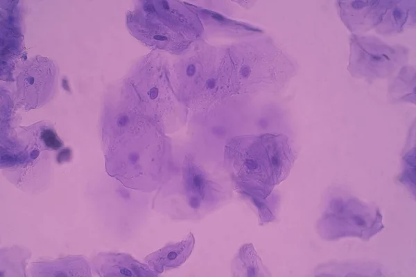
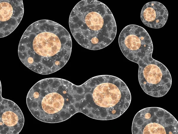
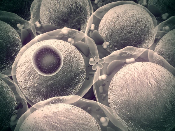
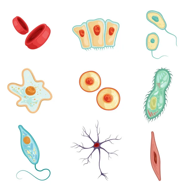
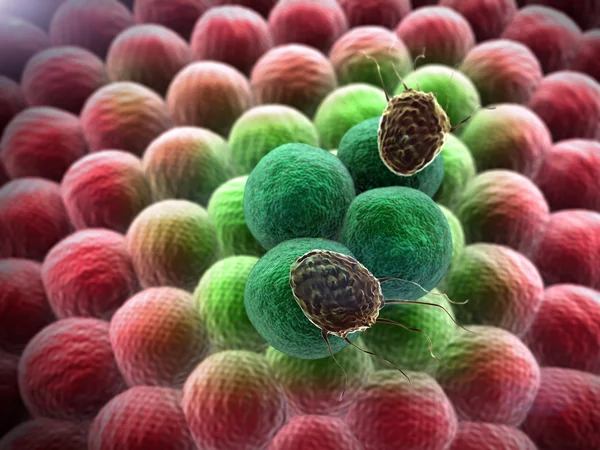
Human Cheek Epithelial Cells. The Tissue That Lines The Inside Of The Mouth Is Known As The Basal Mucosa And Is Composed Of Squamous Epithelial Cells. Education Pathology.
Image, 14.21MB, 6000 × 4000 jpg
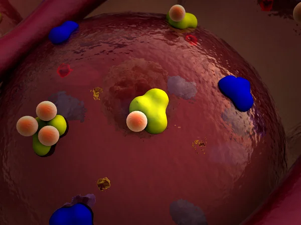
Human Cheek Epithelial Cells. The Tissue That Lines The Inside Of The Mouth Is Known As The Basal Mucosa And Is Composed Of Squamous Epithelial Cells. Education Pathology.
Image, 16.77MB, 6000 × 4000 jpg
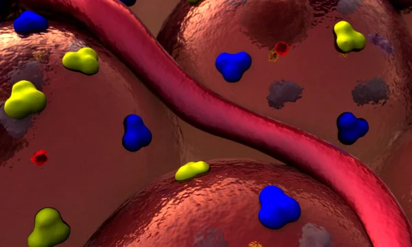


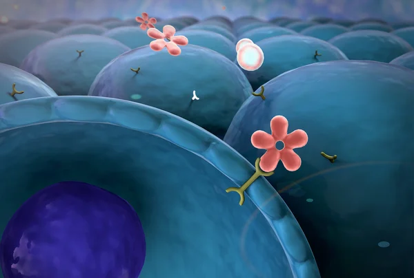


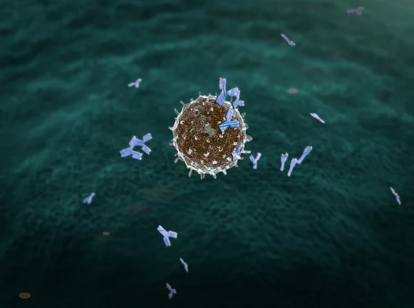


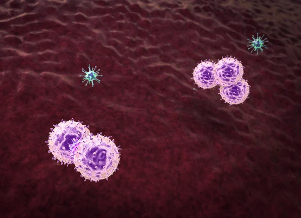
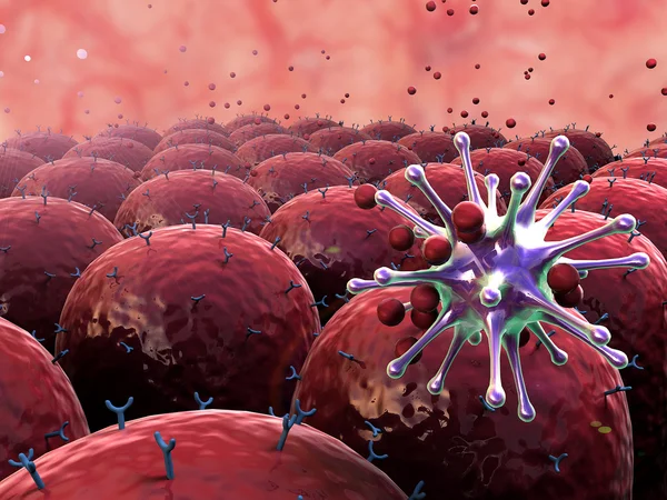
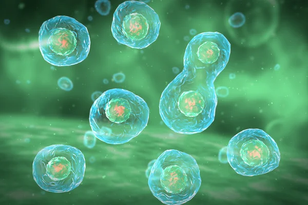
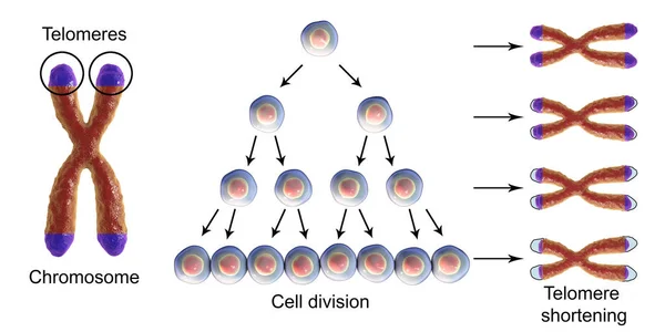
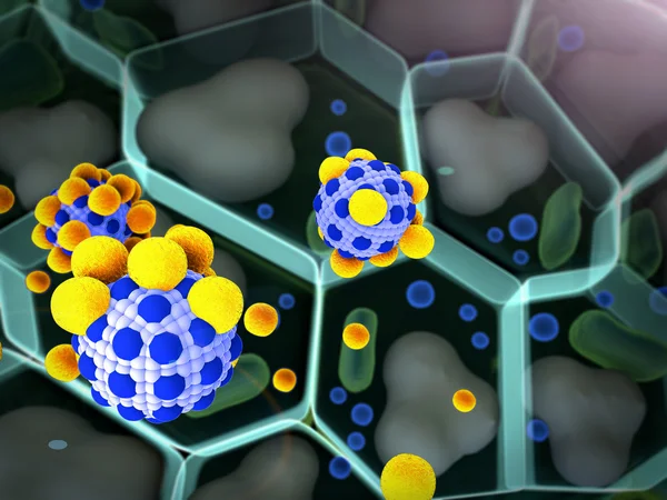
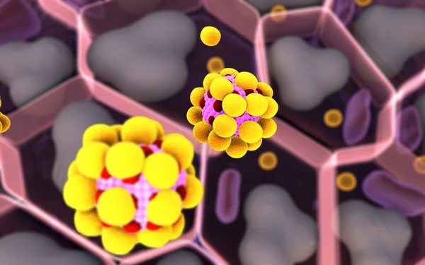
T Cells Are Involved In Cell-mediated Immunity, Whereas B Cells Are Primarily Responsible For Humoral Immunity 3D Render
Image, 1.32MB, 3840 × 2160 jpg

Previous << Page 2 >> Next