photo free
Home Images Vectors Videos

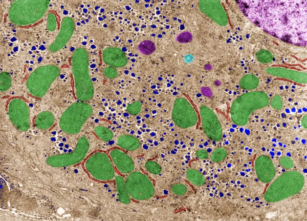
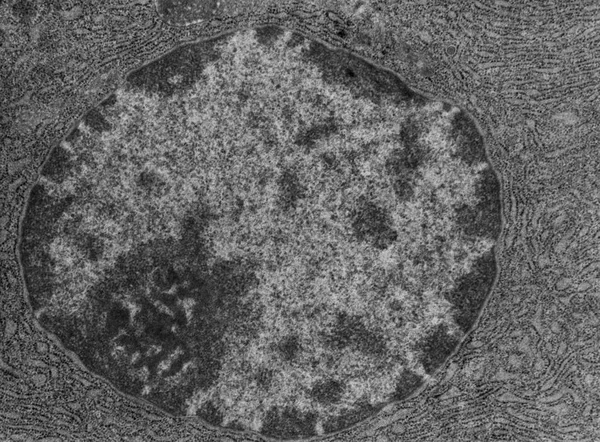
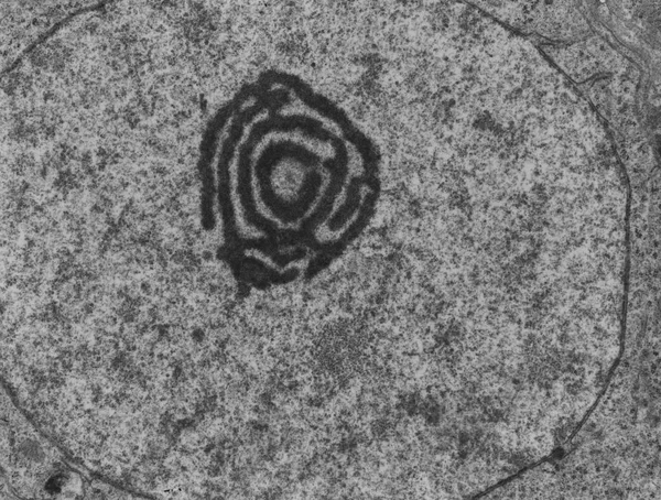
Stock image Cell Ultrastructure

stock image Flexible solar cells from ruthenium.

Flexible Solar Cells From Ruthenium.

Image, 2.24MB, 3000 × 2000 jpg
stock image Lysosomes and lipofuscin. False colour TEM

Lysosomes And Lipofuscin. False Colour TEM

Image, 5.63MB, 3772 × 2716 jpg
stock image Lysosomes and lipofuscin. False colour TEM

Lysosomes And Lipofuscin. False Colour TEM

Image, 8.95MB, 3772 × 2716 jpg
stock image Oogonium. Nucleolus, TEM

Oogonium. Nucleolus, TEM

Image, 8.96MB, 4285 × 3072 jpg
stock image Activated B lymphocyte. False colour TEM

Activated B Lymphocyte. False Colour TEM

Image, 11.24MB, 3616 × 2692 jpg
stock image Melanin granules in skin cell, TEM

Melanin Granules In Skin Cell, TEM

Image, 4.64MB, 3630 × 2693 jpg
stock image Melanin granules in skin cell. False colour TEM

Melanin Granules In Skin Cell. False Colour TEM

Image, 6.29MB, 3630 × 2693 jpg
stock image Oogonium. Nucleolus. FaLse colour TEM

Oogonium. Nucleolus. FaLse Colour TEM

Image, 13.14MB, 4285 × 3072 jpg
stock image Activated B lymphocyte, TEM

Activated B Lymphocyte, TEM

Image, 7.35MB, 3616 × 2692 jpg
stock image Cytoplasm. Cell organelles. False colour TEM

Cytoplasm. Cell Organelles. False Colour TEM

Image, 8.95MB, 3570 × 2705 jpg
stock image Cytoplasm. Cell organelles. False colour TEM

Cytoplasm. Cell Organelles. False Colour TEM

Image, 9.92MB, 3594 × 2586 jpg
stock image Cytoplasm. Cell organelles, TEM

Cytoplasm. Cell Organelles, TEM

Image, 6.08MB, 3594 × 2586 jpg
stock image Gonadotropic cell. False colour TEM

Gonadotropic Cell. False Colour TEM

Image, 11.76MB, 3840 × 2697 jpg
stock image Transmission electron micrograph (TEM) showing a desmosome (macula adherens) with prominent dense plaques where keratin intermediate filaments were attached.

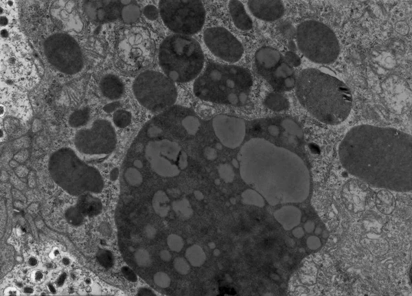
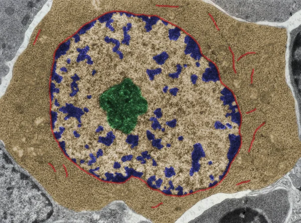
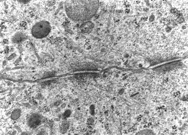
Transmission Electron Micrograph (TEM) Showing A Desmosome (macula Adherens) With Prominent Dense Plaques Where Keratin Intermediate Filaments Were Attached.

Image, 9.87MB, 4792 × 3438 jpg
stock image Nucleus and nucleolus, TEM

Nucleus And Nucleolus, TEM

Image, 7.37MB, 3640 × 2688 jpg
stock image Nucleolus, TEM

Nucleolus, TEM

Image, 7.21MB, 3621 × 2707 jpg
stock image Nucleolus. False colour TEM

Nucleolus. False Colour TEM

Image, 12.46MB, 3621 × 2707 jpg
stock image Nucleus and nucleolus. False colour TEM

Nucleus And Nucleolus. False Colour TEM

Image, 11.89MB, 3640 × 2688 jpg
stock image Nucleolus, TEM

Nucleolus, TEM

Image, 6.83MB, 3556 × 2692 jpg
stock image Neuron. Cell organelles. False colour TEM

Neuron. Cell Organelles. False Colour TEM

Image, 14.74MB, 4108 × 3072 jpg
stock image Cytoplasm. Cell organelles, TEM

Cytoplasm. Cell Organelles, TEM

Image, 5.62MB, 3570 × 2705 jpg
stock image Neuron. Cell organelles, TEM

Neuron. Cell Organelles, TEM

Image, 9.4MB, 4108 × 3072 jpg
stock image Transmission electron micrograph (TEM) showing two desmosomes (maculae adherentes) with prominent dense plaques where keratin intermediate filaments were attached.

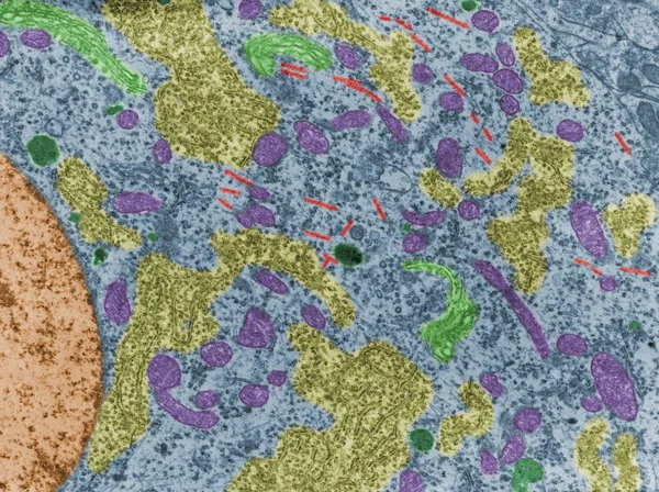
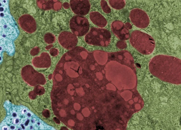
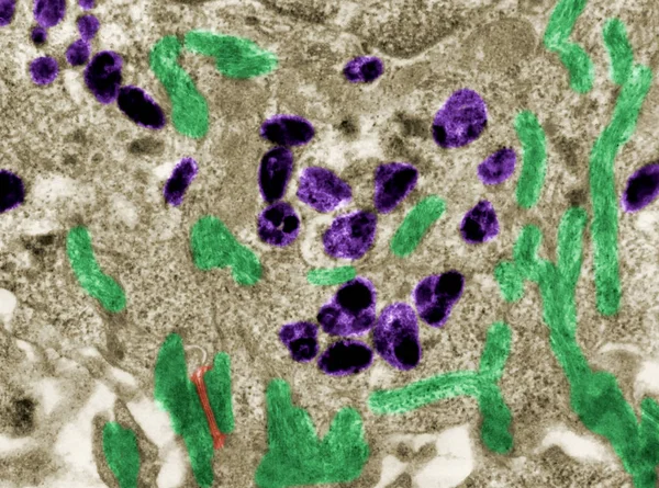
Transmission Electron Micrograph (TEM) Showing Two Desmosomes (maculae Adherentes) With Prominent Dense Plaques Where Keratin Intermediate Filaments Were Attached.

Image, 11.45MB, 4702 × 4020 jpg
stock image Rough endoplasmic reticulum. False colour TEM

Rough Endoplasmic Reticulum. False Colour TEM

Image, 12.91MB, 3840 × 2673 jpg
stock image Rough endoplasmic reticulum, TEM

Rough Endoplasmic Reticulum, TEM

Image, 7.8MB, 3840 × 2673 jpg
stock image An interesting photo taken with a microscope. Unmyelinated fibers in peripheral nerves. Longitudinal section. Hematoxylin and Eosin Stainit.

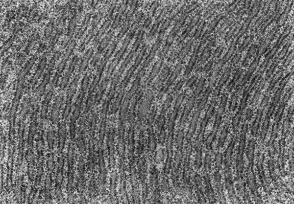
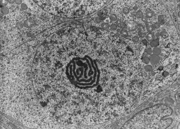
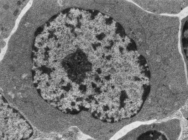
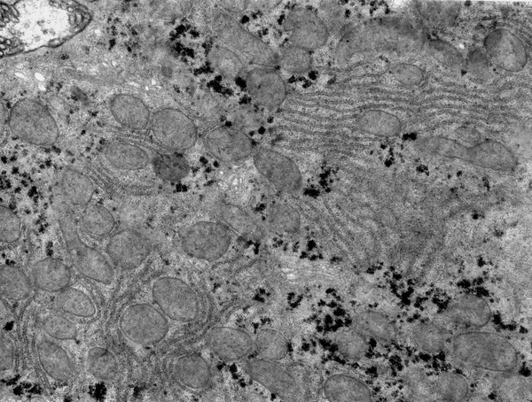
An Interesting Photo Taken With A Microscope. Unmyelinated Fibers In Peripheral Nerves. Longitudinal Section. Hematoxylin And Eosin Stainit.

Image, 2.42MB, 3000 × 2248 jpg
stock image Ultrastructure of centrosomes in cell, basic cell structure

Ultrastructure Of Centrosomes In Cell, Basic Cell Structure

Image, 0.25MB, 3025 × 2025 jpg
Page 1 >> Next