
Stock image Cervical Region page 3
Joint Pain Can Be Caused By Injury Affecting Any Of The Ligaments, Bursae, Or Tendons Surrounding The Joint.
Image, 7.73MB, 8192 × 8192 jpg

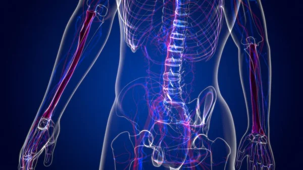
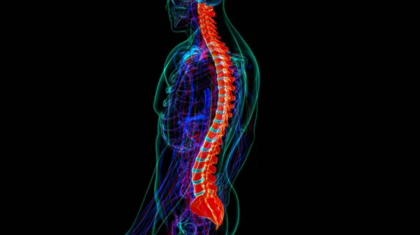
Human Skeleton Vertebral Column Cervical Vertebrae Anatomy 3D Illustration
Image, 1.94MB, 3840 × 2160 jpg
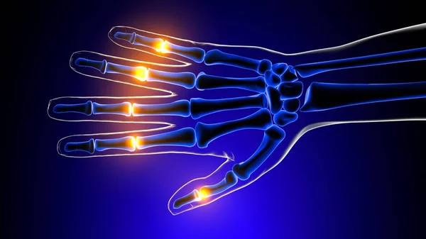
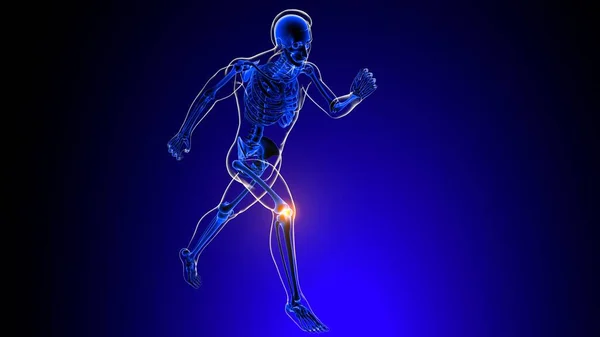
Proximal Interphalangeal Joints Pain Anatomy For Medical Concept 3D Illustration
Image, 2.34MB, 3840 × 2160 jpg


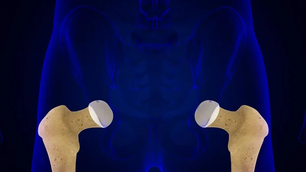
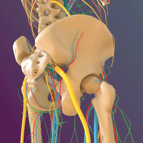
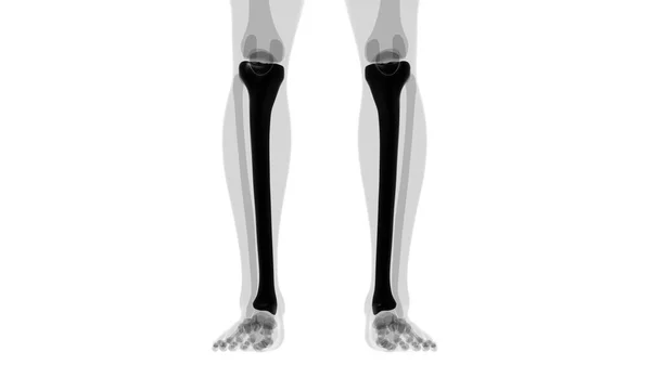
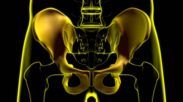
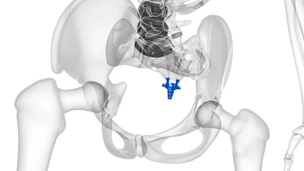
Human Skeleton Hip Or Pelvic Bone Anatomy For Medical Concept 3D Illustration
Image, 1.68MB, 3840 × 2160 jpg
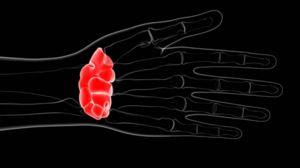
Human Skeleton Hand Wrist Carpals Bone Anatomy For Medical Concept 3D Illustration
Image, 1.01MB, 3840 × 2160 jpg
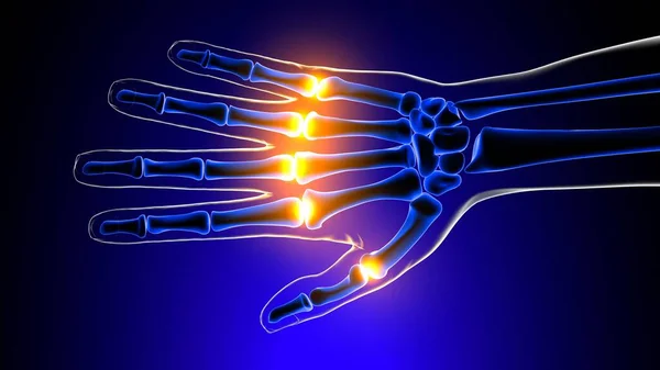
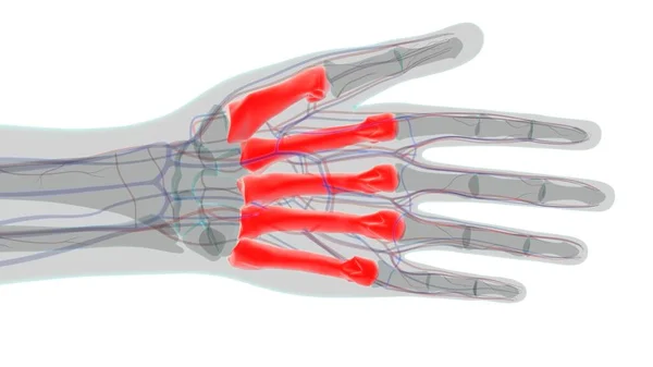
Metacarpophalangeal Joints Pain Anatomy For Medical Concept 3D Illustration
Image, 2.28MB, 3840 × 2160 jpg
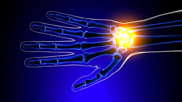
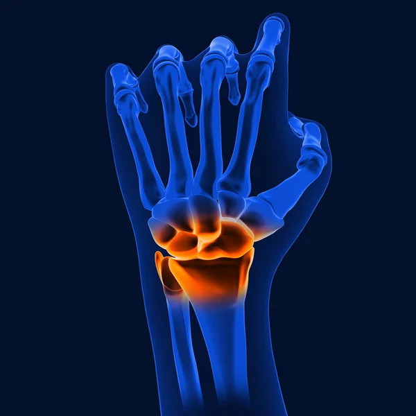
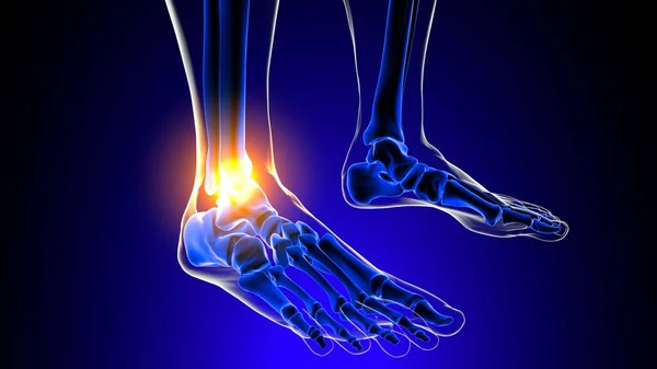
Wrist Or Carpal Joint Pain Anatomy For Medical Concept 3D Illustration
Image, 2.13MB, 3840 × 2160 jpg
Joint Pain Can Be Caused By Injury Affecting Any Of The Ligaments, Bursae, Or Tendons Surrounding The Joint.
Image, 6.96MB, 8192 × 8192 jpg

Human Skeleton Skull Sphenoid Bone Anatomy For Medical Concept 3D Illustration
Image, 1.48MB, 3840 × 2160 jpg

Human Skeleton Skull Mandible Bone Anatomy For Medical Concept 3D Illustration
Image, 1.52MB, 3840 × 2160 jpg
Human Skeleton Hand Matacarapls Bone Anatomy For Medical Concept 3D Illustration
Image, 1.66MB, 3840 × 2160 jpg
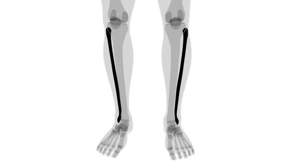
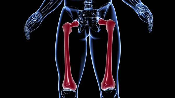
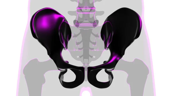
Human Skeleton Hip Or Pelvic Bone Anatomy For Medical Concept 3D Illustration
Image, 1.83MB, 3840 × 2160 jpg

Joint Pain Can Be Caused By Injury Affecting Any Of The Ligaments, Bursae, Or Tendons Surrounding The Joint.
Image, 8.19MB, 8192 × 8192 jpg

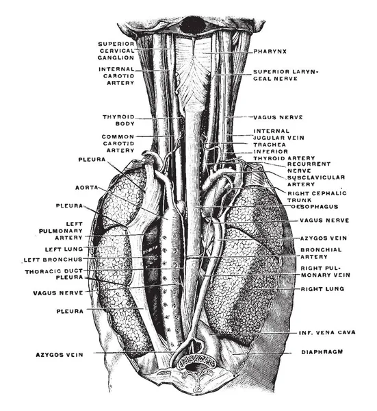
This Diagram Represents Position Of The Esophagus In The Cervical Region, Vintage Line Drawing Or Engraving Illustration.
Vector, 9.71MB, 7564 × 8229 eps

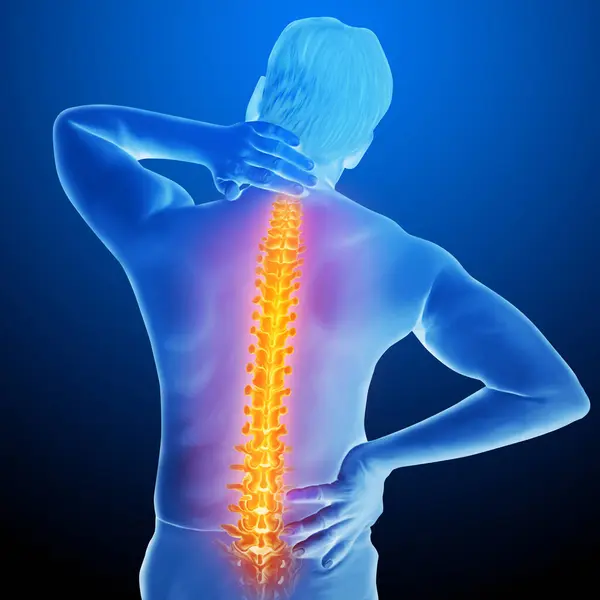
Spine Pain In Sacral And Cervical Region, Human Holding His Back In Area Pain, Logo Design. Spine Medicine And Backbone Health, Vector Design And Illustration
Vector, 0.72MB, 10000 × 8333 eps

Health Care, Medical Cross, Leaves And Running Man, Logo Design. Rehabilitation, Treatment, Musculoskeletal And Spine, Vector Design And Illustration
Vector, 0.71MB, 10000 × 8333 eps

Osteopath Performing Cervical Region Traction On Female Patient Lying On Table In Supine Position
Image, 15.05MB, 6913 × 4609 jpg

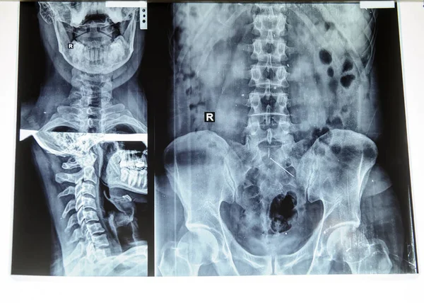
Multiple Side-view X-rays Of Human Necks, Displaying Cervical Vertebrae And Skulls
Image, 9.1MB, 5824 × 3264 jpg

Human Skeleton Vertebral Column Coccyx Or Tail Bone Anatomy 3D Illustration
Image, 1.34MB, 3840 × 2160 jpg
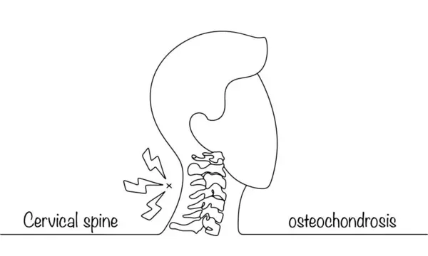
Silhouette Of A Man With Osteochodrosis Of The Cervical Spine. A Disease Of The Musculoskeletal System That Causes Discomfort. Simple Black And White Illustration.
Vector, 0.32MB, 5745 × 3448 eps

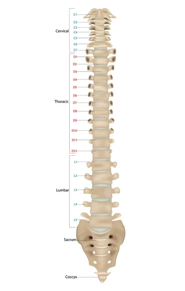
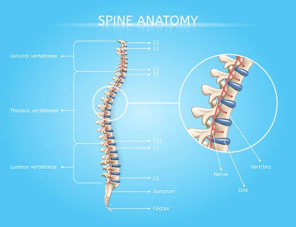
Human Skeleton Anatomy.Vertebral Column Of Human Body Anatomy Infograpic Diagram Including All Vertebra Cervical Thoracic Lumbar Sacral And Coccygeal
Vector, 15.88MB, 4000 × 5000 eps
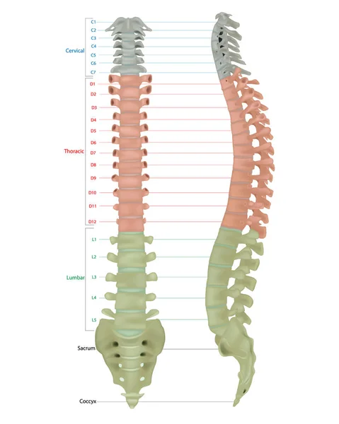
The Vertebral Column, Also Known As The Backbone Or Spine. The Human Vertebral Column And Its Regions Coccyx, Sacrum, Lumbar, Thoracic, . Spine Vertebrae. Anterior View. Front View.
Vector, 5.9MB, 3850 × 6050 eps
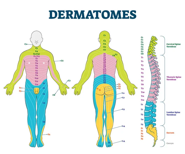
Dermatomes Vector Illustration. Labeled Educational Anatomical Skin Parts.
Vector, 6.15MB, 4800 × 4000 eps

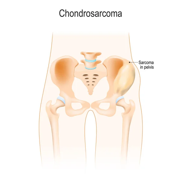
Chondrosarcoma Is A Cancer From Cartilage Cells. Malignant Neoplasm. Lumbar Region. Anatomy Of The Hip, Vertebra, And Pelvis. Vector Illustration For Biological, Science, Medical Use.
Vector, 5.22MB, 5400 × 5399 eps

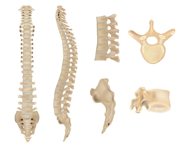
Anatomy Of Vertebral Column And Vertebrae. Human Spine Vertebral Bones. Detailed Medical Illustration. Skeletal System
Vector, 14.02MB, 6000 × 4800 eps

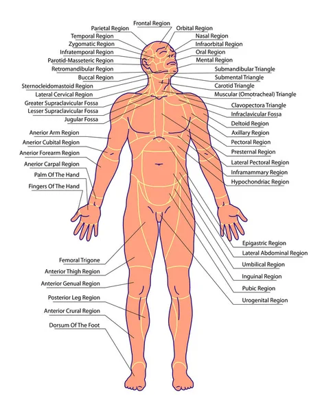
Drawing, Medical, Didactic Board Of General Anatomy Of Anatomy Surface Of The Human Body, Landmarks And Reference Lines, Body Region, Regional Anatomy, Anterior View
Image, 3.49MB, 3772 × 4817 jpg


Scoliosis Awareness Day Is Observed Every Year In June, It Is An Abnormal Lateral Curvature Of The Spine. It Is Most Often Diagnosed In Childhood Or Early Adolescence. Vector Illustration.
Vector, 14.19MB, 4000 × 4000 eps


Previous << Page 3 >> Next