
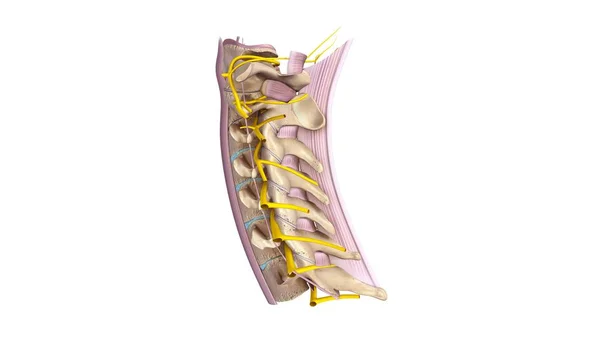
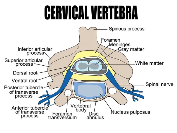
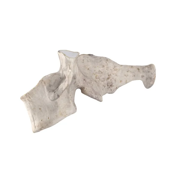
Stock image Cervical Vertebra
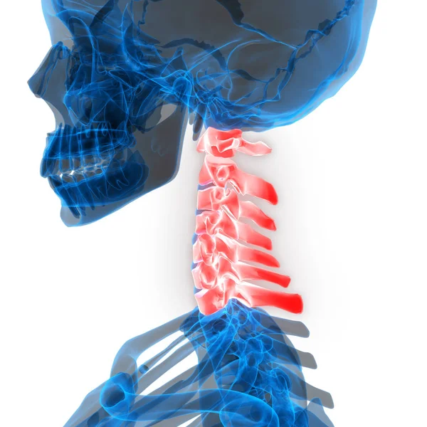
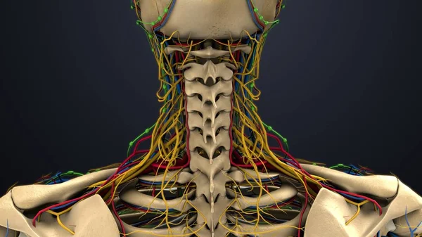
Human Skeleton System Vertebral Column Cervical Vertebrae Anatomy. 3D - Illustration
Image, 3.88MB, 4096 × 4096 jpg


Cervical Vertebrae Vector Illustration. Scheme With Skull, C1 Atlas, C2 Axis, C3, C4, C5, C6 And C7 Vertebra. Intervertebral Disc And Anterior Tubercle Diagram.
Vector, 2.67MB, 5000 × 6538 eps

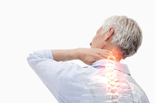
Young Muscular Fitness Man Touching And Grabbing His Neck And Upper Back Suffering Cervical Pain Isolated On Neutral Background. In Sport Injury Incorrect Posture Problems And Body Health Care.
Image, 6.85MB, 5289 × 3657 jpg


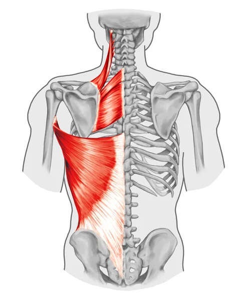
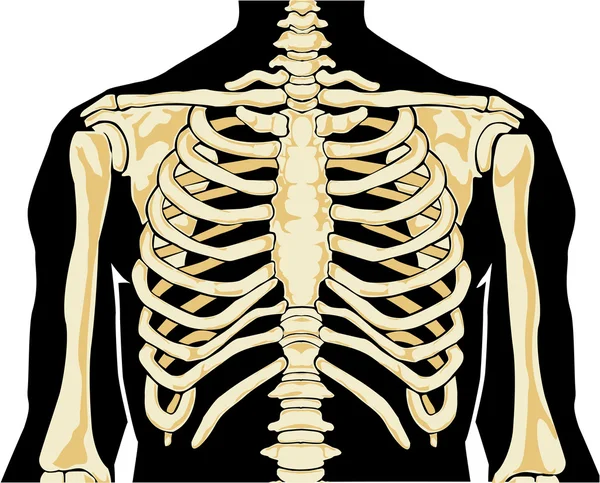
Rhomboid Minor And Rhomboid Major, Levator Scapulae And Latissimus Dorsi Muscles - Didactic Board Of Anatomy Of Human Bony And Muscular System, Posterior View
Image, 3.44MB, 4128 × 5000 jpg
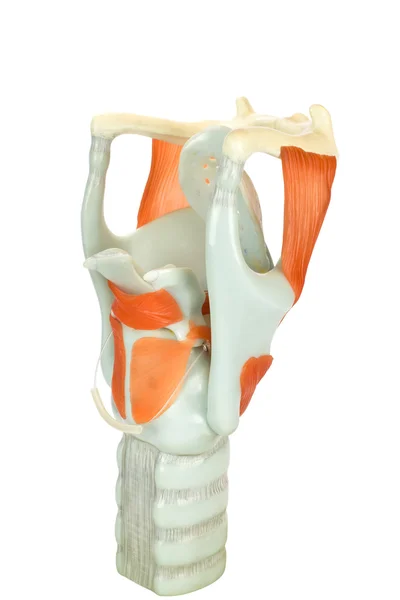
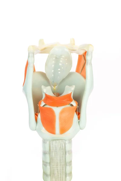
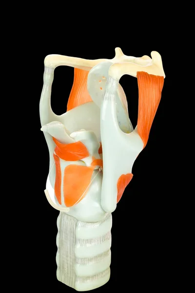
Artificial Model Of Human Larynx Or Voice Box Isolated On White Background
Image, 2.38MB, 3648 × 5472 jpg
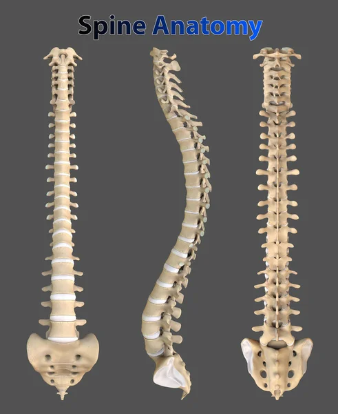
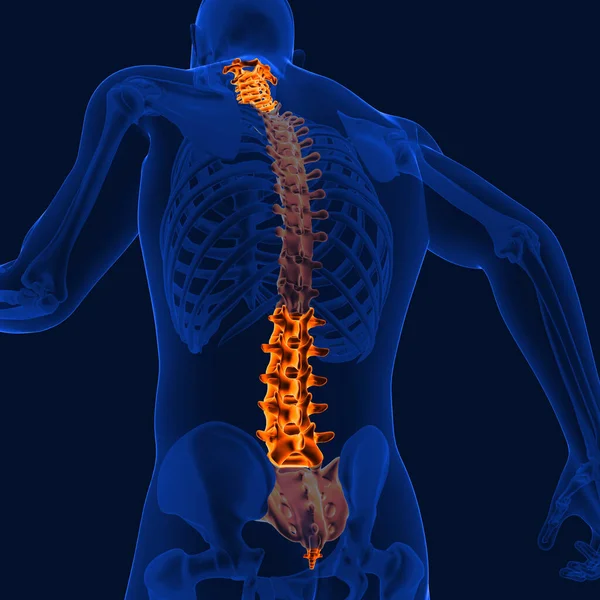
Spine Is Made Of 33 Individual Bones Stacked One On Top Of The Other. This Spinal Column Provides The Main Support For Human Body
Image, 5.43MB, 4600 × 5622 jpg
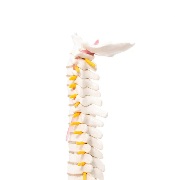
Cervical And Thoracic Spine On A White Background, Isolate. Osteochondrosis And Degenerative Changes In The Spine, Microspondylia
Image, 0.98MB, 3311 × 3399 jpg


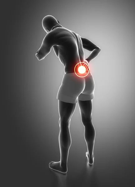
Joint Pain Can Be Caused By Injury Affecting Any Of The Ligaments, Bursae, Or Tendons Surrounding The Joint.
Image, 10.11MB, 8192 × 8192 jpg
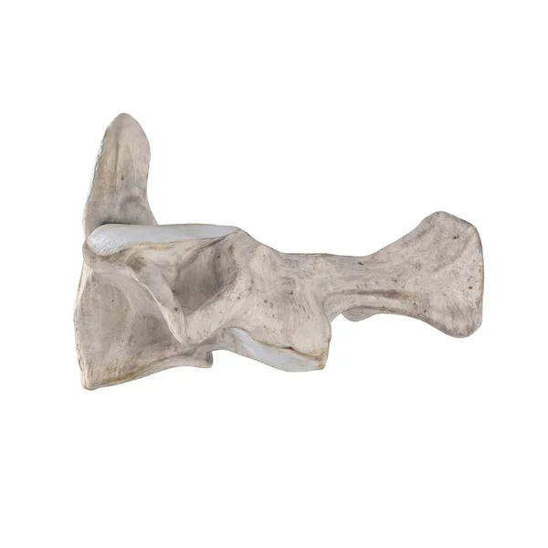
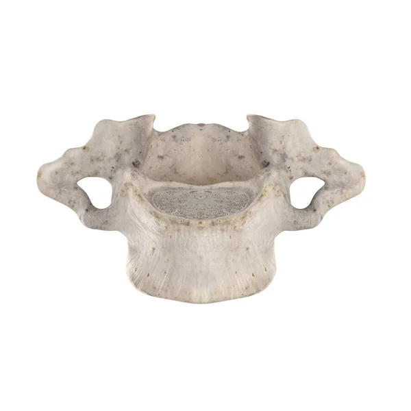
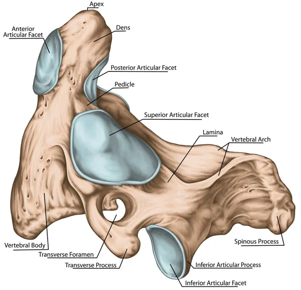
Didactic Board, Cervical Spine, Vertebral Morphology, Second Cervical Vertebra, Axis, Cervical Vertebrae, Dens, Odontoid, Anterior View
Image, 5.47MB, 5906 × 4880 jpg
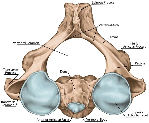
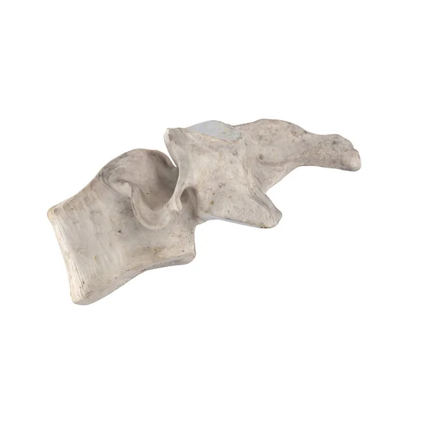
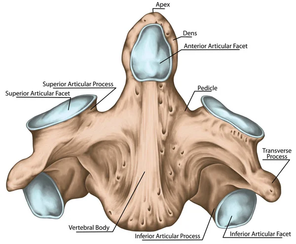
Didactic Board, Cervical Spine, Vertebral Morphology, Second Cervical Vertebra, Axis, Cervical Vertebrae, Transverse Foramen, Vertebral Foramen, Dens, Odontoid, Anterior And Posterior Articular Facet, Superior View
Image, 5.47MB, 5906 × 4884 jpg

X-ray C-spine Or X-ray Image Of Cervical Spine AP And Lateral View For Diagnostic Intervertebral Disc Herniation.
Image, 2.04MB, 5008 × 2325 jpg
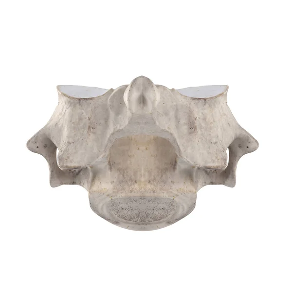
Didactic Board, Cervical Spine, Common Vertebral Morphology, Sixth Cervical Vertebra, Cervical Vertebrae, Anterior View
Image, 3.37MB, 5906 × 2588 jpg
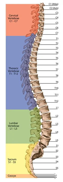
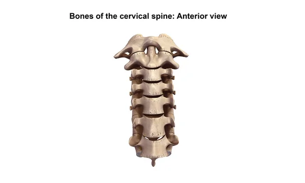
Didactic Board, Anatomy, Human Skeletal System, The Skeleton, Spine, The Bony Spinal Column, Columna Vertebralis, Vertebral Column, Vertebral Bones, Trunk Wall, Anatomical Body, Lateral View
Image, 6.92MB, 3295 × 9449 jpg


Didactic Board, Cervical Spine, Vertebral Morphology, Second Cervical Vertebra, Axis, Cervical Vertebrae, Anterior, Lateral And Superior View
Image, 4.52MB, 5906 × 3032 jpg
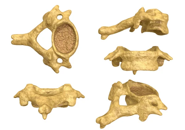
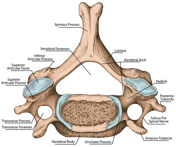
Morphology Of The Cervical Vertebra, Sixth Cervical Vertebra, Multiple Angles And Views
Image, 3.41MB, 3508 × 2480 jpg

Joint Pain Can Be Caused By Injury Affecting Any Of The Ligaments, Bursae, Or Tendons Surrounding The Joint.
Image, 8.19MB, 8192 × 8192 jpg
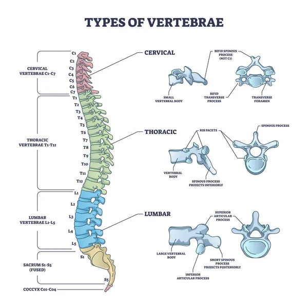
Types Of Vertebrae And Cervical, Thoracic And Lumbar Division Outline Diagram
Vector, 7.73MB, 4000 × 4000 eps

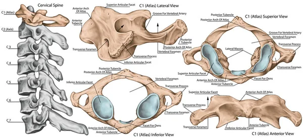
Didactic Board, Cervical Spine, Vertebral Morphology, First Cervical Vertebra, Atlas, Cervical Vertebrae, Anterior, Lateral, Superior And Inferior View
Image, 4.12MB, 5906 × 3405 jpg
Didactic Board, Cervical Spine, Vertebral Morphology, First Cervical Vertebra, Atlas, Cervical Vertebrae, Anterior, Lateral, Superior And Inferior View
Image, 4.48MB, 5906 × 2715 jpg
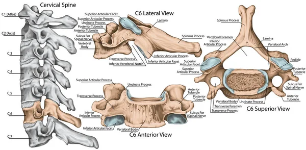
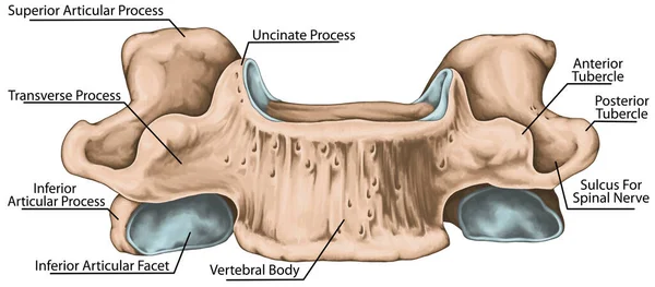
Didactic Board, Cervical Spine, Common Vertebral Morphology, Sixth Cervical Vertebra, Cervical Vertebrae, Anterior, Lateral And Superior View
Image, 4.61MB, 5906 × 2902 jpg
Didactic Board, Cervical Spine, Vertebral Morphology, Second Cervical Vertebra, Axis, Cervical Vertebrae, Transverse Foramen, Vertebral Foramen, Dens, Odontoid, Anterior And Posterior Articular Facet, Lateral View
Image, 6.33MB, 5906 × 5688 jpg
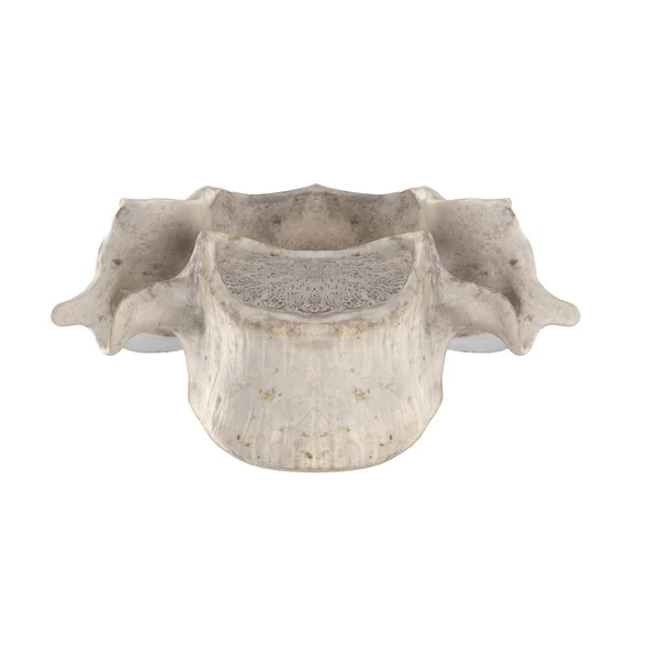
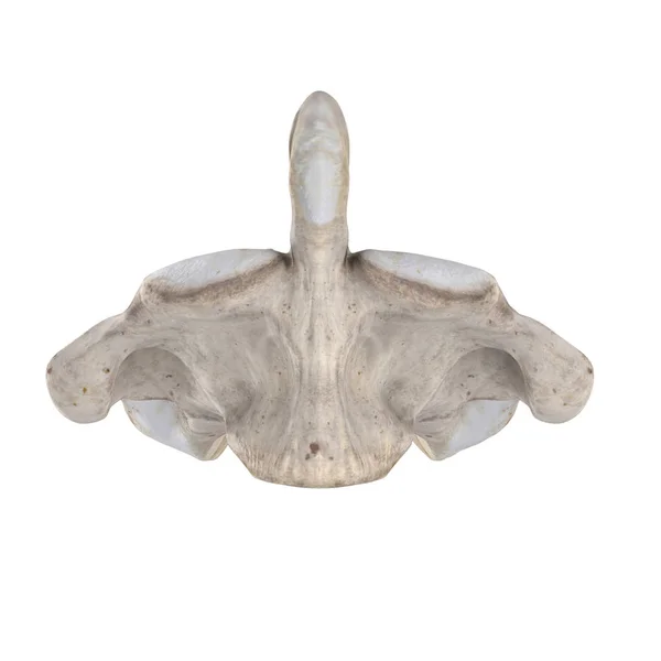
Didactic Board, Cervical Spine, Common Vertebral Morphology, Sixth Cervical Vertebra, Cervical Vertebrae, Transverse Foramen, Vertebral Foramen, Superior View
Image, 5.48MB, 5906 × 4962 jpg
Page 1 >> Next