
Stock image Corneal
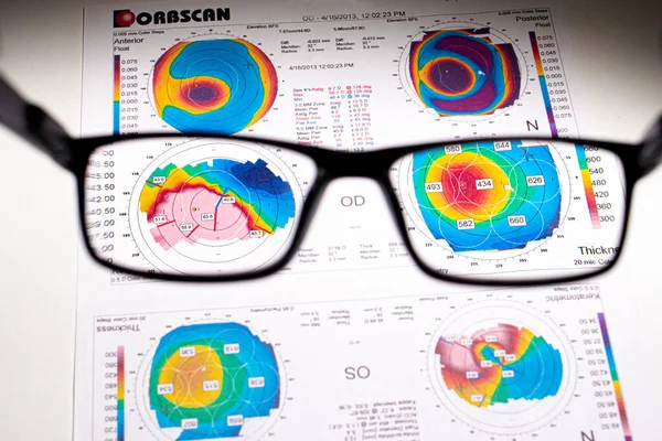
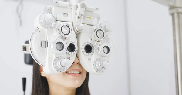
25.07.2020, Zarorizhzhya. Topography Of The Cornea Of The Eye, Layout. Keratoconus And Glasses. Keratotopography - Ophthalmological Examination.
Image, 8.75MB, 4720 × 3147 jpg
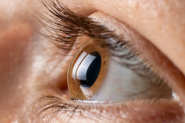
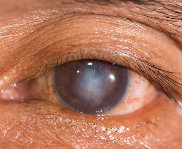
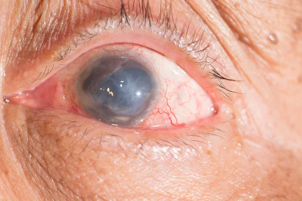
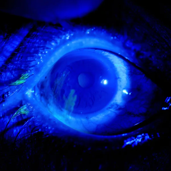
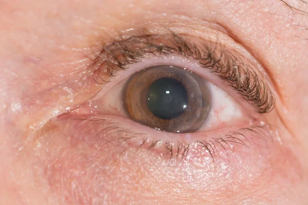
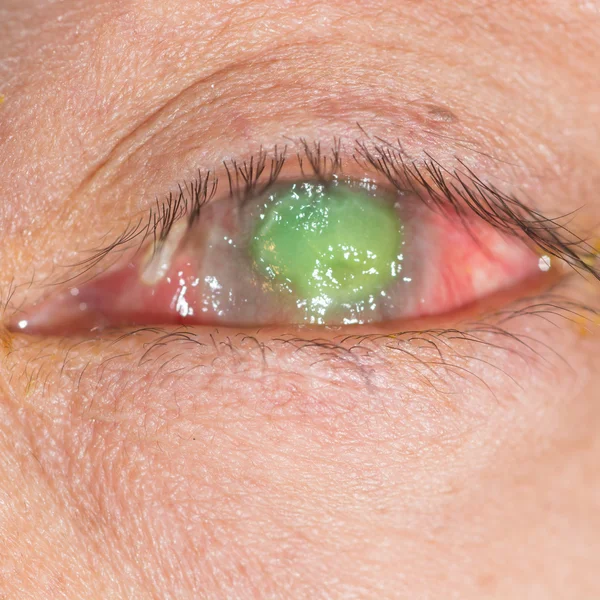
Keratoconus Of Eye, 3th Degree. Contortion Of The Cornea In The Form Of A Cone, Deterioration Of Vision, Astigmatism. Macro Close Up
Image, 16.59MB, 6000 × 4000 jpg


25.07.2020, Zarorizhzhya. Topography Of The Cornea Of The Eye, Layout. Keratoconus And Glasses. Keratotopography - Ophthalmological Examination.
Image, 10.41MB, 6000 × 4000 jpg


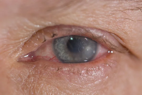
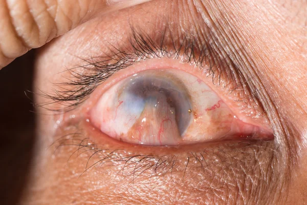
Keratoconus Of Eye, 4th Degree. Contortion Of The Cornea In The Form Of A Cone, Deterioration Of Vision, Astigmatism. Macro Close Up.
Image, 13.7MB, 12604 × 4000 jpg

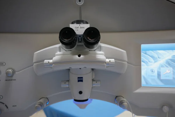
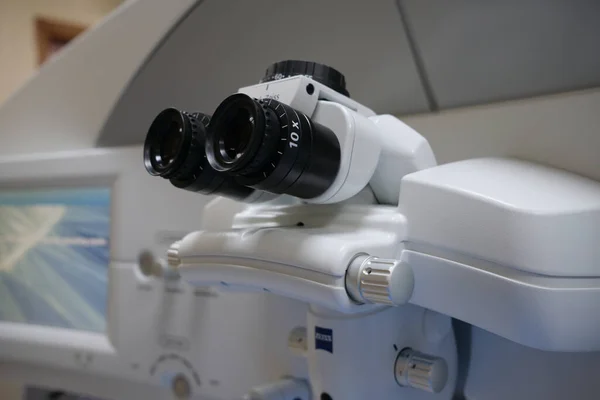
Basra, Iraq - MAY 25, 2021: Microscope Of Femto Smile Machine For Refractive Surgery Operations
Image, 12.05MB, 6240 × 4160 jpg

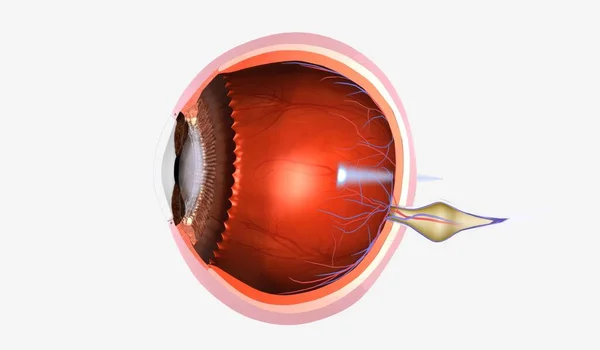
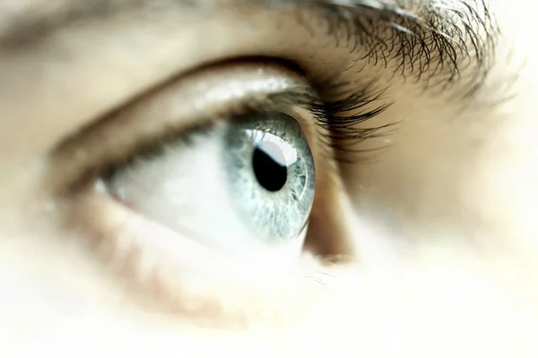
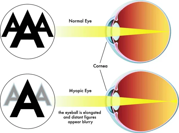
Presbyopia Is A Degenerative Eye Condition Characterized By The Gradual Inability To See Objects Up Close. 3D Rendering
Image, 5.08MB, 7191 × 4200 jpg

Basra, Iraq - MAY 25, 2021: Microscope Of Femto Smile Machine For Refractive Surgery Operations
Image, 12.25MB, 6240 × 4160 jpg

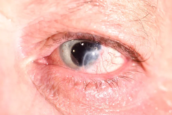
A Man Suffers From Pain In The Eye. Patient With Ophthalmic Disease, Uveitis, Optic Neuritis, Conjunctivitis, Or Eye Injury
Image, 6.02MB, 5425 × 3617 jpg
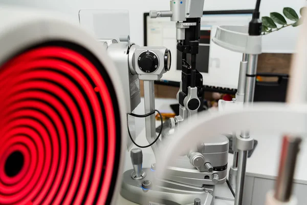
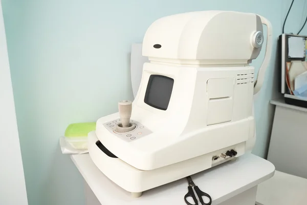
Corneal Topographer Shines Red Light And Slit Lamp For Topography Examination. Corneal Topography Eye Vision Test For Visual Description Of The Shape And Power Of The Cornea
Image, 6.52MB, 5655 × 3770 jpg
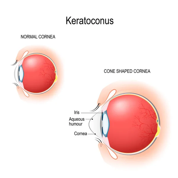
Keratoconus. Normal Cornea And Cone Shaped Cornea. Anatomy Of The Human Eye. Vertical Section Of The Eye And Eyelids. Schematic Diagram. Detailed Illustration. For Biological, Science, And Medical Use.
Vector, 4.19MB, 5120 × 5120 eps

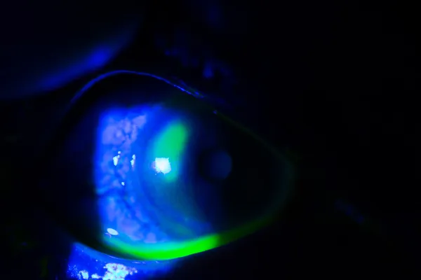
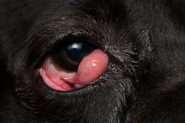
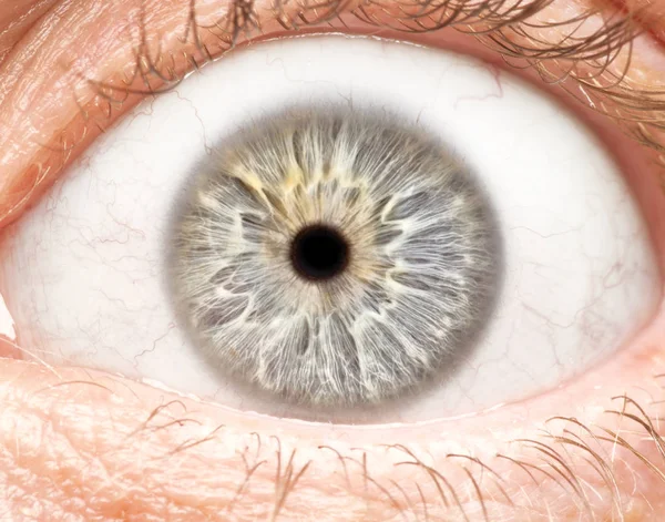
Woman Eye With Corneal Dystrophy, Keratoconus, Thinning Of The Cornea
Image, 3.29MB, 3087 × 2058 jpg
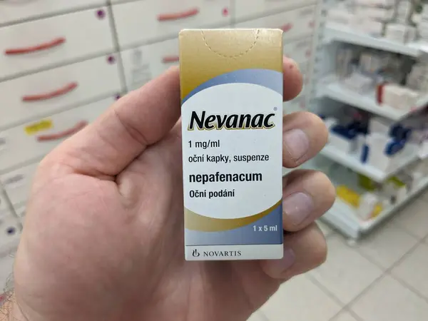
Prague, Czech Republic - July 10 2024: NEVANAC Box Of Medication With Nepafenac Active Substance By Alcon, Used For Treatment Of Postoperative Pain, Inflammation, Cataract Surgery.Eye Drops
Image, 2.09MB, 4032 × 3024 jpg

Results Of The Examination Of The Thickness And Curvature Of The Cornea In A Patient With Keratoconus
Image, 15.9MB, 6240 × 4160 jpg


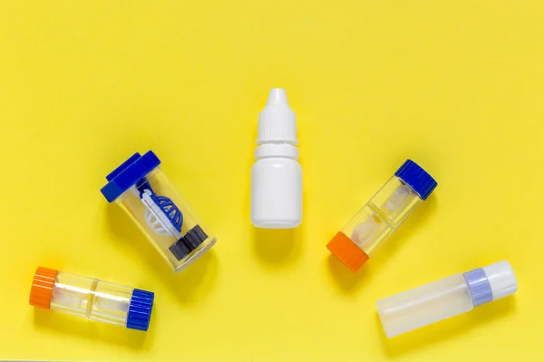
Colorful Flat Lay Of Vision Correction Lenses Equipment On Yellow Background : Eye Drops, Containers, Lenses, Twizzlers, Liquids
Image, 5.98MB, 4000 × 2666 jpg


Page 1 >> Next