photo free
Home Images Vectors Videos

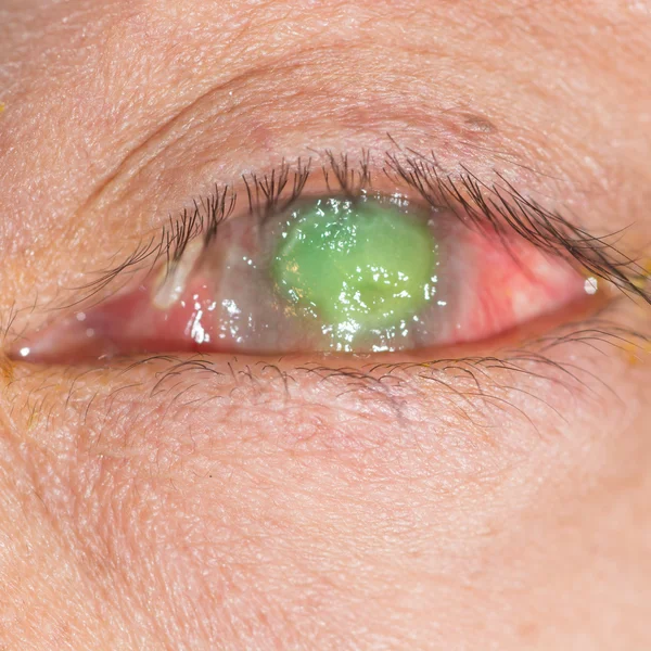
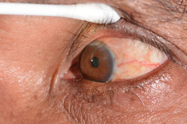
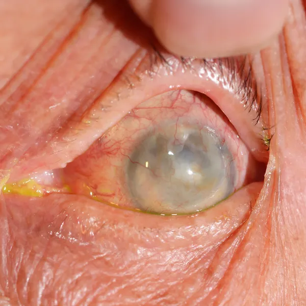
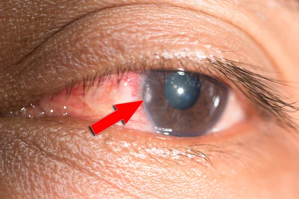
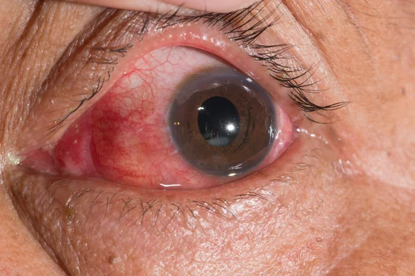
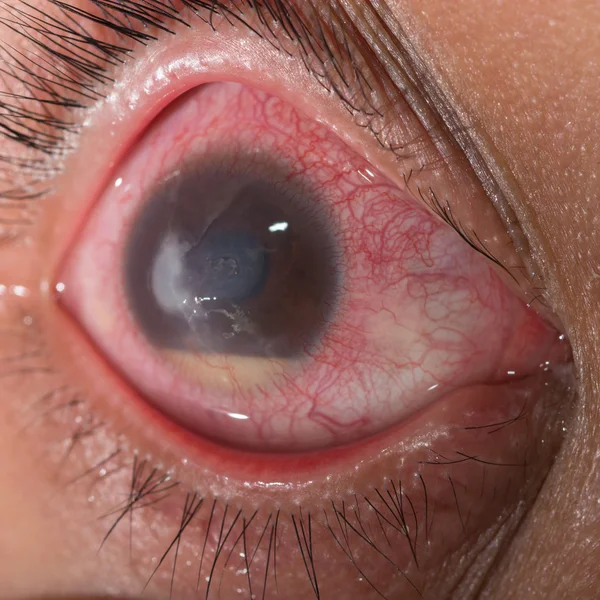
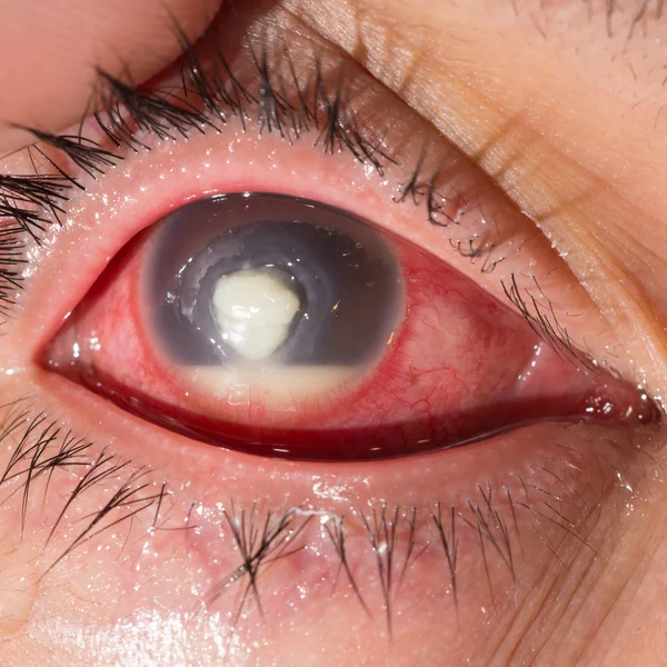
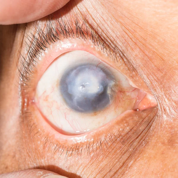
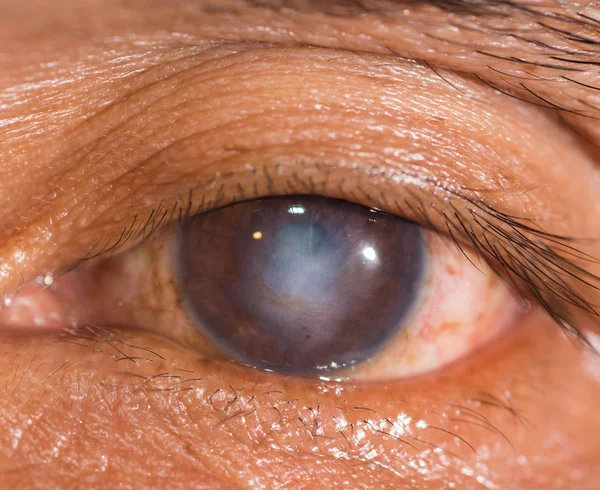
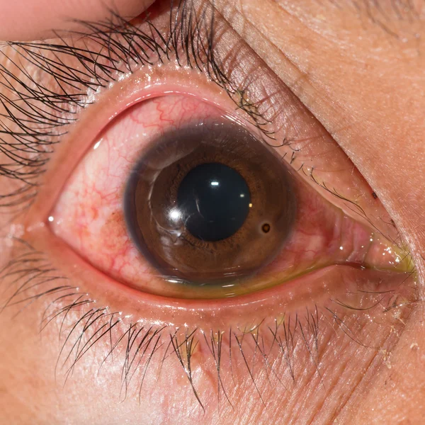
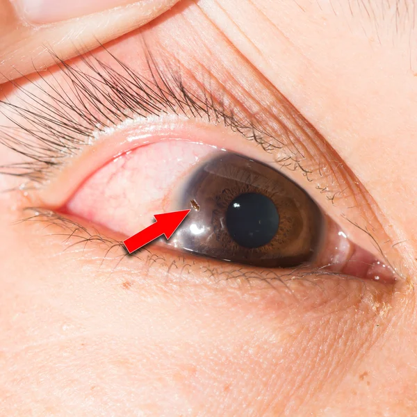
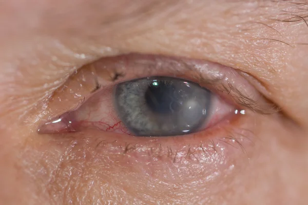
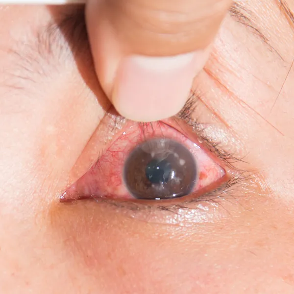
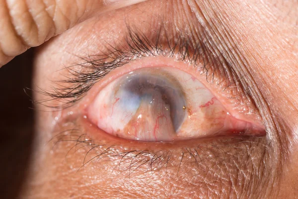
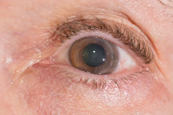
Stock image Corneal Disease

stock image Man checking condition of his eye

Man Checking Condition Of His Eye

Image, 6.47MB, 4786 × 3190 jpg
stock image phthisis bulbi at eye test

Phthisis Bulbi At Eye Test

Image, 10.87MB, 6000 × 4000 jpg
stock image Broken blood vessel in eye. Man with the Subconjunctival hemorrhage.

Broken Blood Vessel In Eye. Man With The Subconjunctival Hemorrhage.

Image, 3.67MB, 3008 × 3008 jpg
stock image Broken blood vessel in eye. Man with the Subconjunctival hemorrhage.

Broken Blood Vessel In Eye. Man With The Subconjunctival Hemorrhage.

Image, 2.96MB, 3056 × 3056 jpg
stock image Man checking condition of his eye

Man Checking Condition Of His Eye

Image, 5.91MB, 4800 × 3200 jpg
stock vector Amblyopia (lazy eye)

Amblyopia (lazy Eye)

Vector, 0.53MB, 5000 × 4482 eps
stock vector Medical illustration of the symptoms and consequences of amblyopia

Medical Illustration Of The Symptoms And Consequences Of Amblyopia

Vector, 0MB, 4000 × 3958 zip
stock image Keratoconus of eye, 3th degree. Contortion of the cornea in the form of a cone, deterioration of vision, astigmatism. Macro close up

Keratoconus Of Eye, 3th Degree. Contortion Of The Cornea In The Form Of A Cone, Deterioration Of Vision, Astigmatism. Macro Close Up

Image, 16.59MB, 6000 × 4000 jpg
stock image 25.07.2020, Zarorizhzhya. Topography of the cornea of the eye, layout. Keratoconus and glasses. Keratotopography - ophthalmological examination.

25.07.2020, Zarorizhzhya. Topography Of The Cornea Of The Eye, Layout. Keratoconus And Glasses. Keratotopography - Ophthalmological Examination.

Image, 8.75MB, 4720 × 3147 jpg
stock image eye with iris lost and corneal scar.

Eye With Iris Lost And Corneal Scar.

Image, 13.03MB, 7360 × 4912 jpg
stock image During eye examination

During Eye Examination

Image, 10.07MB, 6000 × 4000 jpg
stock image eye exam

Eye Exam

Image, 7.76MB, 4000 × 4000 jpg
stock image eye exam

Eye Exam

Image, 12.19MB, 6000 × 4000 jpg
stock image Eye exam

Eye Exam

Image, 7.64MB, 4000 × 4000 jpg
stock image During eye examination

During Eye Examination

Image, 6.69MB, 3854 × 3854 jpg
stock image corneal foreign body, eye exam

Corneal Foreign Body, Eye Exam

Image, 13.09MB, 6000 × 4000 jpg
stock image During eye examination

During Eye Examination

Image, 10.53MB, 6000 × 4000 jpg
stock image Eye examination.

Eye Examination.

Image, 9.47MB, 4654 × 4000 jpg
stock image eye exam

Eye Exam

Image, 7.42MB, 4000 × 4000 jpg
stock image eye exam

Eye Exam

Image, 7.03MB, 4000 × 4000 jpg
stock image corneal scar at eye test

Corneal Scar At Eye Test

Image, 9.25MB, 4000 × 4000 jpg
stock image eye exam

Eye Exam

Image, 6.54MB, 4000 × 4000 jpg
stock image corneal scar at eye test

Corneal Scar At Eye Test

Image, 8.57MB, 4000 × 4000 jpg
stock image corneal foreign body, eye exam

Corneal Foreign Body, Eye Exam

Image, 11.46MB, 6000 × 4000 jpg
stock image During eye examination

During Eye Examination

Image, 8.25MB, 4888 × 4000 jpg
stock image eye exam

Eye Exam

Image, 8.06MB, 4000 × 4000 jpg
stock image episcleritis at eye exam

Episcleritis At Eye Exam

Image, 8.68MB, 4000 × 4000 jpg
stock image Eye examination.

Eye Examination.

Image, 10.05MB, 6000 × 4000 jpg
stock image Eye examination.

Eye Examination.

Image, 10.05MB, 6000 × 4000 jpg
stock image eye exam

Eye Exam

Image, 8.15MB, 4865 × 4000 jpg
stock image eye exam

Eye Exam

Image, 8.19MB, 5247 × 4000 jpg
stock image Penetrating Keratoplasty at eye test

Penetrating Keratoplasty At Eye Test

Image, 7.51MB, 4000 × 4000 jpg
stock image eye exam

Eye Exam

Image, 7.4MB, 4599 × 4000 jpg
stock image eye exam

Eye Exam

Image, 8.62MB, 4000 × 4000 jpg
stock image Eye examination.

Eye Examination.

Image, 9.33MB, 4000 × 4000 jpg
stock image During eye examination

During Eye Examination

Image, 7.25MB, 4000 × 4000 jpg
stock image corneal ulcer

Corneal Ulcer

Image, 3.69MB, 3204 × 2592 jpg
stock image eye exam

Eye Exam

Image, 10.31MB, 6000 × 4000 jpg
stock image eye exam

Eye Exam

Image, 8.66MB, 4000 × 4000 jpg
stock image Scar inside eye

Scar Inside Eye

Image, 10.08MB, 6016 × 4000 jpg
stock image eye exam, fungal corneal ulcer

Eye Exam, Fungal Corneal Ulcer

Image, 6.76MB, 4000 × 4000 jpg
stock image corneal scar at eye test

Corneal Scar At Eye Test

Image, 8.85MB, 4000 × 4000 jpg
stock image Herpes keratitis at eye test

Herpes Keratitis At Eye Test

Image, 9.58MB, 4000 × 4000 jpg
stock image Eye exam

Eye Exam

Image, 10.12MB, 6000 × 4000 jpg
stock image During eye examination

During Eye Examination

Image, 10.38MB, 6000 × 4000 jpg
stock image eye exam, fungal corneal ulcer

Eye Exam, Fungal Corneal Ulcer

Image, 7.56MB, 4000 × 4000 jpg
stock image Eye examination

Eye Examination

Image, 2.85MB, 2867 × 2867 jpg
stock image During eye examination

During Eye Examination

Image, 11.47MB, 6000 × 4000 jpg
stock image Penetrating Keratoplasty at eye test

Penetrating Keratoplasty At Eye Test

Image, 10.96MB, 6000 × 4000 jpg
stock image keratitis during eye examination.

Keratitis During Eye Examination.

Image, 9.48MB, 4000 × 4000 jpg
Page 1 >> Next