
Stock image Corneal Epithelium


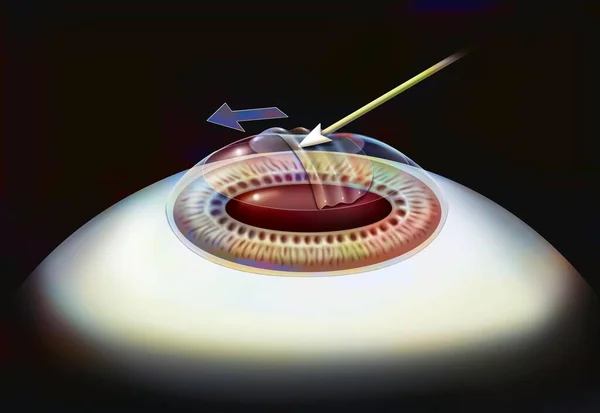
Eye, Surgery, Photokeractectomy Step 3: The Corneal Epithelium Layer Is Detached.
Image, 0.93MB, 4567 × 3150 jpg
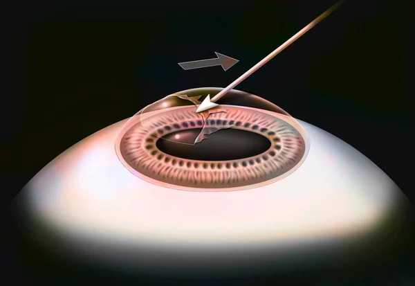
Eye, Refractive Photokeratectomy And Lasek, Step 1: Application Of Alcohol To The Cornea.
Image, 0.86MB, 3651 × 3020 jpg

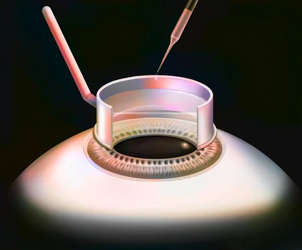
Eye, Surgery, Photokeractectomy Step 2: The Surgeon Scrapes The Surface Layer Of The Cornea.
Image, 1.04MB, 4567 × 3780 jpg

Eye, Surgery, Lasek Step 4: Replacement Of The Corneal Epithelium Layer.
Image, 0.91MB, 4567 × 3150 jpg
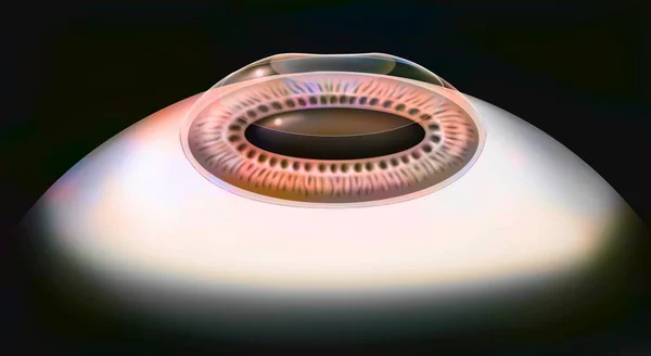
Eye, Surgery, Photokeractectomy Step 2: The Eye After The Refractive Photokeratectomy Operation.
Image, 1.14MB, 5510 × 3020 jpg
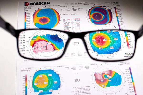
25.07.2020, Zarorizhzhya. Topography Of The Cornea Of The Eye, Layout. Keratoconus And Glasses. Keratotopography - Ophthalmological Examination.
Image, 8.75MB, 4720 × 3147 jpg

25.07.2020, Zarorizhzhya. Topography Of The Cornea Of The Eye, Layout. Keratoconus And Glasses. Keratotopography - Ophthalmological Examination.
Image, 10.41MB, 6000 × 4000 jpg


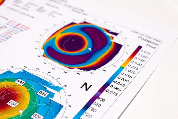
Thinning Corneal Dystrophy, Eye Keratotopography. A Rare Eye Disease.
Image, 13.04MB, 5939 × 3959 jpg

25.07.2020, Zarorizhzhya. Topography Of The Cornea Of The Eye, Layout. Keratoconus Of The Right Eye Is 3rd Degree And The Left Eye Is 2 Degrees.
Image, 13.44MB, 6000 × 4000 jpg
07.25.2020, Zarorizhzhya, Ukraine. Doctor Ophthalmologist Holds A Corneal Topography Examination For Keratoconus.
Image, 11.83MB, 6000 × 4000 jpg
25.07.2020, Zarorizhzhya. Topography Of The Cornea Of The Eye, Layout. Keratoconus Of The Right Eye Is 3rd Degree And The Left Eye Is 2 Degrees.
Image, 13.72MB, 6000 × 4000 jpg

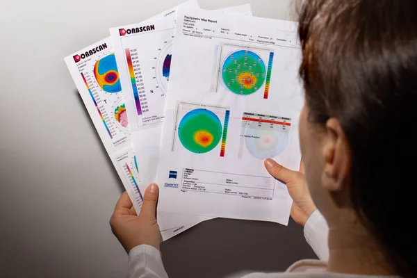
07.25.2020, Zarorizhzhya, Ukraine. A Patient With Keratoconus Holds An Ophthalmologic Examination Report. Keratometry, Corneal Dystrophy, Visual Impairment. A Rare Eye Disease.
Image, 11.49MB, 6000 × 4000 jpg
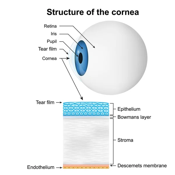
Structure Of The Cornea Medical Vector Illustration On White Background
Vector, 1.5MB, 5000 × 5000 eps
Page 1 >> Next