
Stock image Ct Lumbar

CT Lumbar Spine Or L-S Spine 3D Rendering Image Sagittal View 3D Rendering . Clipping Path.
Image, 1.54MB, 3353 × 4673 jpg

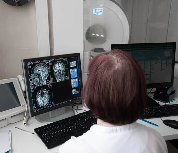
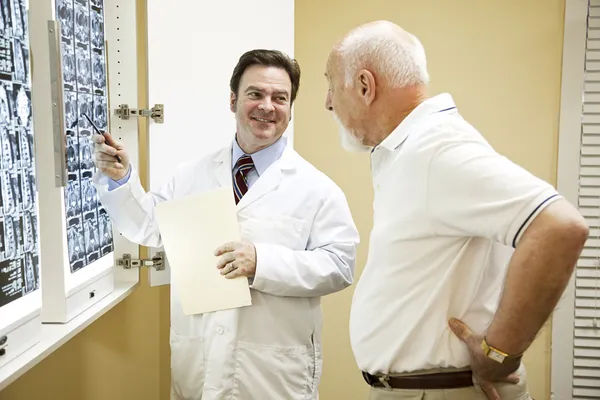
Professional Scientists Work In The Brain Research Laboratory. Neurologists Neuroscientists Surrounded By Monitors Showing CT, MRI Scans Having Discussions And Working On Personal Computers.
Image, 5.14MB, 4635 × 4000 jpg

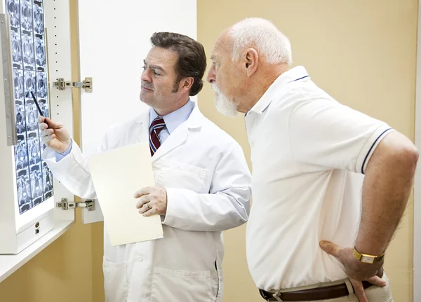
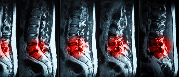
Doctor Analyzes The Results Of Magnetic Resonance Imaging Of A Patient Spine With Chronic Back Pain. The MRI Shows Degenerative Changes Of Spines, Lumbar Discs Herniation And Nerve Roots Compression.
Image, 1.08MB, 3770 × 2121 jpg

Doctor Analyzes The Results Of Magnetic Resonance Imaging Of A Patient Spine With Chronic Back Pain. The MRI Shows Degenerative Changes Of Spines, Lumbar Discs Herniation And Nerve Roots Compression.
Image, 2.17MB, 3840 × 2160 jpg
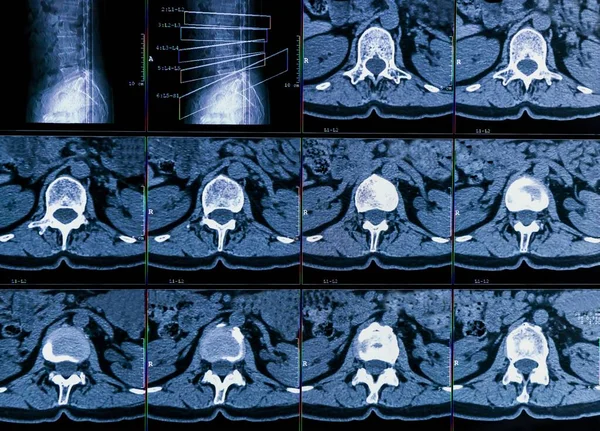
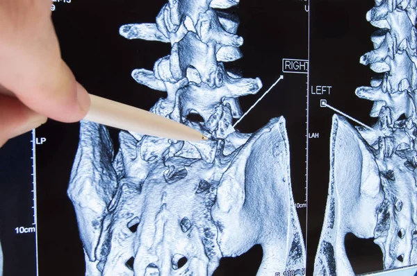
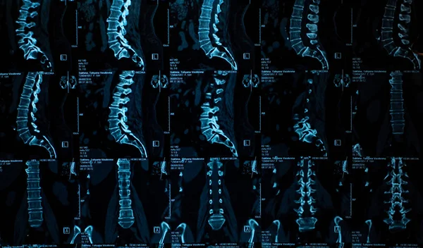
Results Of Computer Tomography Or CT Imaging Of Human Spine Of A Patient With Chronic Back Pain, Shows Degenerative Changes Of Spines, Lumbar Discs Herniation And Nerve Roots Compression
Image, 15.56MB, 6759 × 4861 jpg
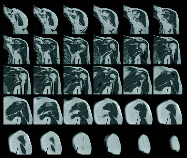
Magnetic Resonance Imaging Of Left Shoulder Rotator Cuff Tear With Suspected Lipoma Of Left Shoulder Science And Education Mri Shoulder Background,Medical.
Image, 10.26MB, 6408 × 5403 jpg
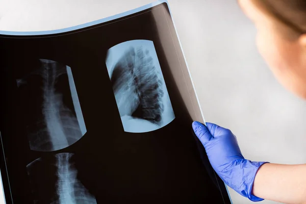
Female Doctor Examining X-ray Film (scan) Thoracic. Concept Of Medical Service, Diagnosis And Treatment. Scoliosis Or Osteochondrosis Detection Test. Copy Space For Text
Image, 6.28MB, 5184 × 3456 jpg

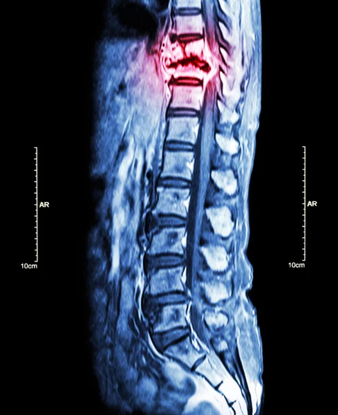
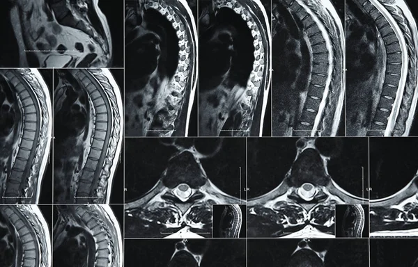
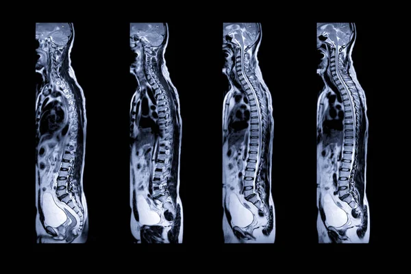
Spine Metastasis ( Cancer Spread To Thoracic Spine ) ( MRI Of Thoracic And Lumbar Spine : Show Thoracic Spine Metastasis And Compress Spinal Cord ( Myelopathy ) ) ( Sagittal Plane )
Image, 9.15MB, 6500 × 4210 jpg
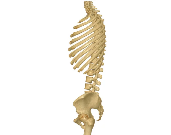
CT Scan Of Whole Spine 3D Rendering Showing Profile Human Spine. Musculoskeletal System Human Body. Structure Spine. Studying Problem Disease And Treatment Methods. Isolated On White Background. Clipping Path.
Image, 1.59MB, 3840 × 3036 jpg
Spine Metastasis ( Cancer Spread To Thoracic Spine ) ( MRI Of Thoracic And Lumbar Spine : Show Thoracic Spine Metastasis And Compress Spinal Cord ( Myelopathy ) ) ( Sagittal Plane )
Image, 5.24MB, 3432 × 4210 jpg
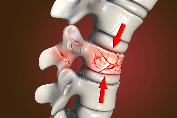
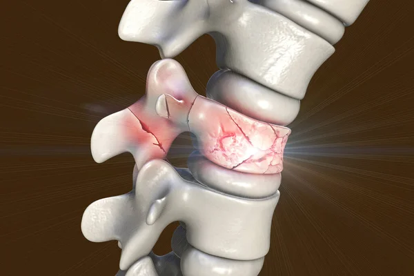
Spinal Fracture, Traumatic Vertebral Injury, 3D Illustration. Compression Fracture Of The Spine
Image, 7.57MB, 6000 × 4000 jpg
Myelography Is Particularly Sensitive At Detecting Small Disk Herniations Compressing Nerves Of The Spine And Spinal Cord Injury.
Image, 3.14MB, 4816 × 3719 jpg
Spine Metastasis ( Cancer Spread To Thoracic Spine ) ( MRI Of Thoracic And Lumbar Spine : Show Thoracic Spine Metastasis And Compress Spinal Cord ( Myelopathy ) ) ( Sagittal Plane )
Image, 5.1MB, 3432 × 4210 jpg

Spinal Fracture, Traumatic Vertebral Injury, 3D Illustration. Compression Fracture Of The Spine
Image, 5.64MB, 6000 × 4000 jpg


Spine Metastasis ( Cancer Spread To Thoracic Spine ) ( MRI Of Thoracic And Lumbar Spine : Show Thoracic Spine Metastasis And Compress Spinal Cord ( Myelopathy ) ) ( Sagittal Plane )
Image, 4.52MB, 3432 × 4210 jpg
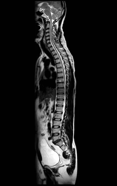
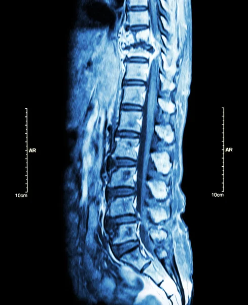
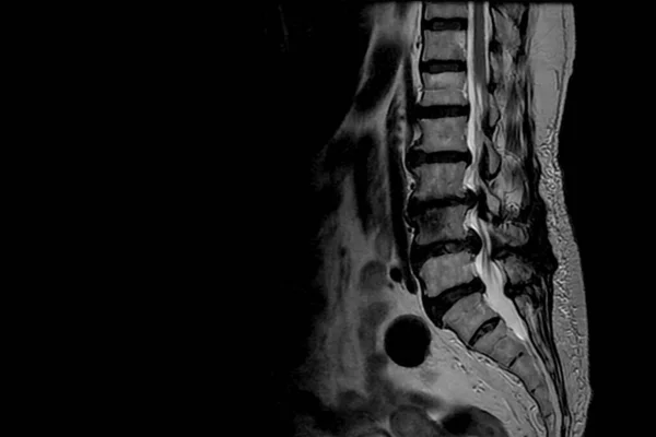
MRI Of Whole Spine T2W Sagittal Plane For Diagnostic Spinal Cord Compression.
Image, 4.7MB, 4624 × 7314 jpg

Spinal Fracture, Traumatic Vertebral Injury, 3D Illustration. Compression Fracture Of The Spine
Image, 11.05MB, 6000 × 4000 jpg

Collection Of CT Lumbar Or L-S Spine 3D Rendering Image Front , Back And Lateral View Showing Compression Fractures At L2. 3D Illustration.
Image, 2.45MB, 3944 × 2680 jpg

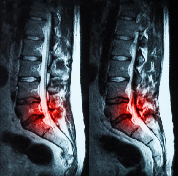
MRI Of Lumbar & Thoracic Spine : Show Fracture Of Thoracic Spine And Compress Spinal Cord ( Myelopathy )
Image, 8.02MB, 4707 × 2542 jpg
Scan Of Lumbosacral Spine. Doctor Pointed On Area Of Lumbar, Where Pathology Is Detected, Such As Radiculitis, Radiculopathy, Hernia, Low Back Pain. Diagnosis Of Spine Diseases By Radiology
Image, 9.72MB, 6016 × 4000 jpg
Close Up Magnetic Resonance Imaging Or Computed Tomography Scan Of Human Lumbar Spine
Image, 8.28MB, 5120 × 3283 jpg
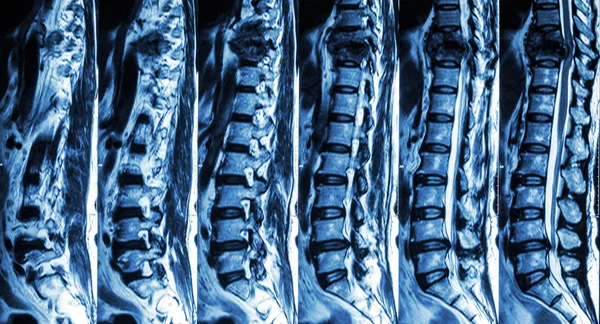
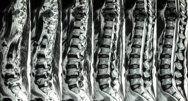
MRI Scans Of The Lumbosacral Spine.. MRI Shows Degenerative Changes In The Spine, Hernia Of The Lumbar Discs And Compression Of The Nerve Roots.
Image, 3.03MB, 3173 × 1861 jpg

Spine Metastasis ( Cancer Spread To Thoracic Spine ) ( MRI Of Thoracic And Lumbar Spine : Show Thoracic Spine Metastasis And Compress Spinal Cord ( Myelopathy ) ) ( Sagittal Plane )
Image, 4.32MB, 3432 × 4210 jpg

Spinal Fracture, Traumatic Vertebral Injury, 3D Illustration. Compression Fracture Of The Spine
Image, 6.13MB, 5752 × 3835 jpg

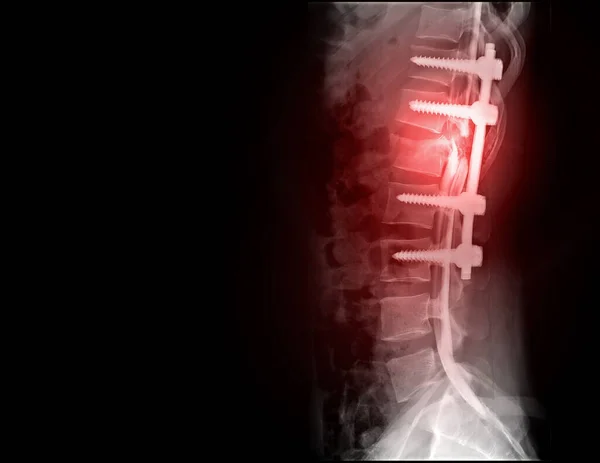
Scan Of Lumbosacral Spine In Lateral Projection Shown Spinal Canal Stenosis .Decrease In Disc Space , Bony Spur Formation And Blank Area At Right Side. Spinal Cord Compression.
Image, 0.28MB, 3000 × 2000 jpg
MRI Of Lumbar & Thoracic Spine : Show Fracture Of Thoracic Spine And Compress Spinal Cord ( Myelopathy )
Image, 8.34MB, 4707 × 2542 jpg
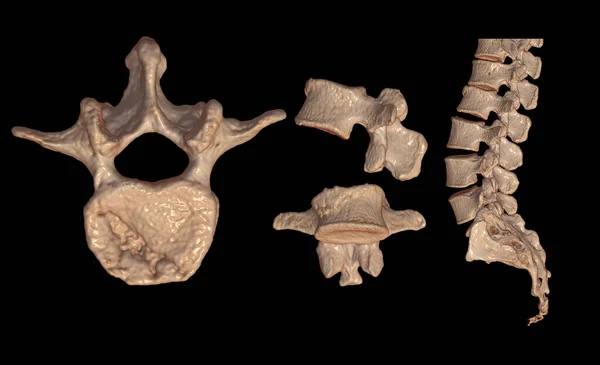
Collection Of CT Lumbar Or L-S Spine 3D Rendering Image Showing Compression Fractures At L2. 3D Illustration.
Image, 2.75MB, 4407 × 2680 jpg
MRI Of Lumbar & Thoracic Spine : Show Fracture Of Thoracic Spine And Compress Spinal Cord ( Myelopathy )
Image, 6.13MB, 4707 × 2542 jpg

Doctor Examines MRI Of Lumbar Spine With Pinched Discs Of Spine And Nerves, Points At Problem Areas By Pen, Close-up
Image, 2.15MB, 3002 × 2001 jpg
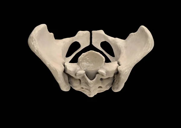
Pelvis, Human Skeleton, Female Pelvic Bone Anatomy, Hip, 3D Artwork, Bones Labeled Anatomy Top View
Image, 0.95MB, 3539 × 2500 jpg


Doctor With Human Spine Anatomy Model. Spinal Cord Disorder And Disease, Back Pain, Lumbar, Sacral Pelvis, Cervical Neck, Thoracic, Coccyx, Orthopedist, Chiropractic, Office Syndrome And Health
Image, 4.83MB, 6963 × 3060 jpg

Adult Female With Muscle Pain On Gray Background. Elderly Woman Having Back Body Ache Due To Piriformis Syndrome, Low Back Pain And Spinal Compression. Office Syndrome And Medical Concept
Image, 7.43MB, 7008 × 3080 jpg

Doctor With Human Spine Anatomy Model. Spinal Cord Disorder And Disease, Back Pain, Lumbar, Sacral Pelvis, Cervical Neck, Thoracic, Coccyx, Orthopedist, Chiropractic, Office Syndrome And Health
Image, 11.45MB, 5336 × 3557 jpg

Doctor With Human Spine Anatomy Model. Spinal Cord Disorder And Disease, Back Pain, Lumbar, Sacral Pelvis, Cervical Neck, Thoracic, Coccyx, Orthopedist, Chiropractic, Office Syndrome And Health
Image, 4.87MB, 6936 × 3048 jpg

Doctor With Human Spine Anatomy Model. Spinal Cord Disorder And Disease, Back Pain, Lumbar, Sacral Pelvis, Cervical Neck, Thoracic, Coccyx, Orthopedist, Chiropractic, Office Syndrome And Health
Image, 8.59MB, 6831 × 4554 jpg

Woman With Human Spine Anatomy Model. Spinal Cord Disorder And Disease, Back Pain, Lumbar, Sacral Pelvis, Cervical Neck, Thoracic, Coccyx, Orthopedist, Chiropractic, Office Syndrome And Health
Image, 6.45MB, 4672 × 4672 jpg

Magnetic Resonance Imaging Of Left Shoulder Rotator Cuff Tear With Suspected Lipoma Of Left Shoulder Science And Education Mri Shoulder Background,Medical.
Image, 7.69MB, 5724 × 4000 jpg
Page 1 >> Next