
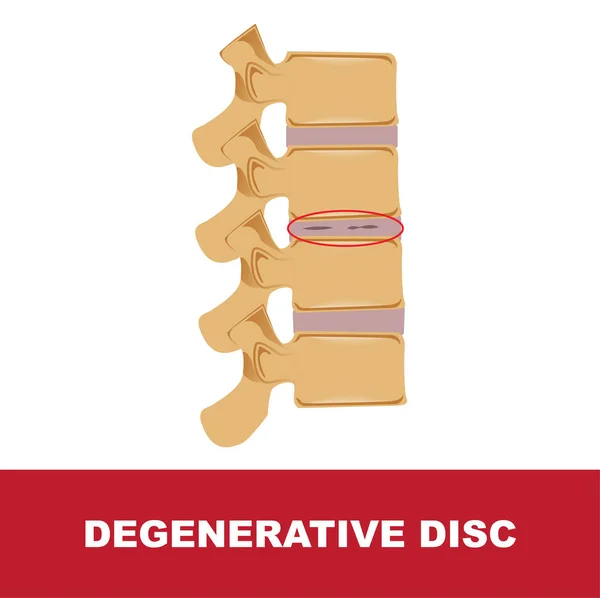
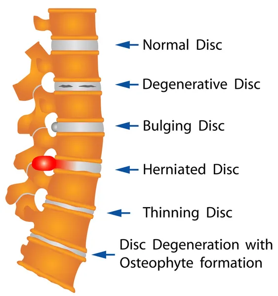
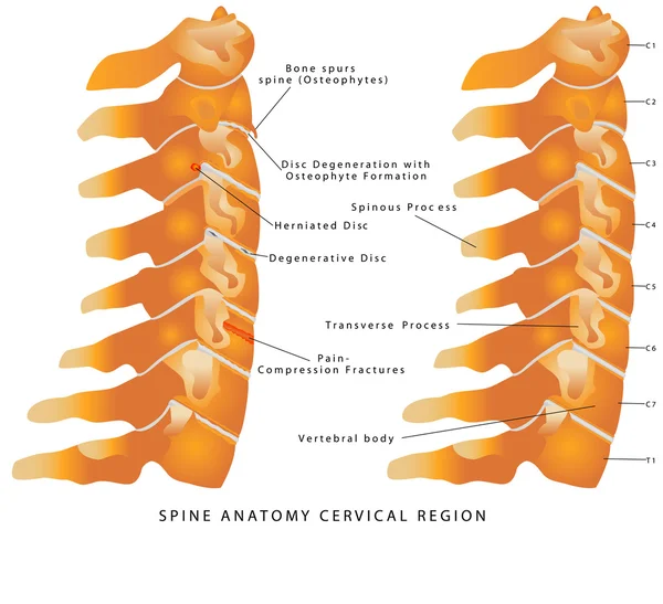
Stock image Degenerative Disc

Pain In The Spine, Woman With Backache At Home, Injury In The Lower Back, Photo With Highlighted Skeleton
Image, 7.91MB, 6048 × 4032 jpg
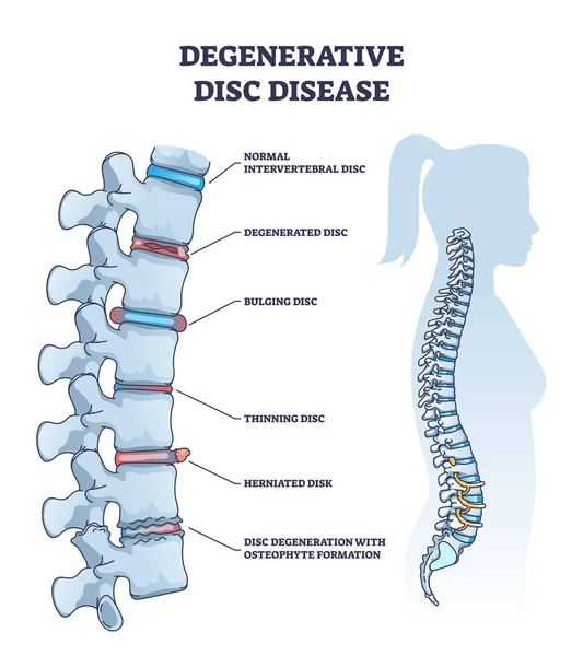
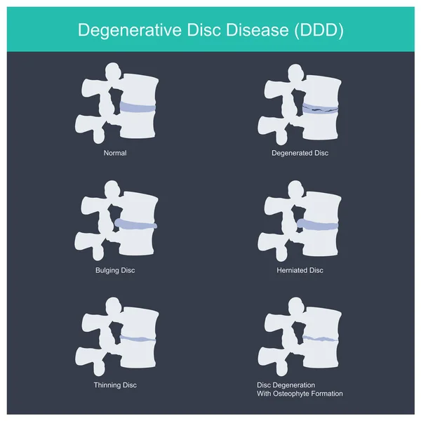
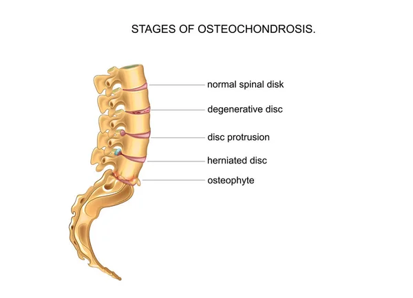
Degenerative Disc Disease With Spine And Vertebra Trauma Outline Diagram. Labeled Educational Normal Intervertebral, Degenerated, Bulging, Thinning And Herniated Problem Example Vector Illustration.
Vector, 7.54MB, 4000 × 4500 eps
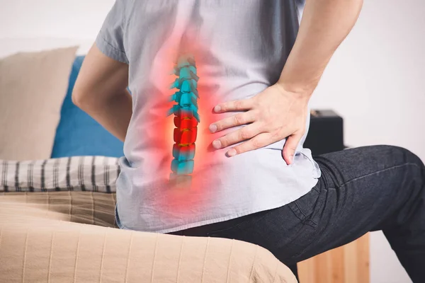
Pain In The Spine, A Man With Backache At Home, Injury In The Lower Back, Photo With Highlighted Skeleton
Image, 12.16MB, 6048 × 4032 jpg
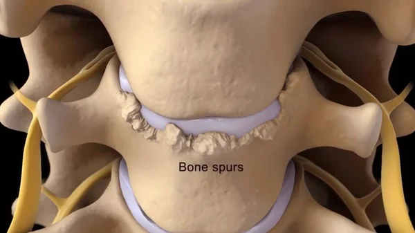
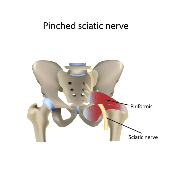
Spinal Stenosis As A Degenerative Illness In The Human Vertebrae Causing Compressed Spine Nerves Medical Concept As A 3D Illustration.
Image, 0.82MB, 3840 × 2160 jpg
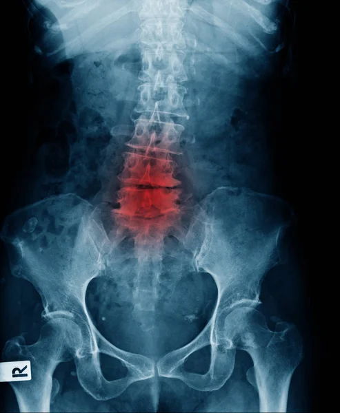
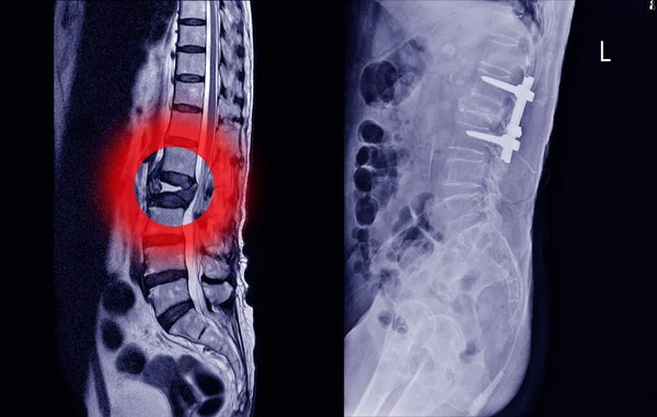
X-ray Image Of Lower Back Show Lumbar Spodylosis And Compression Fracture Of L4-5 Lumbar Spine Level
Image, 1.94MB, 1996 × 2428 jpg
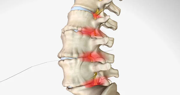
Degenerative Disc Disease Is Characterized By The Gradual Degeneration Of The Intervertebral Discs. 3D Rendering
Image, 2.12MB, 7340 × 3884 jpg

3d Illustration Of A Degenerated, Prolapsed Intervertebral Disc Of The Dog Spine. A Common Disease In Dachshunds But Also In Other Dogs.
Image, 5.07MB, 6000 × 4000 jpg
Pain In The Spine, Man With Backache On Black Background, Intervertebral Hernia Or Disc Injury Concept
Image, 16.61MB, 7952 × 5304 jpg
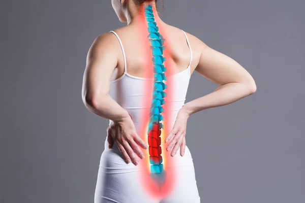
Pain In The Spine, Woman With Backache On Gray Background, Back Injury, Photo With Highlighted Skeleton
Image, 9.44MB, 6048 × 4032 jpg

Pain In The Spine, Woman With Backache At Home, Back Injury, Photo With Highlighted Skeleton
Image, 10.11MB, 6048 × 4032 jpg

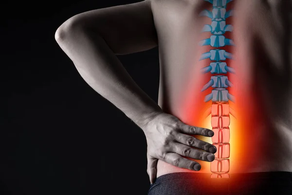
Pain In The Spine, A Man With Backache At Home, Injury In The Lower Back, Photo With Highlighted Skeleton
Image, 9.62MB, 4032 × 6048 jpg
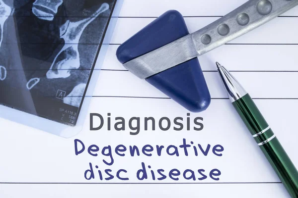
Diagnosis Degenerative Disc Disease. Medical Health History Written With Diagnosis Of Lumbar Disc Disease, MRI Image Sacral Spine And Neurological Hammer. Medical Concept For Neurology, Neuroscience
Image, 5.36MB, 4608 × 3072 jpg

Lumbar Intervertebral Spine Hernia, Woman With Back Pain At Home, Spinal Disc Disease, Health Problems Concept
Image, 21.37MB, 7952 × 5304 jpg

Lumbar Spine Hernia, Man With Back Pain At Home, Compression Injury Of The Intervertebral Disc In The Lower Back, Photo With Highlighted Skeleton
Image, 23.33MB, 7952 × 5304 jpg
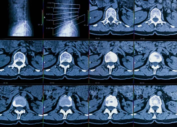
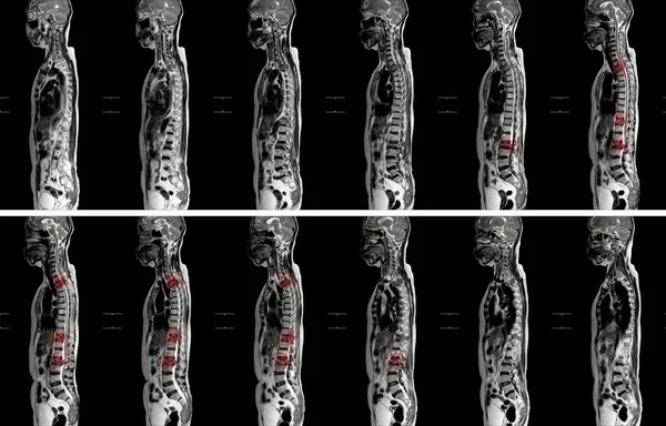
Results Of Computer Tomography Or CT Imaging Of Human Spine Of A Patient With Chronic Back Pain, Shows Degenerative Changes Of Spines, Lumbar Discs Herniation And Nerve Roots Compression
Image, 15.56MB, 6759 × 4861 jpg
X-ray Lower Back Of Young Man Post Fixed Of Lumbar Spine Fracture, Compression Fracture Of L4-5 Spine With Post Operation
Image, 2.05MB, 1996 × 2428 jpg
MRI OF THE THORACIC SPINE These Findings Are Suggestive Of TB Spondylodiscitis Of T11/12 Level With Intraosseous Abscess Formation And Subligamentous Spreading.
Image, 8.31MB, 4852 × 4000 jpg
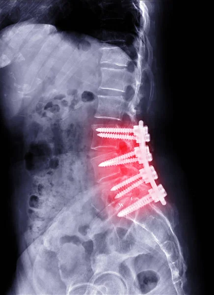
X-ray Image Of L-S Spine Or Lambosacral Spine Lateral Post Operative Lumbar Fixation Plates And Screw.
Image, 7.13MB, 3720 × 5112 jpg

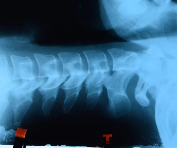
X-ray Image Lumbar Spine And Degenerative Change Of Spine, L-spondylosis X-ray Image In Blue Tone
Image, 2.03MB, 1996 × 2428 jpg

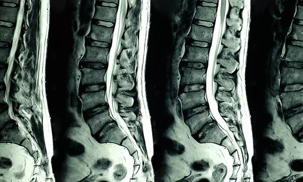
MRI Of Lumbar Spine The Study Reveals Burst Fracture Of L2 Vertebral Body, Appears As Severe Decreased Disc Height And Widening Of Interpedicular Distance.
Image, 2.77MB, 3096 × 4128 jpg

CT Lumbar Spine Or L-S Spine 3D Rendering Image Sagittal View 3D Rendering . Clipping Path.
Image, 1.54MB, 3353 × 4673 jpg


Medical X-ray And MRI Of Lumbar Spine Compression Fracture Bulging Of L1-2. On Arrow Point..Lumbar Spondylosis From L1-2 To L5-S1 Discs.Medical Healthcare Concept.
Image, 6.06MB, 5500 × 3500 jpg
Thoracic And Lumbar Degenerative Change X-ray Image, Back Pain In Old Man Show X-ray Image Of Spondylosis, Spur Loss Of Disc Space And Scoliosis Multiple Level
Image, 2.65MB, 2012 × 2446 jpg
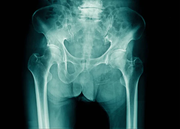
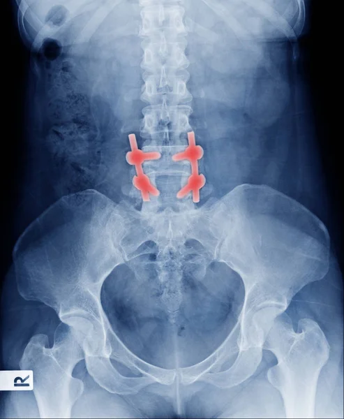
X-ray Image Pelvic Bone And Part Of L-spine With Compression Of Spine Or Degenertive Change
Image, 2.48MB, 2792 × 2010 jpg

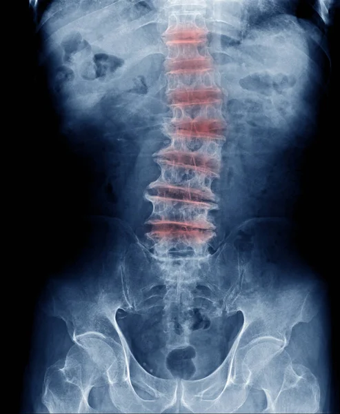
X-ray Image Of Spine Show Degeneration Of Lumbar Spine With Red High Light, Spur Or Calcification At Body Of Lumbar Spine
Image, 1.81MB, 1996 × 2428 jpg
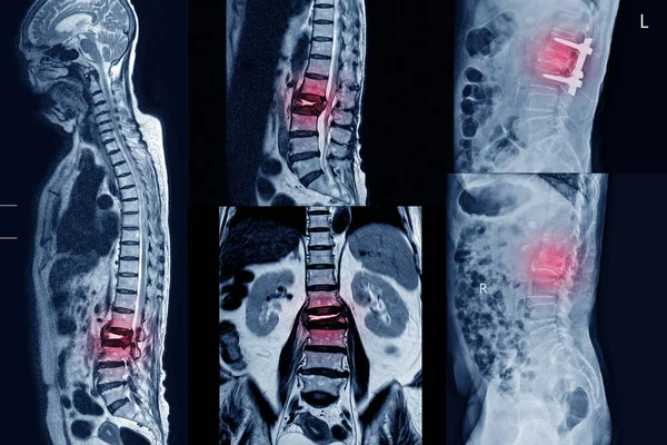
Collection MRI Of Lumbar Spine History Of Fall With Back Pain, Radiate To Leg, Rule Out Spinal Stenosis .Impression:Burst Fracture Of L2 Vertebral Body With Severe Vertebral Collapse.Medical Concept.
Image, 8.31MB, 5820 × 3890 jpg
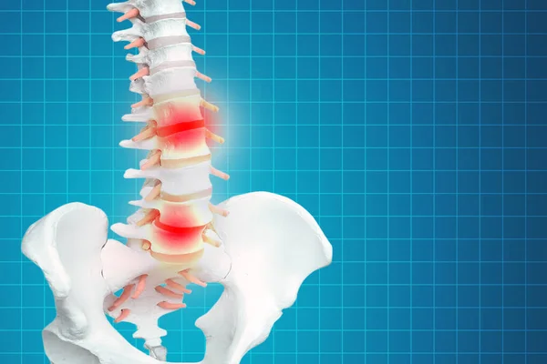
Realistic Skeletal Human Spine And Vertebral Column Or Intervertebral Discs On A Dark Background. Lower Back Pain. Vertebral Column In Glowing Highlight As A Medical Health Care Concept.
Image, 1.26MB, 2611 × 1741 jpg

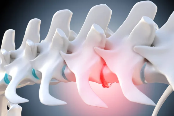
Degenerative Disc Disease Is Characterized By The Gradual Degeneration Of The Intervertebral Discs. 3D Rendering
Image, 1.49MB, 7340 × 3884 jpg
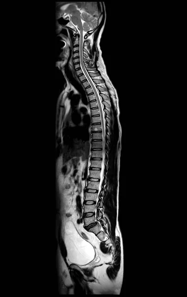
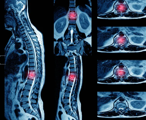
MRI Of Whole Spine T2W Sagittal Plane For Diagnostic Spinal Cord Compression.
Image, 4.7MB, 4624 × 7314 jpg
MRI OF THORACOLUMBAR SPINE IMPRESSION: Moderate Pathological Compression Of T11 And L2 Levels With Enhancing Multiple Marrow Lesions At T1, T10 ToT12, L2, L3 To L5 Levels.
Image, 4.96MB, 5688 × 3643 jpg
MRI Scan Of Lumbar Spines Of A Patient With Chronic Back Pain Showing Degenerative Change Of The L Spines With Right Lateral Protrution Of L4-5 Disc And L4 Nerve Root Compression
Image, 11.99MB, 5967 × 3596 jpg

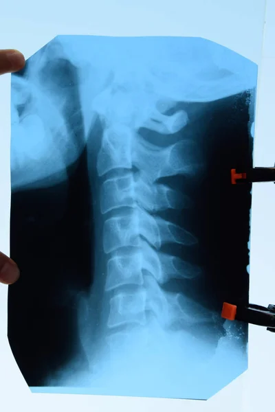
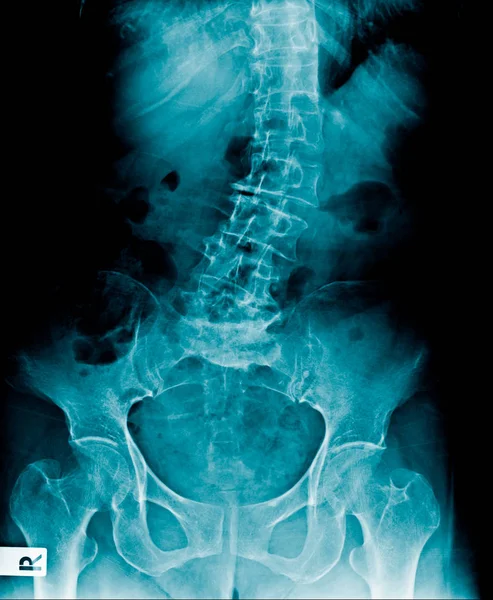
Hight Quality X-ray Lumbar Spine In Blue Tone, Xrau Image Of Old Man Show Spondylosis Or Degeneration Of Spine And Part Og Hip And Pelvic Bone
Image, 1.72MB, 1996 × 2428 jpg
Cardiologist Doctor Holding Stethoscopet, Blurred Electrocardiogram Result On Paper With AICD Pacemaker In Chest X-ray Background.medical Concept
Image, 5.46MB, 5500 × 3500 jpg
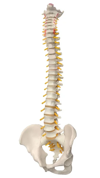
Skeletal Human Spine And Vertebral Column Or Intervertebral Discs. Human Spine Anatomy.
Image, 0.9MB, 1635 × 2900 jpg

Lumbar Spondysis X-ray Image, Back Pain Old Man Show Mild Scoliosis, Spur, And Degenerative Of Disc
Image, 2.65MB, 2012 × 2446 jpg
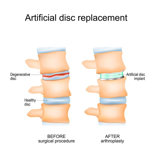
Intervertebral Disc Arthroplasty. Close-up Of A Human Spinal Column Before Surgical Procedure And After Arthroplasty. Degenerative Disc And Artificial Disc Replacement. Vector Poster
Vector, 10.57MB, 4444 × 4444 eps

Page 1 >> Next