
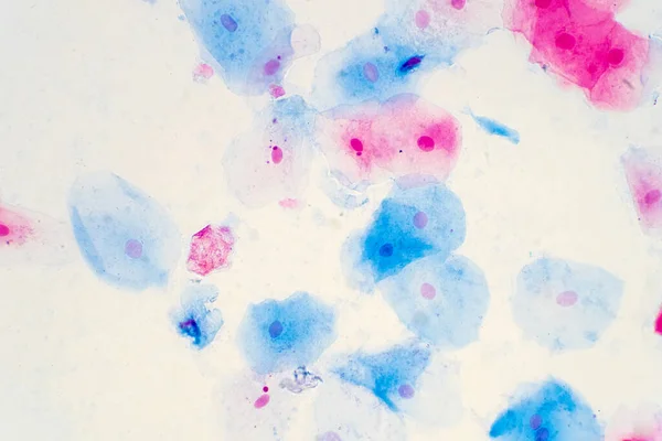
Stock image Education Histology

Squamous Epithelial Cells Of Human Cervix Under The Microscope View. Pap Smear Test Is A Procedure To Test For Cervical Cancer In Women.
Image, 14.68MB, 5385 × 3590 jpg

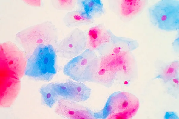
Squamous Epithelial Cells Of Human Cervix Under The Microscope View. Pap Smear Test Is A Procedure To Test For Cervical Cancer In Women.
Image, 18.53MB, 6000 × 4000 jpg
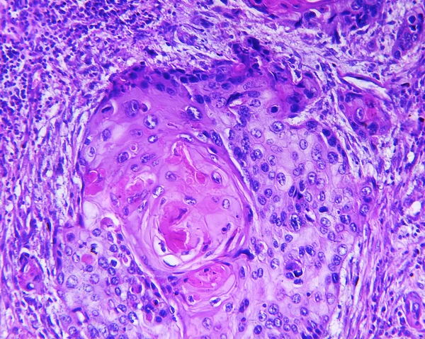
Squamous Epithelial Cells Of Human Cervix Under The Microscope View. Pap Smear Test Is A Procedure To Test For Cervical Cancer In Women.
Image, 17.23MB, 5883 × 3922 jpg
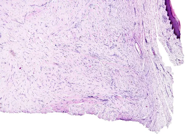
Squamous Epithelial Cells Of Human Cervix Under The Microscope View. Pap Smear Test Is A Procedure To Test For Cervical Cancer In Women.
Image, 11.03MB, 4506 × 3004 jpg
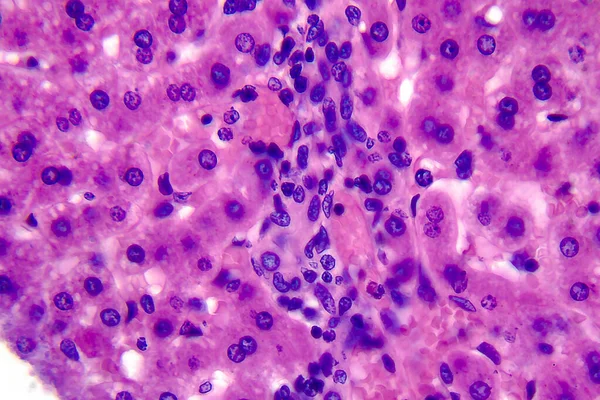
Coccidiosis, Coccidia In Liver, Light Micrograph, Photo Under Microscope
Image, 4.14MB, 4055 × 2703 jpg


Russia - Ekaterinburg, 04.14.2021: Old Black And White Photos Of University Students And Young Scientists In USSR. Stock Footage. Clever Soviet Men And Women On Retro Animated Photos.
Image, 1.49MB, 3840 × 2160 jpg


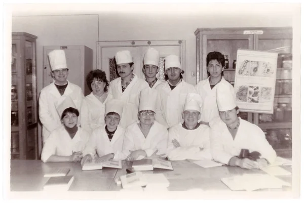
Students Of Vitebsk Medical Institute At Department Of Histology (group Vintage Photo 1987), Belarus
Image, 1.4MB, 2450 × 1639 jpg


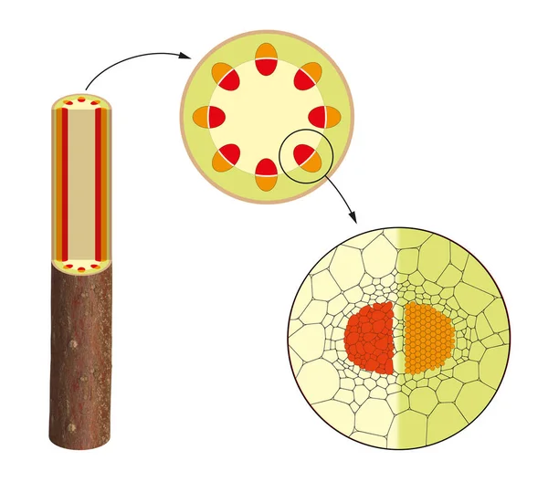
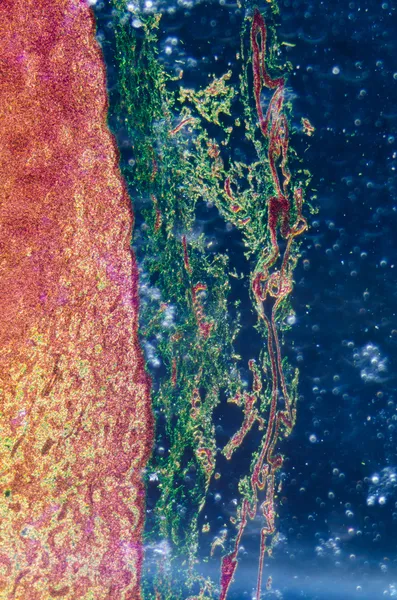
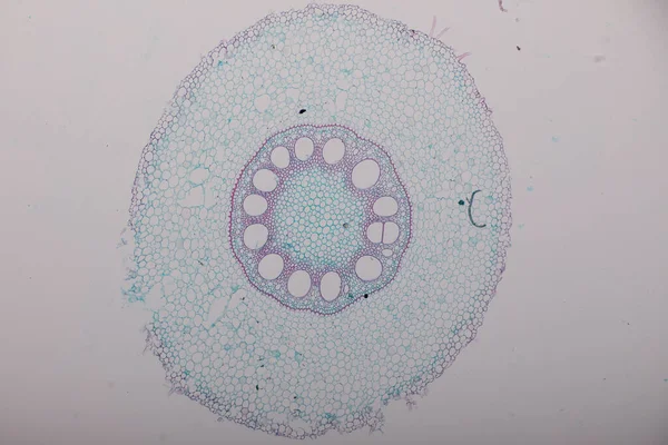
The Study Plant Tissue Of Under The Microscope For Classroom Education.
Image, 14.4MB, 6720 × 4480 jpg

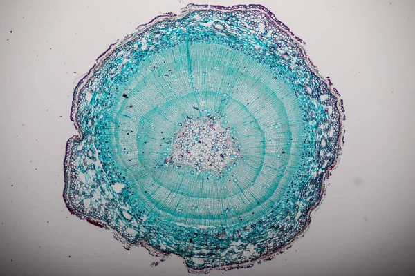
The Study Plant Tissue Of Under The Microscope For Classroom Education.
Image, 17.77MB, 6720 × 4480 jpg


The Study Plant Tissue Of Under The Microscope For Classroom Education.
Image, 10.6MB, 6720 × 4480 jpg


Gastric Mucosa With Hypertrophic Gastritis. Infographics. Vector Illustration On Isolated Background
Vector, 1.75MB, 5000 × 5000 eps

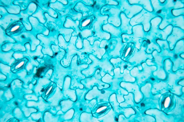
The Study Plant Tissue Of Under The Microscope For Classroom Education.
Image, 25.28MB, 6720 × 4480 jpg


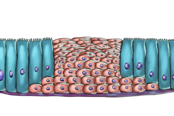
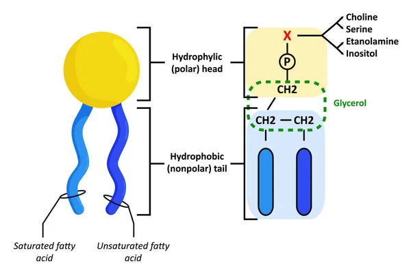
Histological Structure Of Epidermis - Skin Layers Shcematic Vector Illustration Showing Stratum Basale, Spinosum, Granulosum, Lucidum And Corneum
Vector, 7.1MB, 3090 × 3090 eps


The Study Plant Tissue Of Under The Microscope For Classroom Education.
Image, 13.16MB, 6720 × 4480 jpg

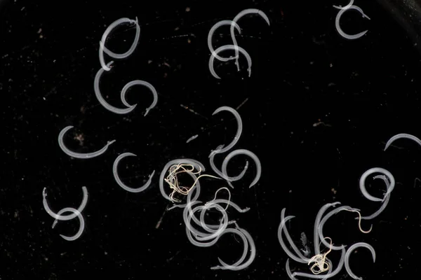
Schistosoma Is A Genus Of Trematodes, Commonly Known As Blood Flukes For Education In Laboratory.
Image, 5MB, 6720 × 4480 jpg

Fish Blood, Smear, 80X Light Micrograph. Fish Blood Erythrocytes With Micronucleus, Under The Light Microscope. Four Individual Shots Combined Into One Overall Picture. Isolated, On White Background.
Image, 9.57MB, 9000 × 5693 jpg


Page 1 >> Next