photo free
Home Images Vectors Videos

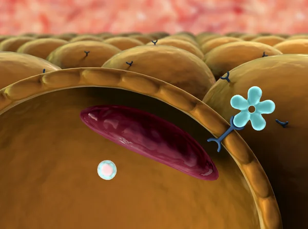
Stock image Enzyme Catalysis

stock vector Detailed Vector Illustration of Enzyme Catalysis Reaction Stages: Biochemical Processes in White Background

Detailed Vector Illustration Of Enzyme Catalysis Reaction Stages: Biochemical Processes In White Background

Vector, 0.34MB, 5000 × 3000 ai
stock vector Detailed and Labeled Vector Illustration of Components of Pyruvate Dehydrogenase Complex for Biochemistry, Molecular Biology, and Health Science Education on White Background

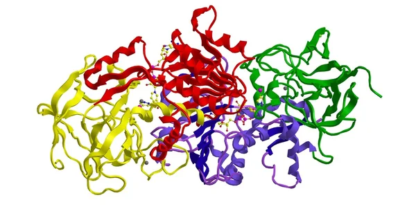
Detailed And Labeled Vector Illustration Of Components Of Pyruvate Dehydrogenase Complex For Biochemistry, Molecular Biology, And Health Science Education On White Background

Vector, 0.38MB, 5000 × 3000 ai
stock image PETase is a bacterial enzyme that breaks down PET-plastic to monomeric molecules. The whole bacterial degradation process yields terephtalic acid and ethylene glycol, which are environmentally harmless.

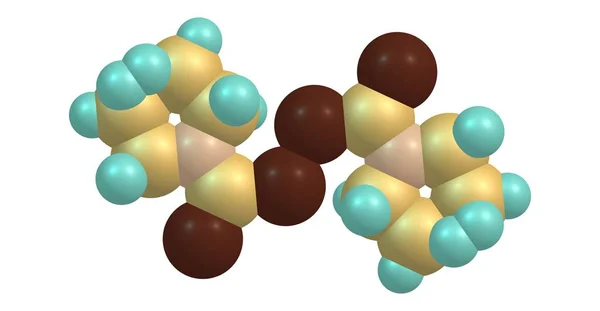
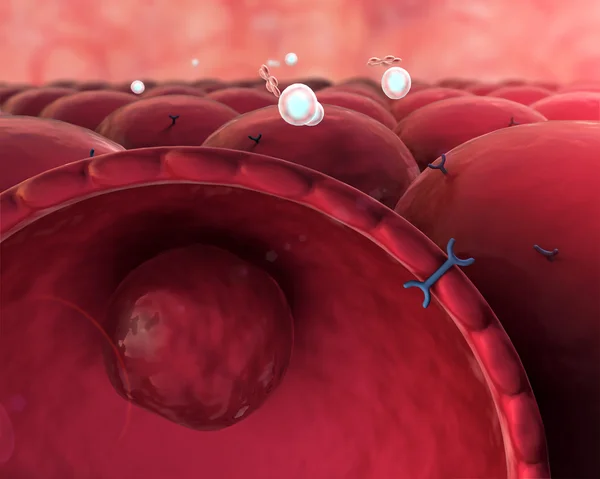
PETase Is A Bacterial Enzyme That Breaks Down PET-plastic To Monomeric Molecules. The Whole Bacterial Degradation Process Yields Terephtalic Acid And Ethylene Glycol, Which Are Environmentally Harmless.

Image, 4.58MB, 8000 × 6000 jpg
stock image Living cell

Living Cell

Image, 22.26MB, 6000 × 4000 jpg
stock image structure of the molecule

Structure Of The Molecule

Image, 8.47MB, 7744 × 5792 jpg
stock image Cell, insulin

Cell, Insulin

Image, 7.15MB, 6000 × 4000 jpg
stock image mechanism of action of insulin

Mechanism Of Action Of Insulin

Image, 9.84MB, 7760 × 5832 jpg
stock image Reactions relate to the free energy of the involved molecules

Reactions Relate To The Free Energy Of The Involved Molecules

Image, 3.53MB, 13235 × 7995 jpg
stock image Molecular structure of human beta 3 alcohol dehydrogenase

Molecular Structure Of Human Beta 3 Alcohol Dehydrogenase

Image, 0.68MB, 2877 × 1450 jpg
stock vector Mechanism of enzyme action with substrate and product complexes. Enzyme Kinetics

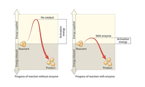
Mechanism Of Enzyme Action With Substrate And Product Complexes. Enzyme Kinetics

Vector, 2.43MB, 6000 × 2700 eps
stock image Disulfiram molecular structure isolated on white

Disulfiram Molecular Structure Isolated On White

Image, 0.83MB, 7000 × 3638 jpg
stock image RNA polymerase (from yeast) is composed of several proteins. It unwinds DNA strands (violet) and builds RNA (red) out of the nucleotides uridine, adenosine, cytlidine and guanosine monophosphate. Source: PDB entry 1i6h

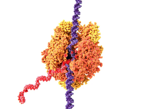
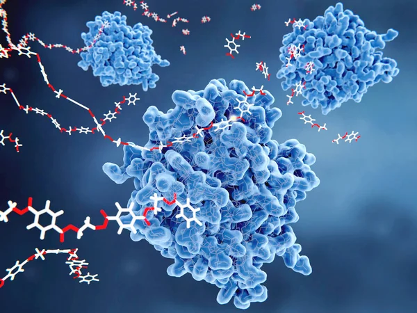
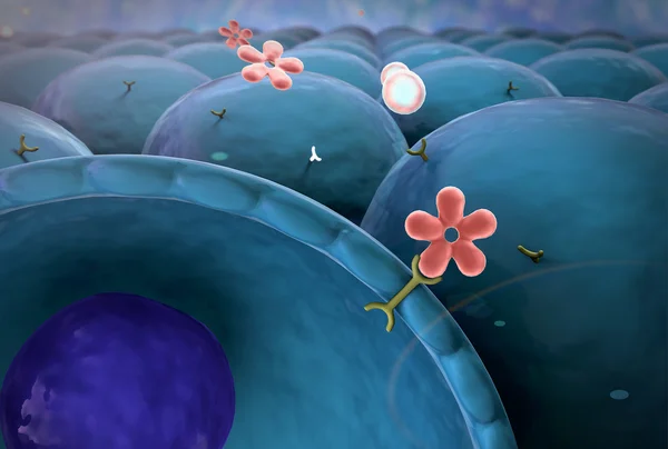
RNA Polymerase (from Yeast) Is Composed Of Several Proteins. It Unwinds DNA Strands (violet) And Builds RNA (red) Out Of The Nucleotides Uridine, Adenosine, Cytlidine And Guanosine Monophosphate. Source: PDB Entry 1i6h

Image, 5.4MB, 8000 × 6000 jpg
stock image Cell, vein

Cell, Vein

Image, 20.81MB, 6000 × 4000 jpg
stock image Insulin, cell

Insulin, Cell

Image, 6.64MB, 5936 × 4000 jpg
stock image Fluconazole interacting with a heme group in the active site of the lanosterol 14 alpha-demethylase

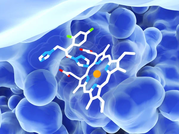
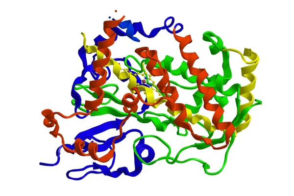
Fluconazole Interacting With A Heme Group In The Active Site Of The Lanosterol 14 Alpha-demethylase

Image, 5.65MB, 8000 × 6000 jpg
stock image Saccharomyces cerevisiae is a highly ethanol tolerant organism 3D

Saccharomyces Cerevisiae Is A Highly Ethanol Tolerant Organism 3D

Image, 0.13MB, 2400 × 2000 jpg
stock image Biochemical term Cofactors written in white bold letters on blue background. 3d illustration.

Biochemical Term Cofactors Written In White Bold Letters On Blue Background. 3d Illustration.

Image, 3.51MB, 3176 × 2000 jpg
stock image Insulin, molecule, cell

Insulin, Molecule, Cell

Image, 5.93MB, 6000 × 4800 jpg
stock image structure of the molecule

Structure Of The Molecule

Image, 8.95MB, 7840 × 5840 jpg
stock image Molecule, insulin

Molecule, Insulin

Image, 5.42MB, 5848 × 3800 jpg
stock image vein, cell

Vein, Cell

Image, 13.74MB, 6000 × 4000 jpg
stock image RNA polymerase unwinds DNA strands (violet) and builds RNA (red) out of the nucleotides  adenosine (magenta), cytlidine (violet), guanosine (yellow) and uridine (white) monophosphate.

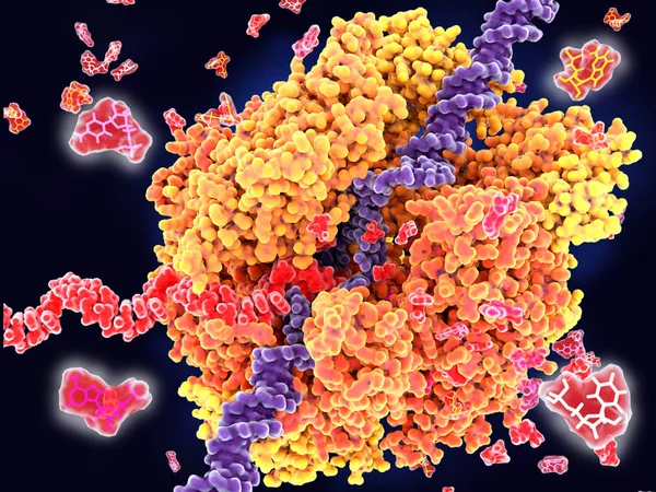
RNA Polymerase Unwinds DNA Strands (violet) And Builds RNA (red) Out Of The Nucleotides Adenosine (magenta), Cytlidine (violet), Guanosine (yellow) And Uridine (white) Monophosphate.

Image, 9.4MB, 8000 × 6000 jpg
stock image Enzyme -  word from metal blocks - concept sepia tone photo on shine background

Enzyme - Word From Metal Blocks - Concept Sepia Tone Photo On Shine Background

Image, 2.11MB, 4328 × 2824 jpg
stock image Molecular structure of cytochrome P450

Molecular Structure Of Cytochrome P450

Image, 0.57MB, 2511 × 1670 jpg
stock image enzymology typography, wordart, wordcloud, enzyme, enzymology, science, chemical

Enzymology Typography, Wordart, Wordcloud, Enzyme, Enzymology, Science, Chemical

Image, 0.92MB, 6400 × 4800 jpg
stock image RNA polymerase (yeast) s composed of several proteins. It unwinds DNA strands (violet) and builds RNA (red) out of the nucleotides uridine, adenosine, cytlidine and guanosine monophosphate.

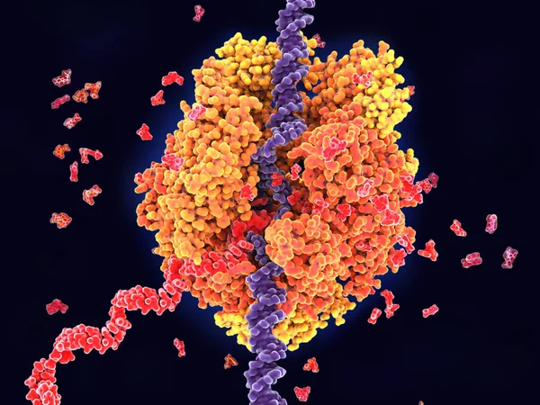
RNA Polymerase (yeast) S Composed Of Several Proteins. It Unwinds DNA Strands (violet) And Builds RNA (red) Out Of The Nucleotides Uridine, Adenosine, Cytlidine And Guanosine Monophosphate.

Image, 7.45MB, 8000 × 6000 jpg
stock image A fungal lanosterol 14-alpha demethylase with fluconazole bound to the active site.

A Fungal Lanosterol 14-alpha Demethylase With Fluconazole Bound To The Active Site.

Image, 6.33MB, 8000 × 6000 jpg
stock image Vein, cell

Vein, Cell

Image, 18.86MB, 6000 × 4000 jpg
stock vector Lock and key model. The correct and incorrect keys. Vector illustration

Lock And Key Model. The Correct And Incorrect Keys. Vector Illustration

Vector, 11.15MB, 4444 × 4444 eps
stock image Insulin, cell

Insulin, Cell

Image, 4.32MB, 5848 × 3800 jpg
Page 1 >> Next