
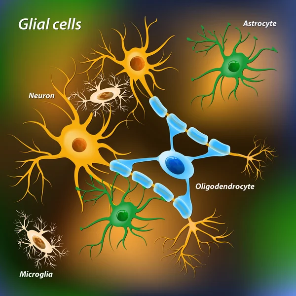
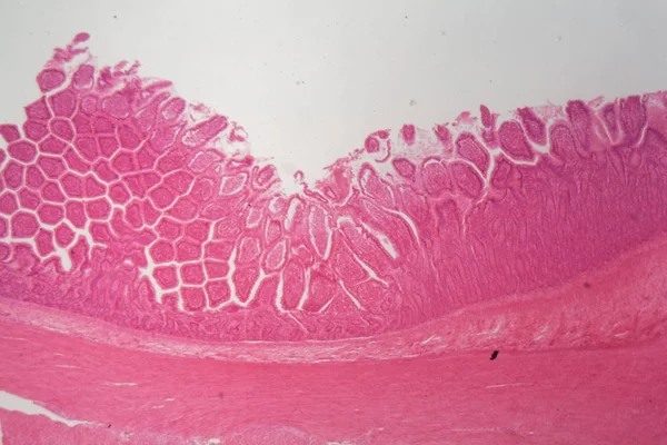
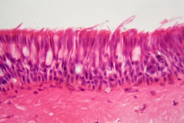
Stock image Ependymal
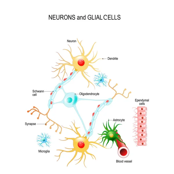
Neurons And Glial Cells (Neuroglia) In Brain (oligodendrocyte, Microglia, Astrocytes And Schwann Cells), Ependymal Cells (ependymocytes). Vector Diagram For Educational, Medical, Biological And Science Use
Vector, 13.41MB, 4444 × 4444 eps
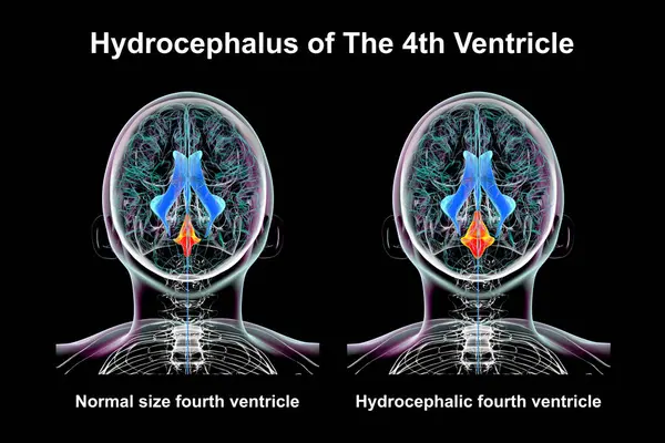
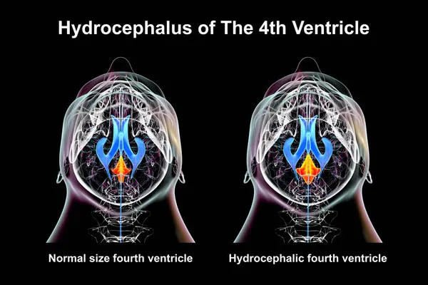
A 3D Scientific Illustration Depicting Isolated Enlargement Of The Fourth Brain Ventricle (right) Compared To The Normal Size Fourth Ventricle (left), Top View.
Image, 8.49MB, 7800 × 5200 jpg
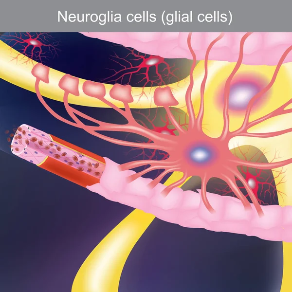
The Duty Is To Treat The Central Nervous System, Bringing Food And Oxygen To The Nerve Cells.
Vector, 21.32MB, 5000 × 5000 eps

A 3D Scientific Illustration Depicting Isolated Enlargement Of The Fourth Brain Ventricle (right) Compared To The Normal Size Fourth Ventricle (left), Side View.
Image, 7.09MB, 7800 × 5200 jpg
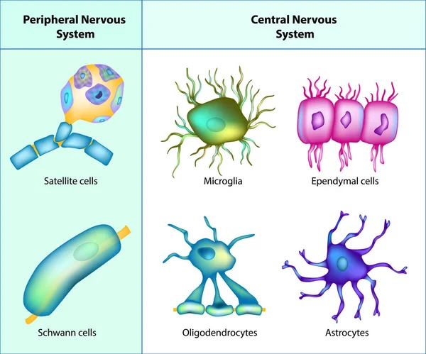
Types Of Neuroglia: Oligodendrocytes, Astrocytes, Microglia, Schwann Cells, Satellite Cells, Ependymal Cells.
Vector, 4.54MB, 5000 × 4165 eps

A 3D Scientific Illustration Depicting Isolated Enlargement Of The Fourth Brain Ventricle (right) Compared To The Normal Size Fourth Ventricle (left).
Image, 7.13MB, 7800 × 5200 jpg
A 3D Scientific Illustration Depicting Isolated Enlargement Of The Fourth Brain Ventricle (right) Compared To The Normal Size Fourth Ventricle (left), Bottom View.
Image, 7.25MB, 7800 × 5200 jpg
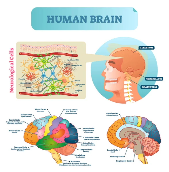
Brain Vector Illustration. Medical Educational Scheme With Neurological Cells Closeup. Silhouette With Cerebrum, Cerebellum And Stem. Cortex And Lobe Diagram.
Vector, 8.39MB, 3983 × 4067 eps
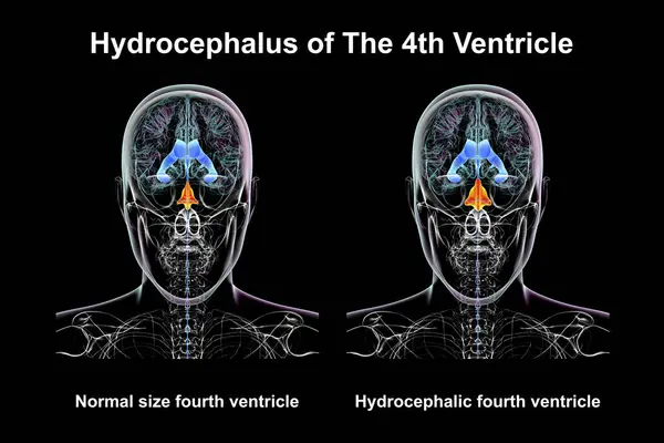
A 3D Scientific Illustration Depicting Isolated Enlargement Of The Fourth Brain Ventricle (right) Compared To The Normal Size Fourth Ventricle (left), Front View.
Image, 6.39MB, 7800 × 5200 jpg

A 3D Scientific Illustration Depicting Isolated Enlargement Of The Fourth Brain Ventricle (right) Compared To The Normal Size Fourth Ventricle (left).
Image, 8.24MB, 7800 × 5200 jpg


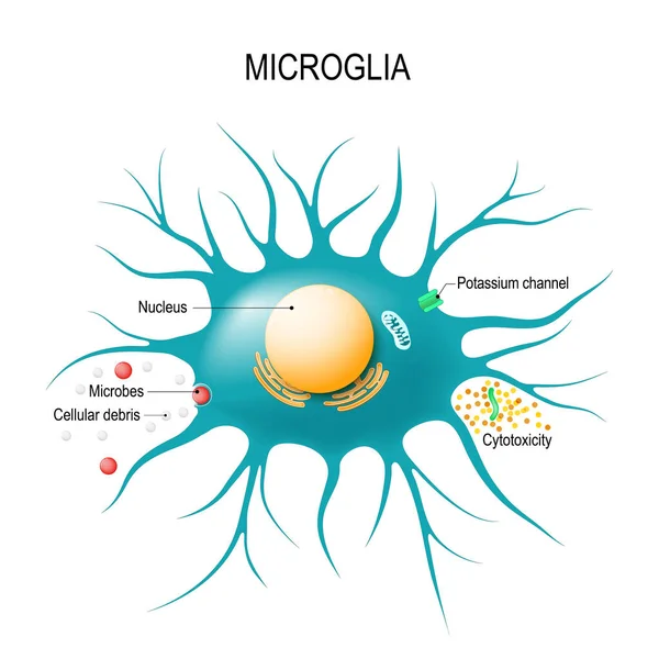
Anatomy Of A Microglial Cell. Glial Cell Is The Macrophage For Immune Defence The Central Nervous System. Vector Diagram For Educational, Medical, Biological And Science Use
Vector, 2.19MB, 5478 × 5479 eps

A 3D Scientific Illustration Depicting Isolated Enlargement Of The Fourth Brain Ventricle (right) Compared To The Normal Size Fourth Ventricle (left).
Image, 7.38MB, 7800 × 5200 jpg

Vector Illustration Of Ependymal Cells, A Type Of Glial Cell. Ependymocytes
Vector, 5.34MB, 9036 × 4923 eps


Glioma Cancer Tumor As Malignant Cells Outbreak As A Brain Disease Attacking Neurons As A Medical Concept Of Neurological Disease With 3D Illustration Elements
Image, 12.7MB, 7316 × 3620 jpg
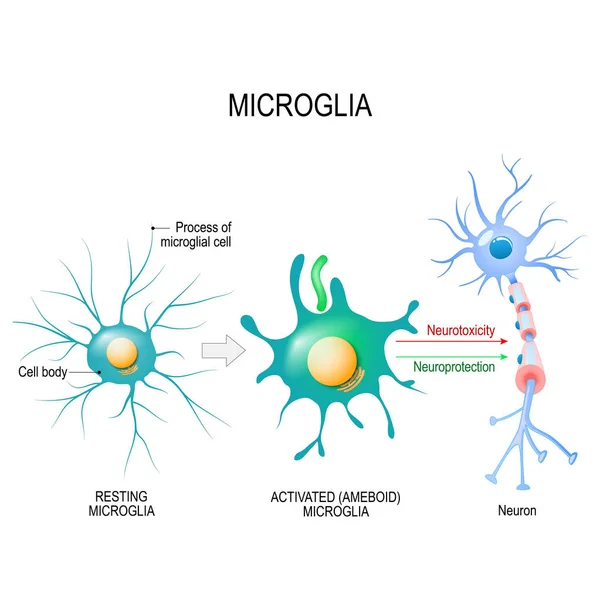
Activation Of A Microglial Cell. Vector Diagram For Educational, Medical, Biological And Science Use
Vector, 1.96MB, 5743 × 5744 eps
Page 1 >> Next