photo free
Home Images Vectors Videos

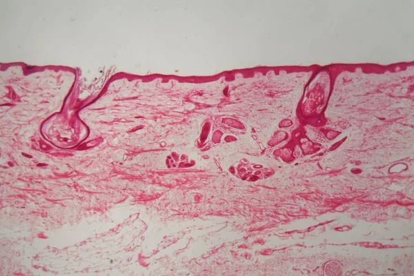
Stock image Epidermis Cells page 2

stock image Close up human skin. Macro epidermis

Close Up Human Skin. Macro Epidermis

Image, 19.14MB, 7360 × 4912 jpg
stock image science medical anthropotomy physiology microscopic human thyroi

Science Medical Anthropotomy Physiology Microscopic Human Thyroi

Image, 5.85MB, 3264 × 3264 jpg
stock image Scalp and hair follicles of human under the microscope in Lab.

Scalp And Hair Follicles Of Human Under The Microscope In Lab.

Image, 30.84MB, 6720 × 4480 jpg
stock image Scalp and hair follicles of human under the microscope in Lab.

Scalp And Hair Follicles Of Human Under The Microscope In Lab.

Image, 17.22MB, 6720 × 4480 jpg
stock vector Cellulite

Cellulite

Vector, 13.45MB, 6141 × 3500 eps
stock vector Cellulite

Cellulite

Vector, 3.05MB, 6906 × 4353 eps
stock image Scalp and hair follicles of human under the microscope in Lab.

Scalp And Hair Follicles Of Human Under The Microscope In Lab.

Image, 41.27MB, 6720 × 4480 jpg
stock image Scalp and hair follicles of human under the microscope in Lab.

Scalp And Hair Follicles Of Human Under The Microscope In Lab.

Image, 25.95MB, 6720 × 4480 jpg
stock image microscopic section of liver tissue

Microscopic Section Of Liver Tissue

Image, 5.75MB, 3168 × 3168 jpg
stock image science medical anthropotomy physiology microscopic human thyroi

Science Medical Anthropotomy Physiology Microscopic Human Thyroi

Image, 8.53MB, 4928 × 3264 jpg
stock image 3D illustration of spherical and rod-shaped bacteria on skin with hairs, microscopic view of skin microflora, bacteria Staphylococcus, Streptococcus, Propionibacterium on skin

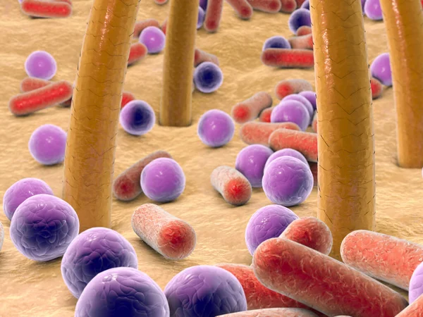
3D Illustration Of Spherical And Rod-shaped Bacteria On Skin With Hairs, Microscopic View Of Skin Microflora, Bacteria Staphylococcus, Streptococcus, Propionibacterium On Skin

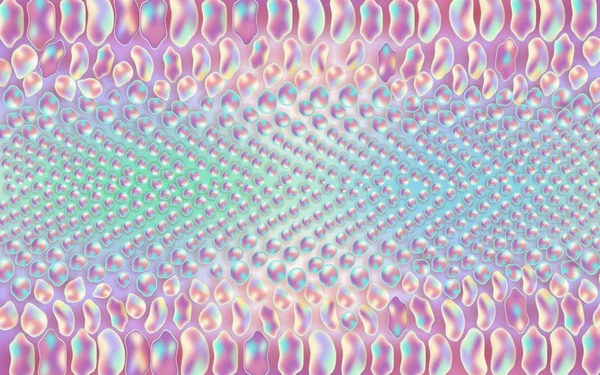
Image, 1.62MB, 4000 × 3000 jpg
stock image microscopic section of human pancreas

Microscopic Section Of Human Pancreas

Image, 6.59MB, 3264 × 3264 jpg
stock image Scalp and hair follicles of human under the microscope in Lab.

Scalp And Hair Follicles Of Human Under The Microscope In Lab.

Image, 25.57MB, 6720 × 4480 jpg
stock image Onion epidermis under light microscope. Purple colored, large epidermal cells of an onion, Allium cepa, in a single layer. Each cell with wall, membrane, cytoplasm, nucleus and large vacuole.

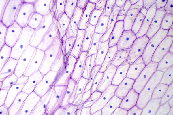
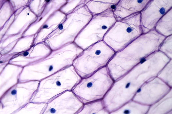
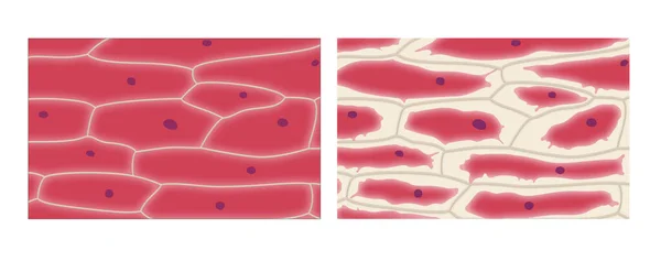
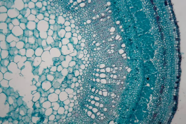
Onion Epidermis Under Light Microscope. Purple Colored, Large Epidermal Cells Of An Onion, Allium Cepa, In A Single Layer. Each Cell With Wall, Membrane, Cytoplasm, Nucleus And Large Vacuole.

Image, 3.62MB, 3786 × 2524 jpg
stock image Amphibian skin with ulcer under a microscope

Amphibian Skin With Ulcer Under A Microscope

Image, 1.18MB, 5616 × 3744 jpg
stock image Onion epidermis with large cells under light microscope

Onion Epidermis With Large Cells Under Light Microscope

Image, 12.07MB, 4873 × 3228 jpg
stock vector Human skin anatomy

Human Skin Anatomy

Vector, 1.86MB, 6900 × 4119 eps
stock vector Skin cross section showing cellulite

Skin Cross Section Showing Cellulite

Vector, 2.82MB, 5688 × 3505 eps
stock image Human Skin Anatomy 3d illustration isolated white background.

Human Skin Anatomy 3d Illustration Isolated White Background.

Image, 6.34MB, 5252 × 3939 jpg
stock image science medical anthropotomy physiology microscopic human thyroi

Science Medical Anthropotomy Physiology Microscopic Human Thyroi

Image, 3.64MB, 3000 × 1987 jpg
stock image Schematic illustration of the skin. It provides protection and isolation of the environmental organism. There are three main layers that theThey make up: epidermis, dermis, subcutaneous tissue and skin accessories such as hair, sebaceous glands and

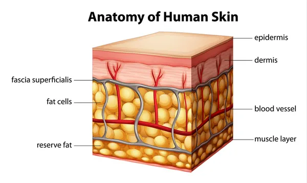
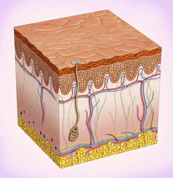
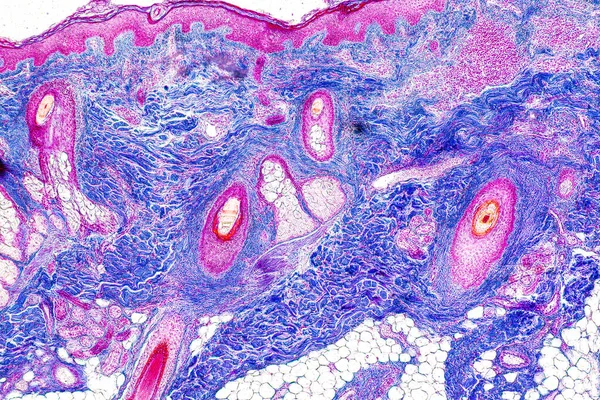
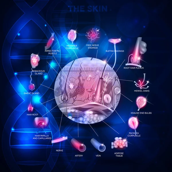
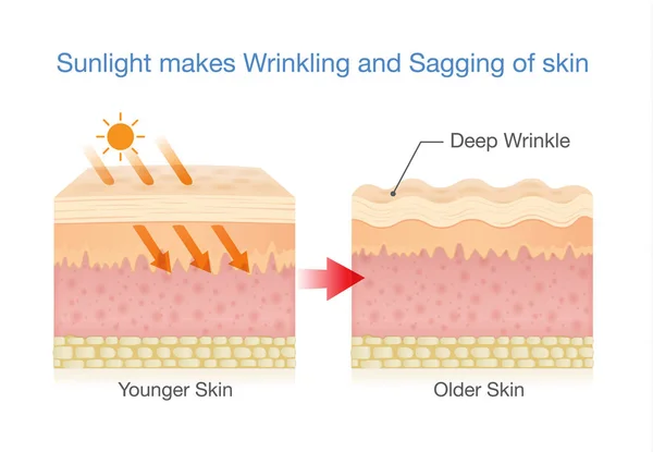
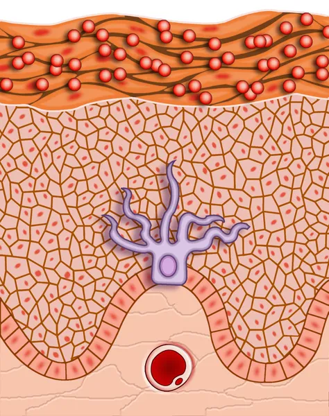
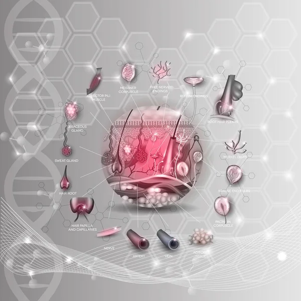
Schematic Illustration Of The Skin. It Provides Protection And Isolation Of The Environmental Organism. There Are Three Main Layers That TheThey Make Up: Epidermis, Dermis, Subcutaneous Tissue And Skin Accessories Such As Hair, Sebaceous Glands And

Image, 5.89MB, 3149 × 3236 jpg
stock image micrograph of blood vessel, artery and vein

Micrograph Of Blood Vessel, Artery And Vein

Image, 10.36MB, 4752 × 3168 jpg
stock image Skin with follicle under the microscope

Skin With Follicle Under The Microscope

Image, 1.08MB, 4500 × 3000 jpg
stock image Detail of skin under the microscope.

Detail Of Skin Under The Microscope.

Image, 1.07MB, 4500 × 3000 jpg
stock image microscopic section of human kidney tissue

Microscopic Section Of Human Kidney Tissue

Image, 7.29MB, 2839 × 2839 jpg
stock image micrograph of small intestinum tenue tissue

Micrograph Of Small Intestinum Tenue Tissue

Image, 8.38MB, 3168 × 3168 jpg
stock vector Phases of the wound healing process. poster with capillary, red

Phases Of The Wound Healing Process. Poster With Capillary, Red

Vector, 9.63MB, 4444 × 4444 eps
stock image Skin with follicle under the microscope

Skin With Follicle Under The Microscope

Image, 0.78MB, 4500 × 3000 jpg
stock image Squamous cell carcinoma

Squamous Cell Carcinoma

Image, 2.14MB, 2504 × 2250 jpg
stock image Skin with follicle under the microscope

Skin With Follicle Under The Microscope

Image, 1.2MB, 4500 × 3000 jpg
stock vector Skin anatomy abstract scientific design

Skin Anatomy Abstract Scientific Design

Vector, 12.88MB, 5000 × 5000 eps
stock image Scalp and hair follicles of human under the microscope in Lab.

Scalp And Hair Follicles Of Human Under The Microscope In Lab.

Image, 13.81MB, 6720 × 4480 jpg
stock image micrograph of stomach tissue

Micrograph Of Stomach Tissue

Image, 6.84MB, 3168 × 3168 jpg
stock image Detail of skin under the microscope.

Detail Of Skin Under The Microscope.

Image, 0.81MB, 4500 × 3000 jpg
stock image Detail of skin under the microscope.

Detail Of Skin Under The Microscope.

Image, 0.79MB, 4500 × 3000 jpg
stock vector Sunlight makes sagging skin.

Sunlight Makes Sagging Skin.

Vector, 1.2MB, 5900 × 4085 eps
stock image Plasmolysis in onion epidermis cells

Plasmolysis In Onion Epidermis Cells

Image, 8.22MB, 12139 × 4809 jpg
stock vector Skin anatomy and Sensory receptors in the skin

Skin Anatomy And Sensory Receptors In The Skin

Vector, 9.79MB, 5000 × 5000 eps
stock vector root structure. monocot and dicot stems.

Root Structure. Monocot And Dicot Stems.

Vector, 1.38MB, 4444 × 4444 eps
stock vector Colorful ornate snakeskin pattern

Colorful Ornate Snakeskin Pattern

Vector, 7.74MB, 6325 × 3953 eps
stock image skin depigmentation

Skin Depigmentation

Image, 0.7MB, 2000 × 2529 jpg
stock image Stem cells of a lentil plant under the microscope.

Stem Cells Of A Lentil Plant Under The Microscope.

Image, 1.31MB, 4500 × 3000 jpg
stock image Skin anatomy

Skin Anatomy

Image, 3.24MB, 3454 × 3400 jpg
stock vector Skin anatomy structure in the round shape detailed anatomy illus

Skin Anatomy Structure In The Round Shape Detailed Anatomy Illus

Vector, 13.52MB, 5000 × 5000 eps
stock image microscopic section of human artery vein

Microscopic Section Of Human Artery Vein

Image, 5.76MB, 4928 × 3264 jpg
stock image Cellulite skin cross-section layers. 3d illustration medical concept. Fat cells inside skin

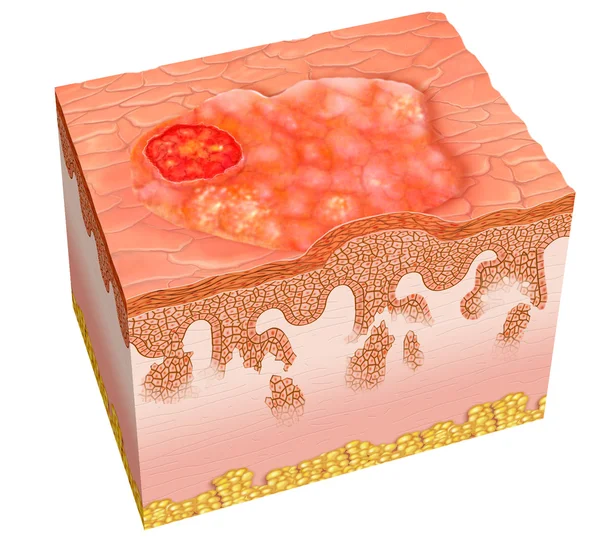
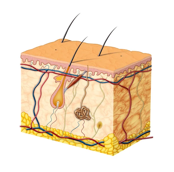
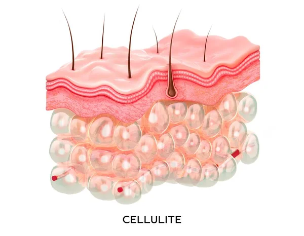
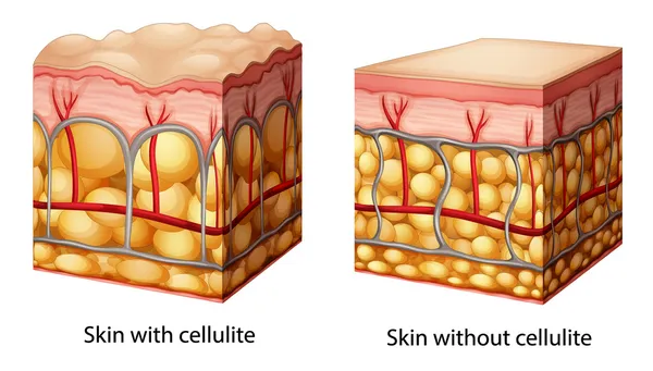
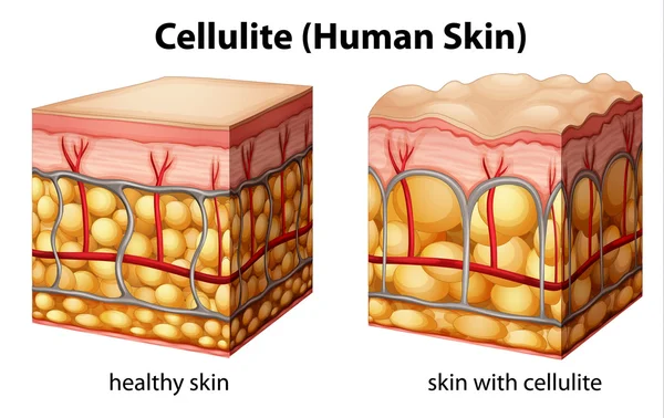
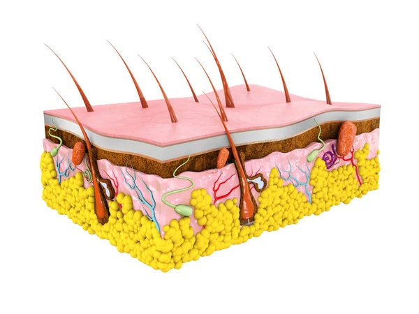
Cellulite Skin Cross-section Layers. 3d Illustration Medical Concept. Fat Cells Inside Skin

Image, 0.89MB, 6214 × 4837 jpg
stock image Scalp and hair follicles of human under the microscope in Lab.

Scalp And Hair Follicles Of Human Under The Microscope In Lab.

Image, 13.63MB, 6720 × 4480 jpg
stock image Skin anatomy

Skin Anatomy

Image, 5.07MB, 6120 × 4096 jpg
stock vector Skin anatomy in round shape

Skin Anatomy In Round Shape

Vector, 6.25MB, 4970 × 5025 eps
stock vector Skin anatomy, skin burn classification

Skin Anatomy, Skin Burn Classification

Vector, 11.45MB, 6465 × 3499 eps
Previous << Page 2 >> Next