
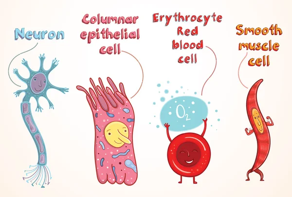
Stock image Epithelial Cells
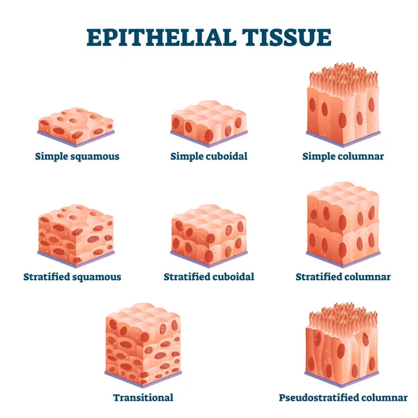
Epithelial Tissue With Labeled Squamous, Cuboidal And Columnar Examples.
Vector, 8.25MB, 4000 × 4000 eps
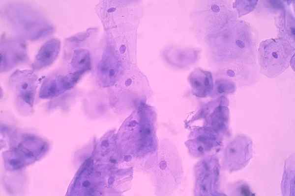
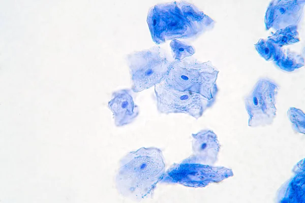
Human Cheek Epithelial Cells. The Tissue That Lines The Inside Of The Mouth Is Known As The Basal Mucosa And Is Composed Of Squamous Epithelial Cells. Education Pathology.
Image, 14.21MB, 6000 × 4000 jpg
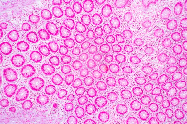
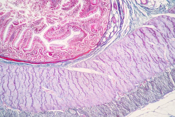
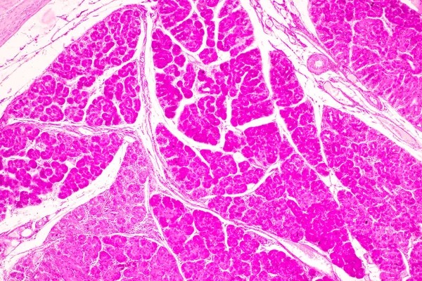
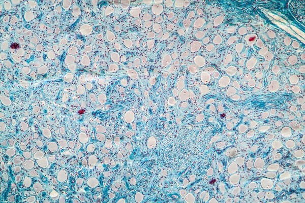
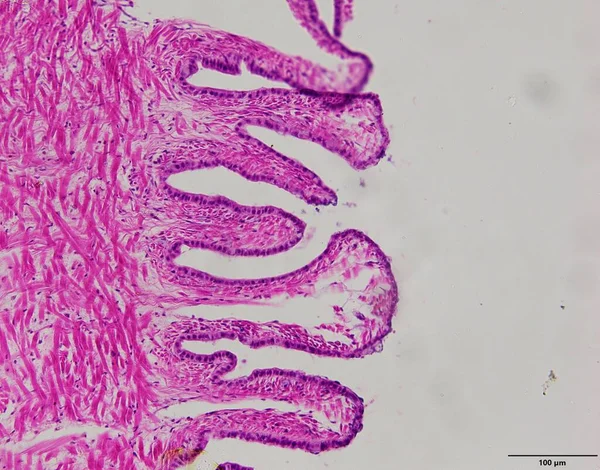
Backgrounds Of Characteristics Tissue Of Stomach Human, Small Intestine Human, Pancreas Human And Large Intestine Human Under The Microscope In Lab.
Image, 20.92MB, 6720 × 4480 jpg
Human Cheek Epithelial Cells. The Tissue That Lines The Inside Of The Mouth Is Known As The Basal Mucosa And Is Composed Of Squamous Epithelial Cells. Education Pathology.
Image, 16.77MB, 6000 × 4000 jpg
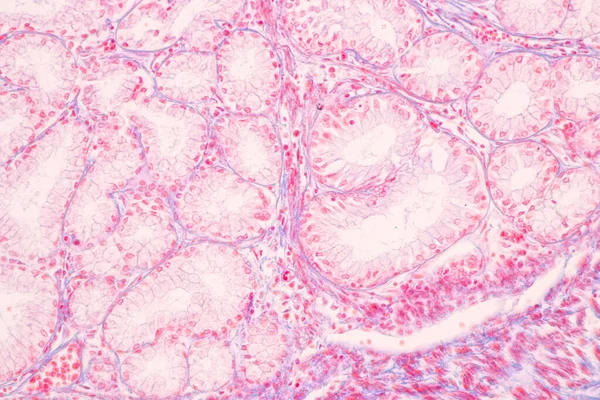
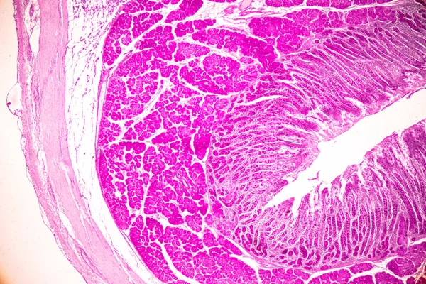
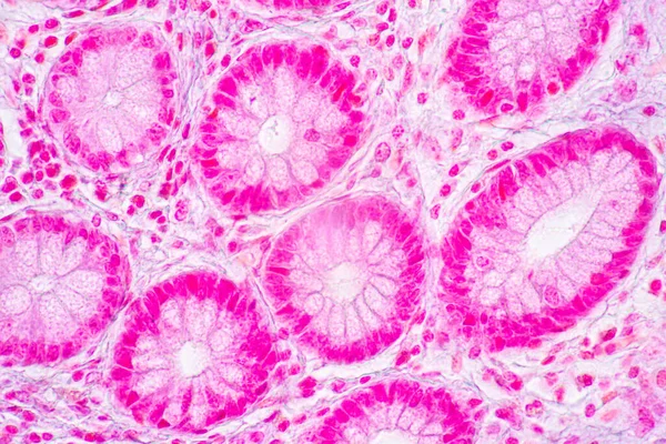
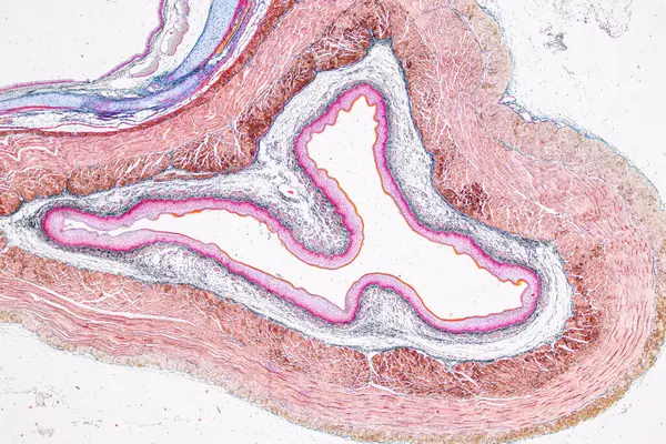
Tissue Of Small Intestine (Duodenum), Large Intestine Human And Stomach Human Under The Microscope In Lab.
Image, 17.29MB, 8192 × 5461 jpg

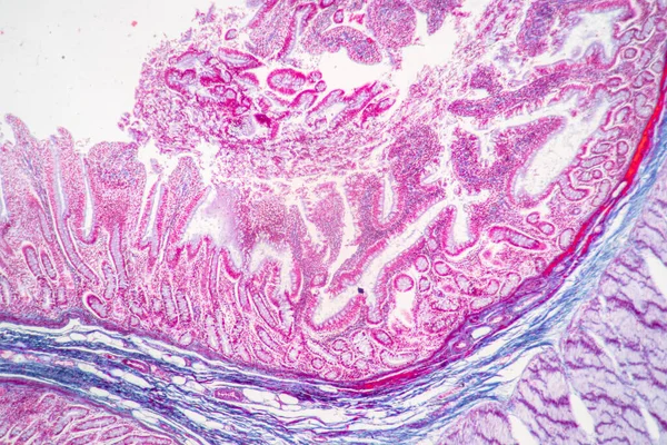
Backgrounds Of Characteristics Tissue Of Stomach Human, Small Intestine Human, Pancreas Human And Large Intestine Human Under The Microscope In Lab.
Image, 19.31MB, 6720 × 4480 jpg
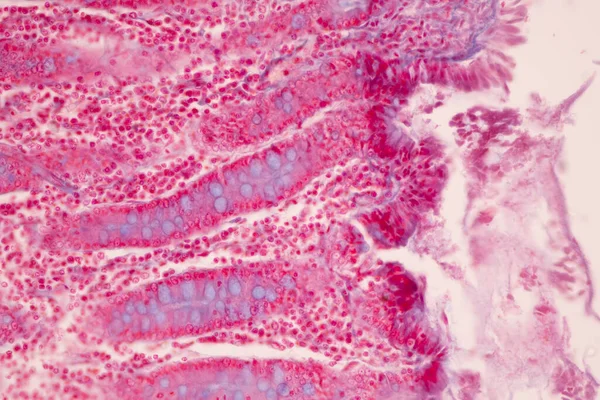
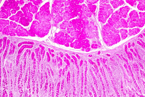
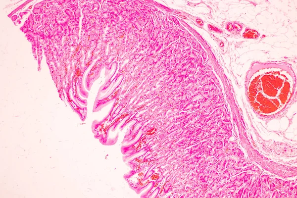
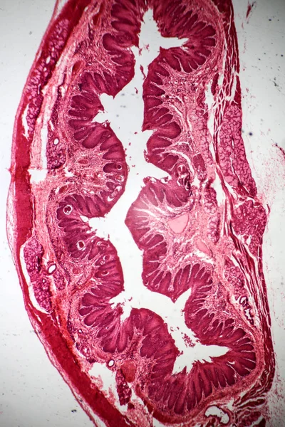
Tissue Of Small Intestine (Duodenum) And Vermiform Appendix Human Under The Microscope In Lab.
Image, 18.99MB, 6000 × 4000 jpg
Backgrounds Of Characteristics Tissue Of Stomach Human, Small Intestine Human, Pancreas Human And Large Intestine Human Under The Microscope In Lab.
Image, 20.16MB, 6720 × 4480 jpg


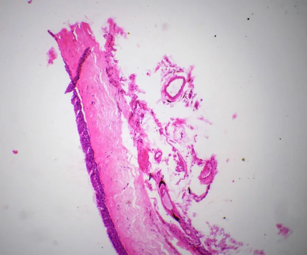
Tissue Of Small Intestine (Duodenum) And Vermiform Appendix Human Under The Microscope In Lab.
Image, 20.31MB, 6000 × 4000 jpg

Tissue Of Small Intestine (Duodenum) And Vermiform Appendix Human Under The Microscope In Lab.
Image, 22.69MB, 6000 × 4000 jpg
Tissue Of Small Intestine (Duodenum) And Vermiform Appendix Human Under The Microscope In Lab.
Image, 21.54MB, 6000 × 4000 jpg
Backgrounds Of Characteristics Tissue Of Stomach Human, Small Intestine Human, Pancreas Human And Large Intestine Human Under The Microscope In Lab.
Image, 20.75MB, 6720 × 4480 jpg


Tissue Of Small Intestine (Duodenum) And Vermiform Appendix Human Under The Microscope In Lab.
Image, 17.16MB, 6000 × 4000 jpg

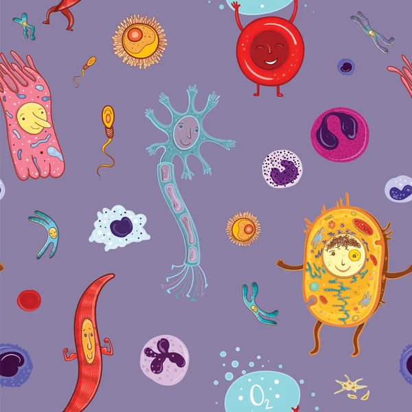
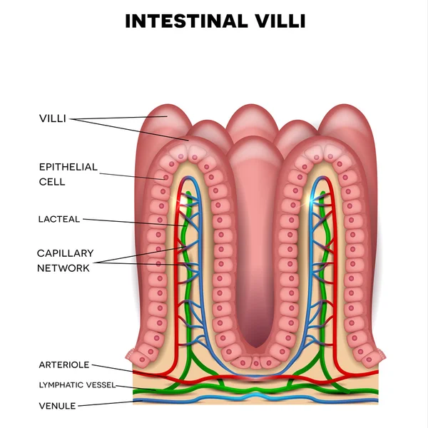
Structure Of The Enterocyte. Absorptive Cells Intestine. Infographics. Vector Illustration On Isolated Background.
Vector, 0.62MB, 5000 × 5000 eps
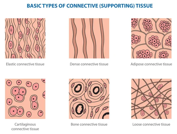

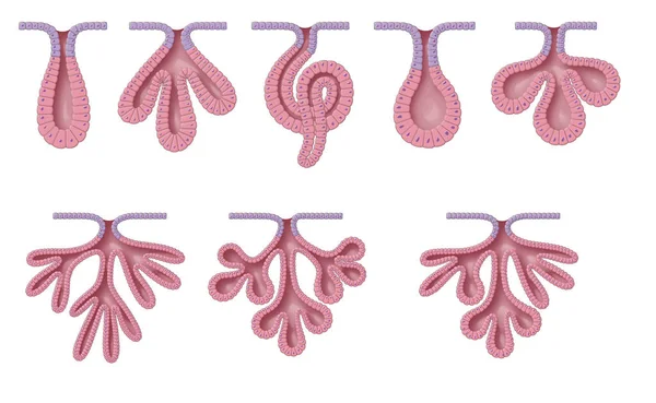
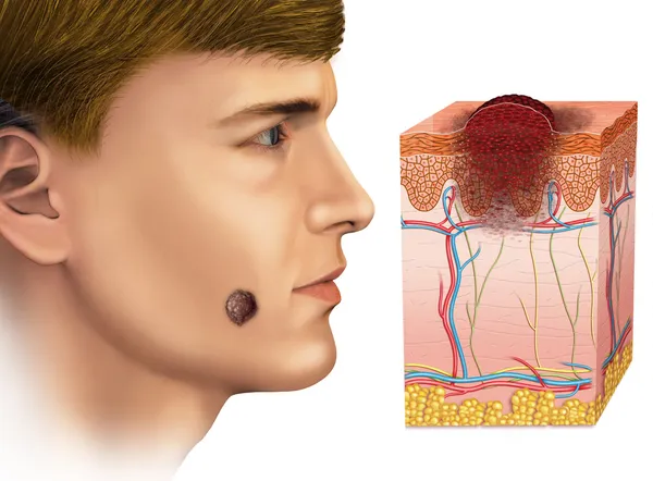
Exocrine Glands Have Two Structural Classifications, Unicellular (one Cell Layer) And Multicellular (many Cell Layers)
Image, 10.06MB, 9449 × 5809 jpg

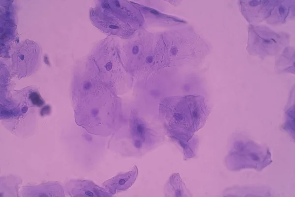
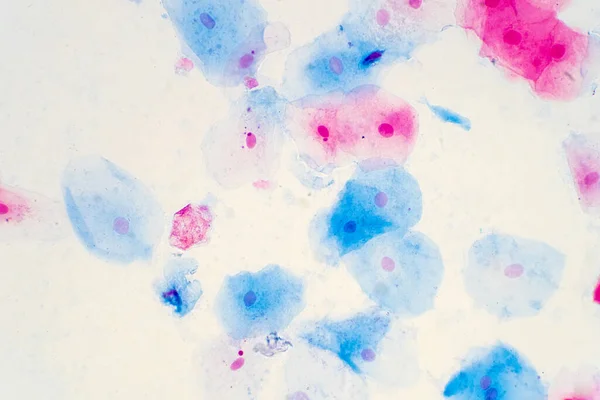
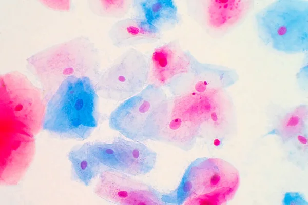
Squamous Epithelial Cells Of Human Cervix Under The Microscope View. Pap Smear Test Is A Procedure To Test For Cervical Cancer In Women.
Image, 14.68MB, 5385 × 3590 jpg


Squamous Epithelial Cells Of Human Cervix Under The Microscope View. Pap Smear Test Is A Procedure To Test For Cervical Cancer In Women.
Image, 18.53MB, 6000 × 4000 jpg
Squamous Epithelial Cells Of Human Cervix Under The Microscope View. Pap Smear Test Is A Procedure To Test For Cervical Cancer In Women.
Image, 17.23MB, 5883 × 3922 jpg
Human Cheek Epithelial Cells. The Tissue That Lines The Inside Of The Mouth Is Known As The Basal Mucosa And Is Composed Of Squamous Epithelial Cells. Education Pathology.
Image, 10.51MB, 6000 × 4000 jpg
Squamous Epithelial Cells Of Human Cervix Under The Microscope View. Pap Smear Test Is A Procedure To Test For Cervical Cancer In Women.
Image, 11.03MB, 4506 × 3004 jpg


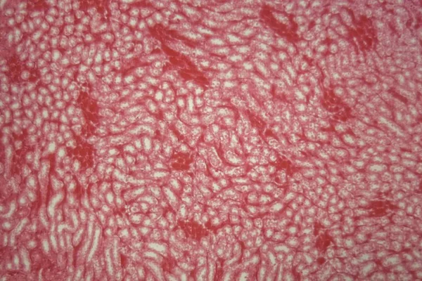
Pathology And Histology Tissue Of Mouse, Rabbit, Cat And Cow Under Microscope.
Image, 32.75MB, 6000 × 4000 jpg

Pathology And Histology Tissue Of Mouse, Rabbit, Cat And Cow Under Microscope.
Image, 6.85MB, 2667 × 4000 jpg
Pathology And Histology Tissue Of Mouse, Rabbit, Cat And Cow Under Microscope.
Image, 18.37MB, 6000 × 4000 jpg

Pathology And Histology Tissue Of Mouse, Rabbit, Cat And Cow Under Microscope.
Image, 8.03MB, 6000 × 3245 jpg
Page 1 >> Next