
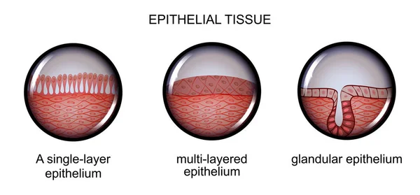
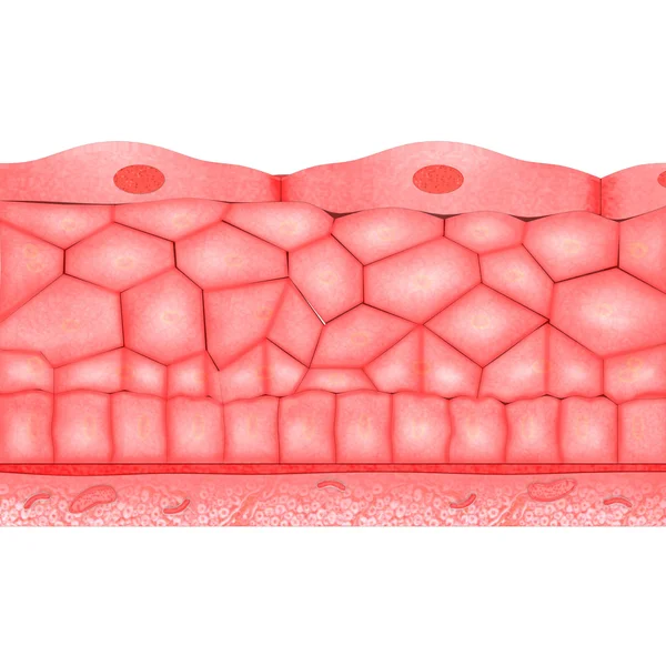
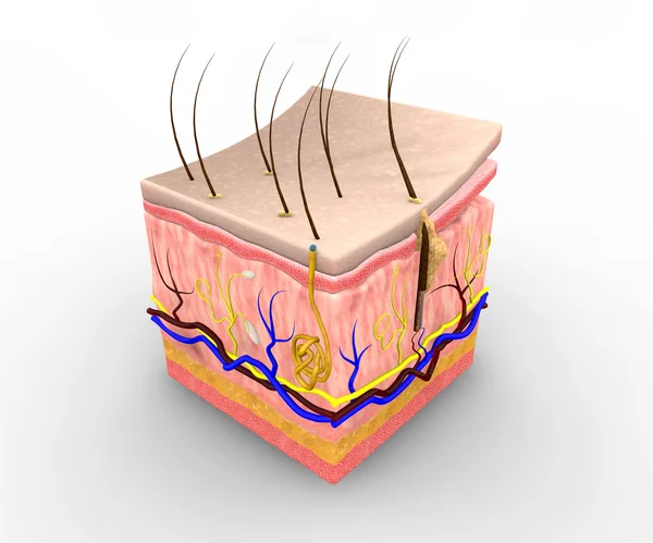
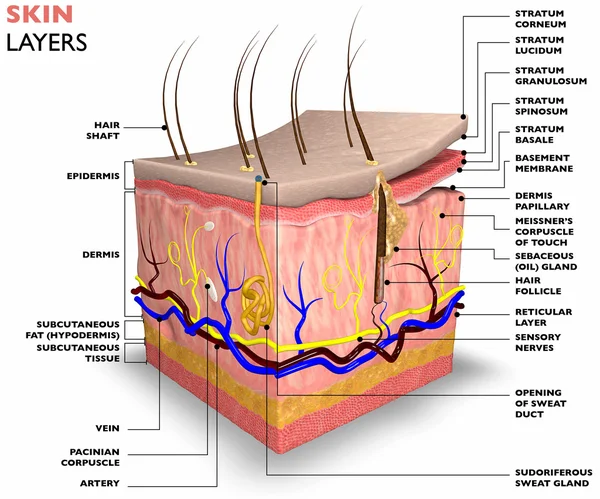
Stock image Epithelial Tissue
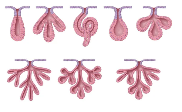
Exocrine Glands Have Two Structural Classifications, Unicellular (one Cell Layer) And Multicellular (many Cell Layers)
Image, 10.06MB, 9449 × 5809 jpg
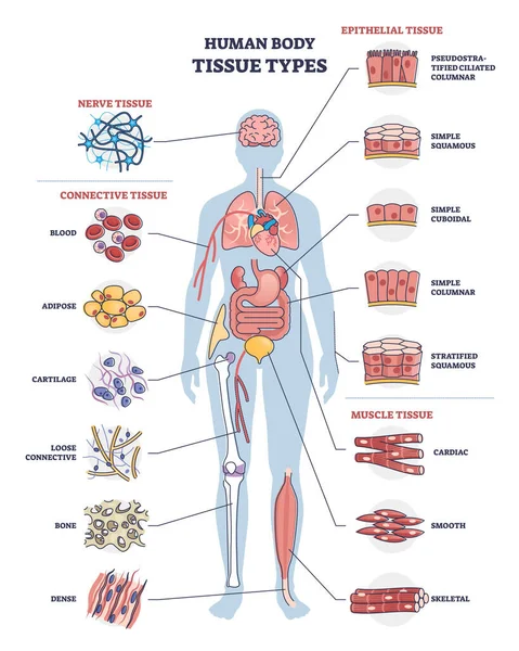
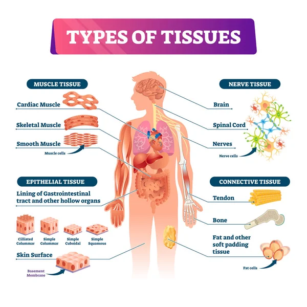
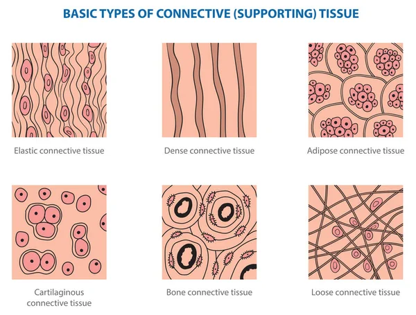
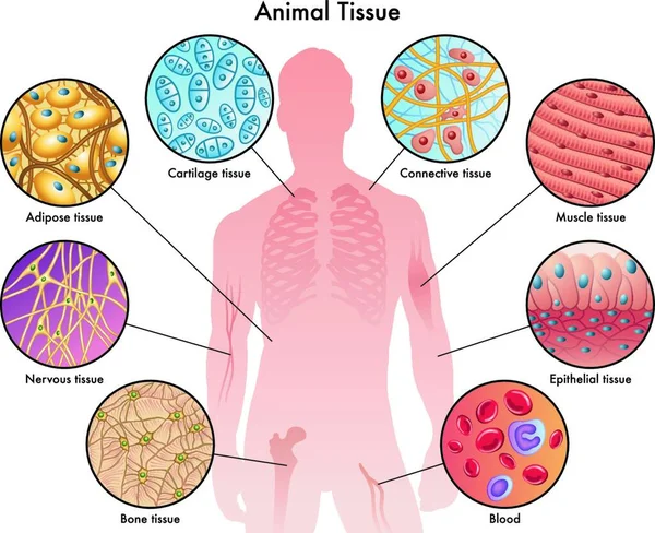
Human Body Tissue Types With Nerve, Connective And Epithelial Outline Diagram
Vector, 7.23MB, 3840 × 4800 eps
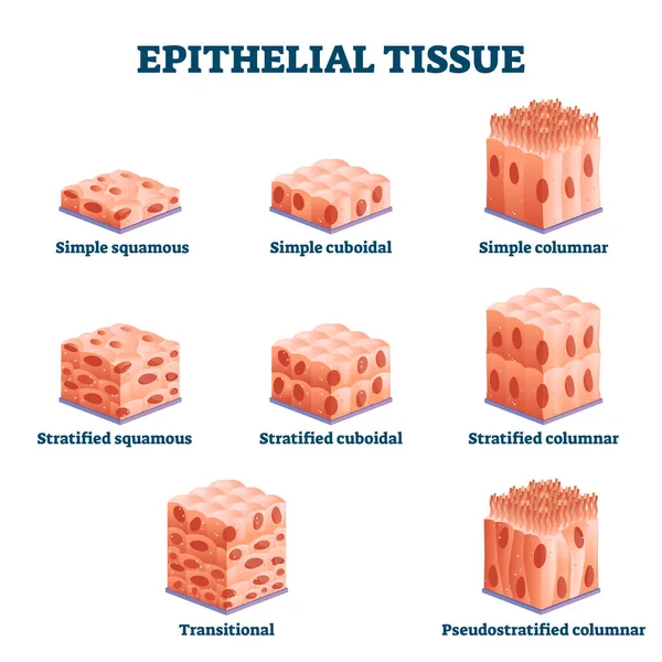
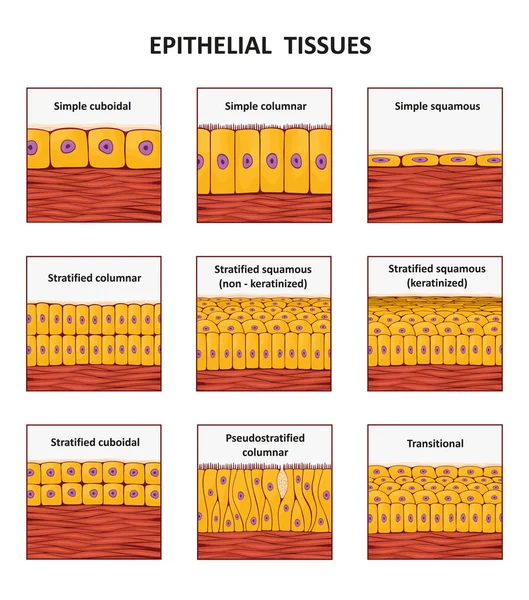
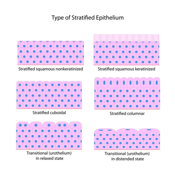
Epithelial Tissue With Labeled Squamous, Cuboidal And Columnar Examples.
Vector, 8.25MB, 4000 × 4000 eps
Types Of Tissues Vector Illustration. Labeled Inner Organ Structure Scheme.
Vector, 11.09MB, 4000 × 4000 eps
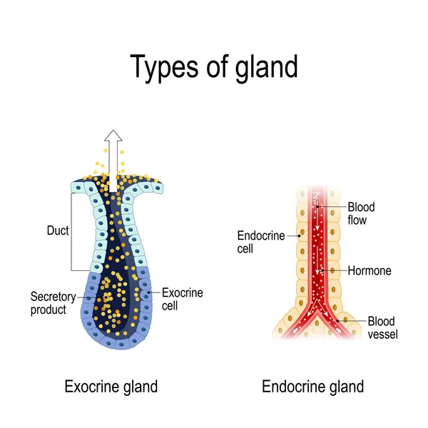
Types Of Gland. Anatomy Of An Endocrine And Exocrine Glands. Different Of Glands Secretion. Cross-section. Vector Diagram For Educational, Medical, Biological And Science Use
Vector, 1.64MB, 4950 × 4950 eps
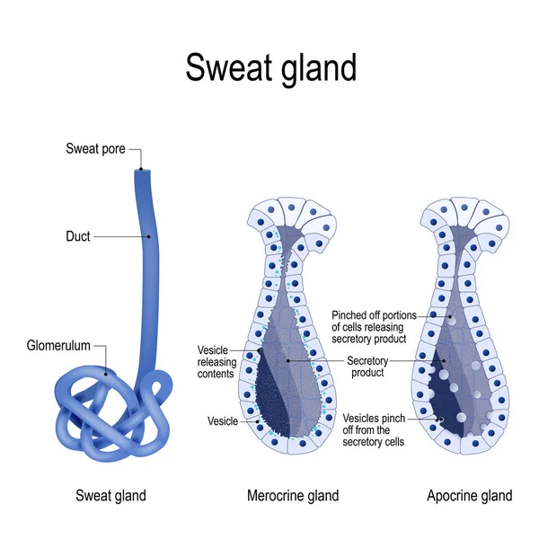
Sweat Gland. Merocrine And Apocrine. Different Of Manner Of Secretion. Cross-section Of The Human Skin, With The Sweat Gland. Close-up Of Dark And Clear Cells, Lumen, Sweat Duct, Glomerulum And Pore. Labeled Vector Diagram For Educational, Medical,
Vector, 3.31MB, 5165 × 5165 eps
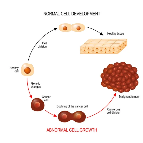
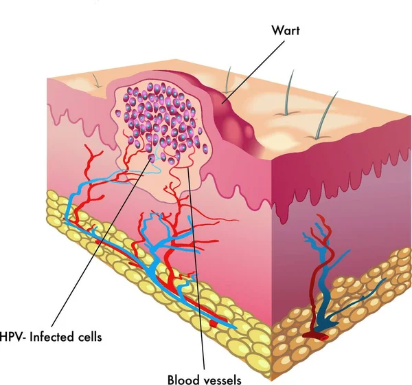
Cancer Cell And Normal Cell. Healthy Tissue And Malignant Tumour. Illustration Showing Cancer Disease Development. Vector Diagram For Your Design, Educational, Biological, Science And Medical Use
Vector, 2.66MB, 6102 × 6102 eps

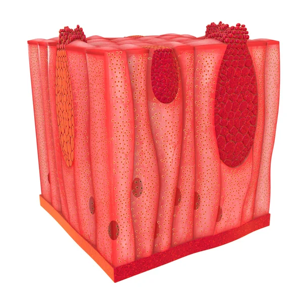
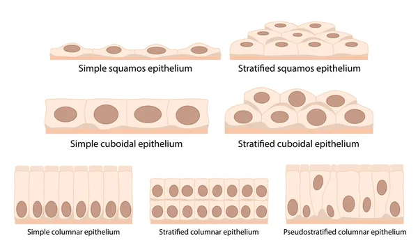
Cells Of Epithelial Tissue: Squamous (flattened And Thin), Cuboidal (boxy, As Wide As It Is Tall), Columnar (rectangular, Taller Than It Is Wide), Pseudostratified.
Vector, 6.24MB, 5209 × 3125 eps
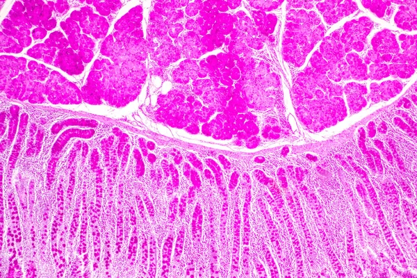
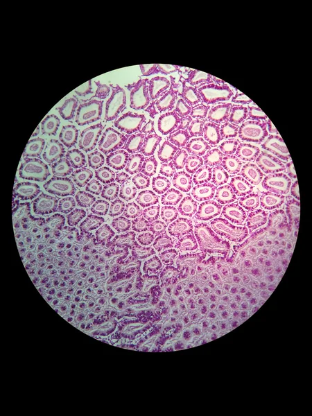
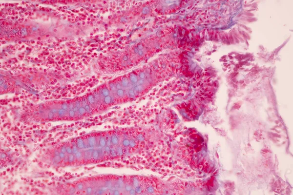
Tissue Of Small Intestine (Duodenum) And Vermiform Appendix Human Under The Microscope In Lab.
Image, 22.69MB, 6000 × 4000 jpg
Tissue Of Small Intestine (Duodenum) And Vermiform Appendix Human Under The Microscope In Lab.
Image, 21.54MB, 6000 × 4000 jpg

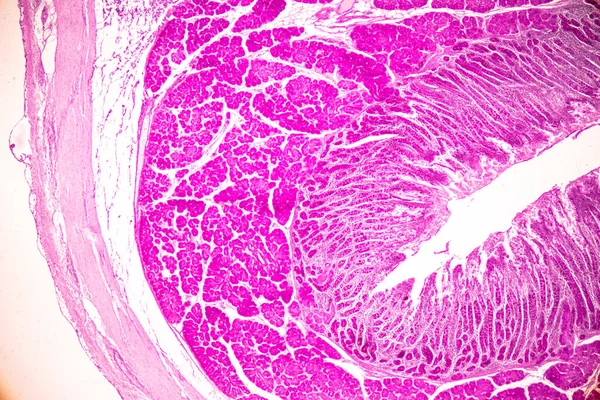
Tissue Of Small Intestine (Duodenum) And Vermiform Appendix Human Under The Microscope In Lab.
Image, 18.99MB, 6000 × 4000 jpg

Tissue Of Small Intestine (Duodenum) And Vermiform Appendix Human Under The Microscope In Lab.
Image, 20.31MB, 6000 × 4000 jpg

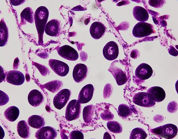
Squamous Epithelial Cells Of Human Cervix Under The Microscope View. Pap Smear Test Is A Procedure To Test For Cervical Cancer In Women.
Image, 14.68MB, 5385 × 3590 jpg
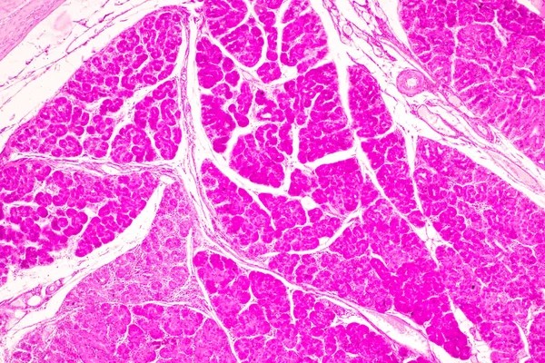
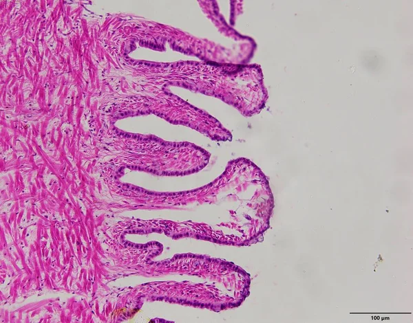
Tissue Of Small Intestine (Duodenum), Large Intestine Human And Stomach Human Under The Microscope In Lab.
Image, 17.29MB, 8192 × 5461 jpg

Tissue Of Small Intestine (Duodenum) And Vermiform Appendix Human Under The Microscope In Lab.
Image, 17.16MB, 6000 × 4000 jpg


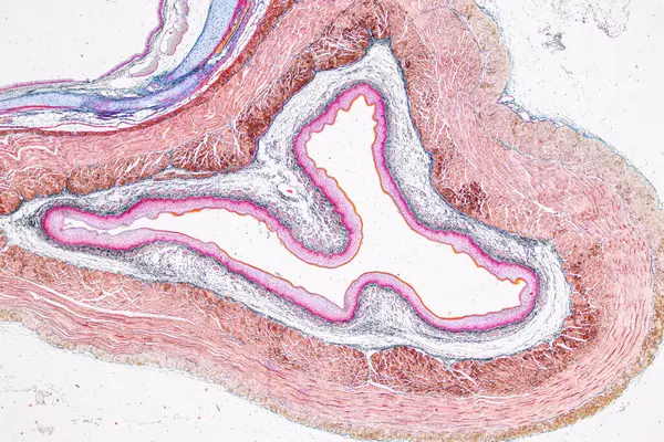
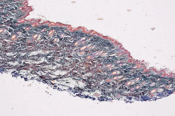
Pathology And Histology Tissue Of Mouse, Rabbit, Cat And Cow Under Microscope.
Image, 32.75MB, 6000 × 4000 jpg

Pathology And Histology Tissue Of Mouse, Rabbit, Cat And Cow Under Microscope.
Image, 6.85MB, 2667 × 4000 jpg
Pathology And Histology Tissue Of Mouse, Rabbit, Cat And Cow Under Microscope.
Image, 18.37MB, 6000 × 4000 jpg

Pathology And Histology Tissue Of Mouse, Rabbit, Cat And Cow Under Microscope.
Image, 8.03MB, 6000 × 3245 jpg

Pathology And Histology Tissue Of Mouse, Rabbit, Cat And Cow Under Microscope.
Image, 34.2MB, 6000 × 4000 jpg

Pathology And Histology Tissue Of Mouse, Rabbit, Cat And Cow Under Microscope.
Image, 18.99MB, 6000 × 4000 jpg

Pathology And Histology Tissue Of Mouse, Rabbit, Cat And Cow Under Microscope.
Image, 34.94MB, 6000 × 4000 jpg

Pathology And Histology Tissue Of Mouse, Rabbit, Cat And Cow Under Microscope.
Image, 10.76MB, 6000 × 4000 jpg

Pathology And Histology Tissue Of Mouse, Rabbit, Cat And Cow Under Microscope.
Image, 16.84MB, 6000 × 4000 jpg

Pathology And Histology Tissue Of Mouse, Rabbit, Cat And Cow Under Microscope.
Image, 20.89MB, 6000 × 4000 jpg
Pathology And Histology Tissue Of Mouse, Rabbit, Cat And Cow Under Microscope.
Image, 18.02MB, 6000 × 4000 jpg

Page 1 >> Next