
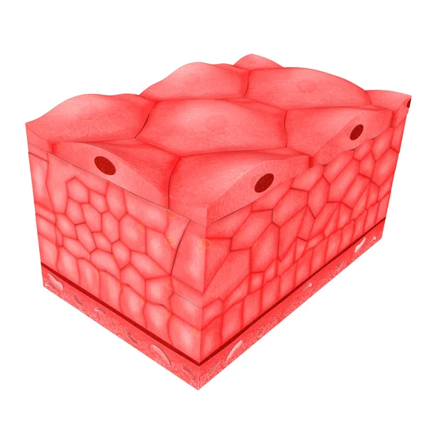
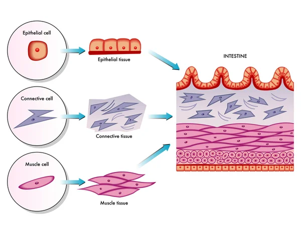
Stock image Epithelium Tissue
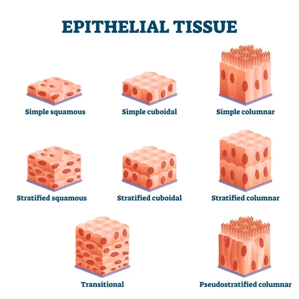
Epithelial Tissue With Labeled Squamous, Cuboidal And Columnar Examples.
Vector, 8.25MB, 4000 × 4000 eps

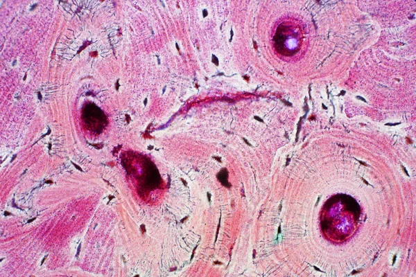
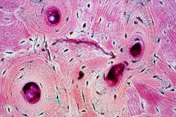
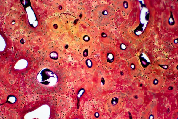
Histology Of Human Compact Bone Tissue Under Microscope View For Education, Muscle Bone Connection And Connective Tissue
Image, 18.19MB, 6000 × 4000 jpg
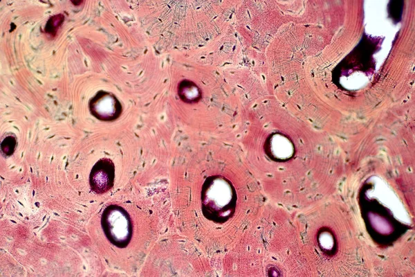
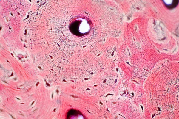
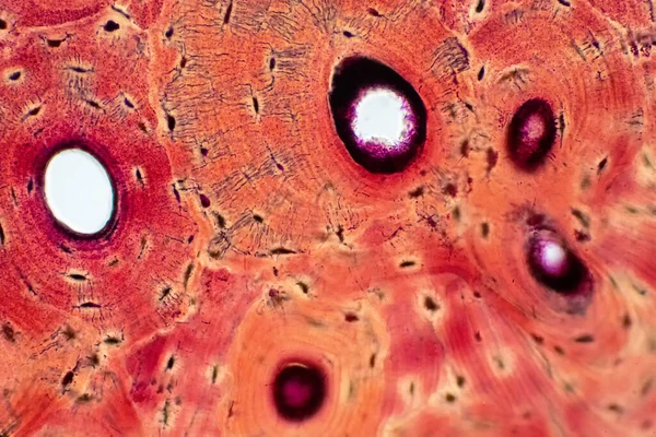
Histology Of Human Compact Bone Tissue Under Microscope View For Education, Muscle Bone Connection And Connective Tissue
Image, 13.74MB, 5786 × 3857 jpg
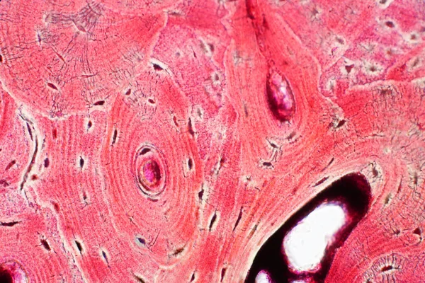
Histology Of Human Compact Bone Tissue Under Microscope View For Education, Muscle Bone Connection And Connective Tissue
Image, 17.36MB, 6000 × 4000 jpg
Histology Of Human Compact Bone Tissue Under Microscope View For Education, Muscle Bone Connection And Connective Tissue
Image, 18.53MB, 6000 × 4000 jpg

Histology Of Human Compact Bone Tissue Under Microscope View For Education, Muscle Bone Connection And Connective Tissue
Image, 20.09MB, 6000 × 4000 jpg
Histology Of Human Compact Bone Tissue Under Microscope View For Education, Muscle Bone Connection And Connective Tissue
Image, 17.56MB, 6000 × 4000 jpg

Histology Of Human Compact Bone Tissue Under Microscope View For Education, Muscle Bone Connection And Connective Tissue
Image, 19.59MB, 6000 × 4000 jpg
Histology Of Human Compact Bone Tissue Under Microscope View For Education, Muscle Bone Connection And Connective Tissue
Image, 17.82MB, 6000 × 4000 jpg
Muscle Bone Connection And Connective Tissue. Histology Of Human Compact Bone Tissue Under Microscope View For Education.
Image, 20.37MB, 6000 × 4000 jpg
Histology Of Human Compact Bone Tissue Under Microscope View For Education, Muscle Bone Connection And Connective Tissue
Image, 16.83MB, 6000 × 4000 jpg
Muscle Bone Connection And Connective Tissue. Histology Of Human Compact Bone Tissue Under Microscope View For Education.
Image, 19.6MB, 6000 × 4000 jpg
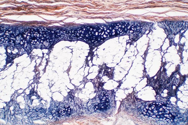
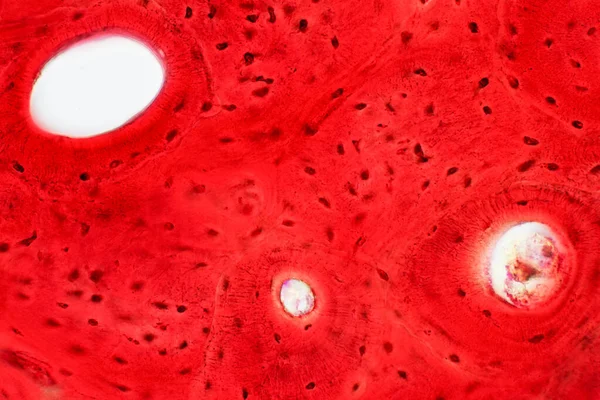
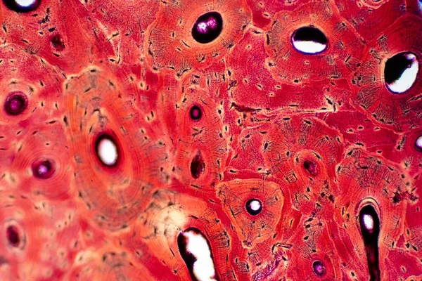
Cartilage Is A Resilient And Smooth Elastic Tissue For Pathology Education, A Rubber-like Padding That Covers And Protects The Ends Of Long Bones At The Joints.
Image, 15.16MB, 6000 × 4000 jpg


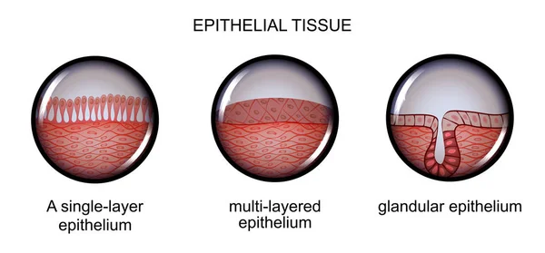
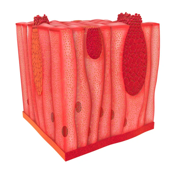
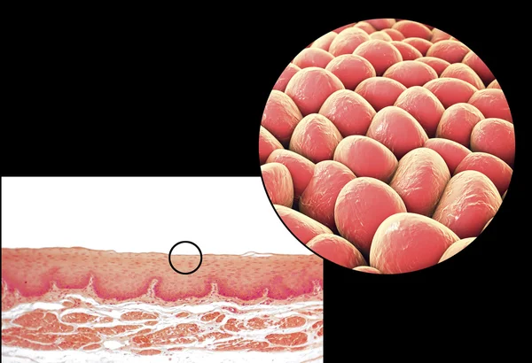
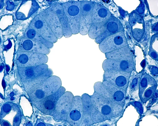
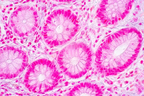
Ciliated Columnar Epithelium. Epithelial Cells Forms The Lining Of The Stomach And Intestines, Duodenum, Fallopian Tubes, Uterus, Central Canal Of The Spinal Cord, Nose, Ears And The Taste Buds.
Vector, 0.98MB, 4444 × 4444 eps
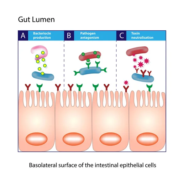
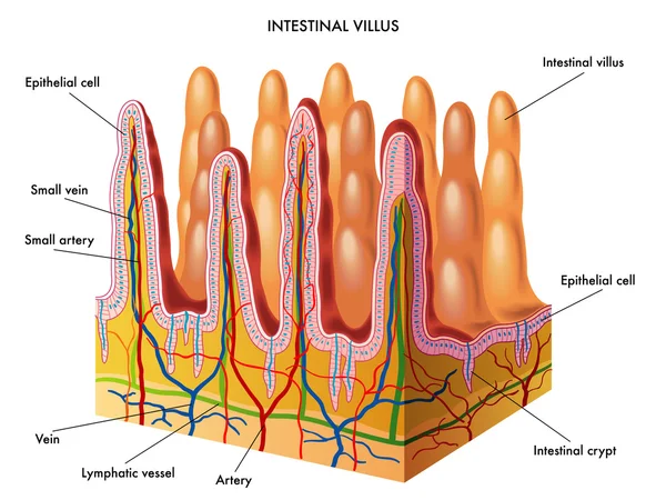
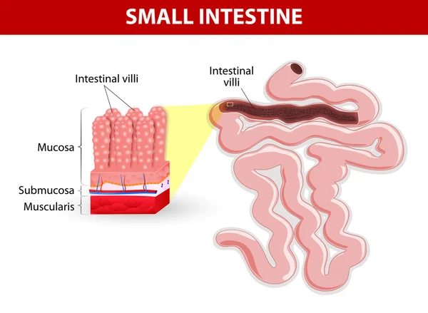
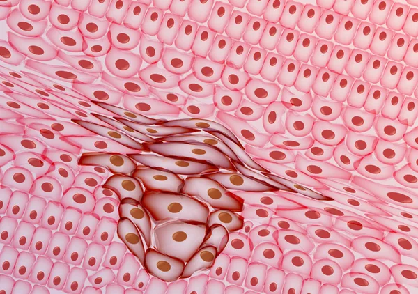
Gut Lumen. Enterocytes, Or Intestinal Absorptive Cells. Small Intestine. Columnar Epithelial Cells
Vector, 1.32MB, 5000 × 5000 eps


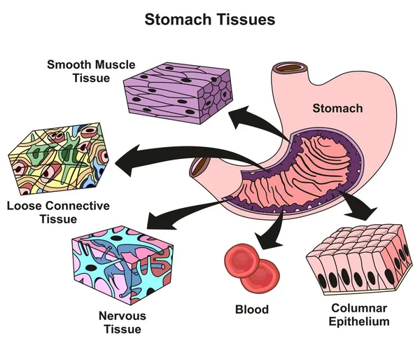
Stomach Tissues Types And Structure Infographic Diagram Including Smooth Muscle Loose Connective Nervous Blood, Columnar Epithelium For Medical Science Education And Health Care
Vector, 0.51MB, 2254 × 1833 eps


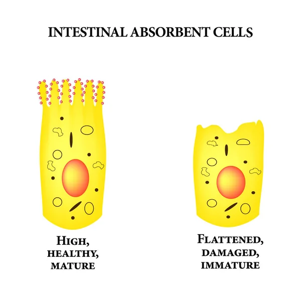
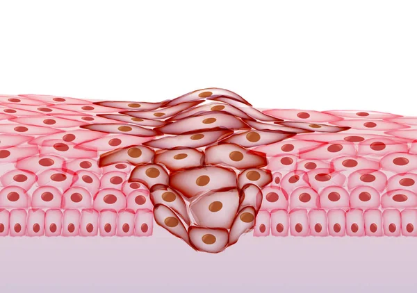
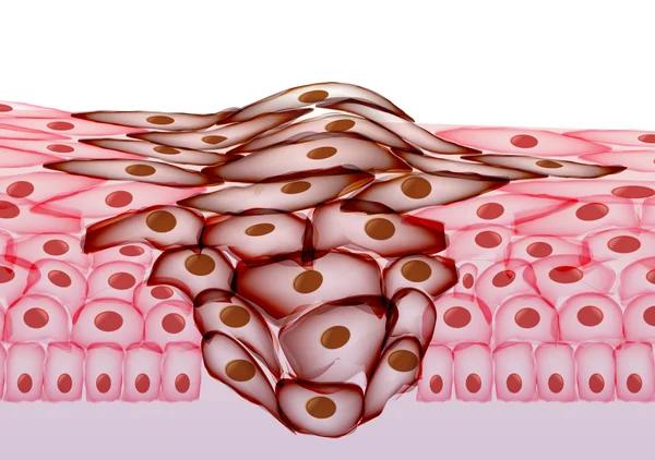
Structure Of The Enterocyte. Deformed, Sick Cell. Absorptive Cells Intestine. Infographics. Vector Illustration On Isolated Background.
Vector, 1.55MB, 5000 × 5000 eps
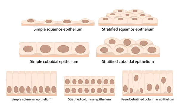
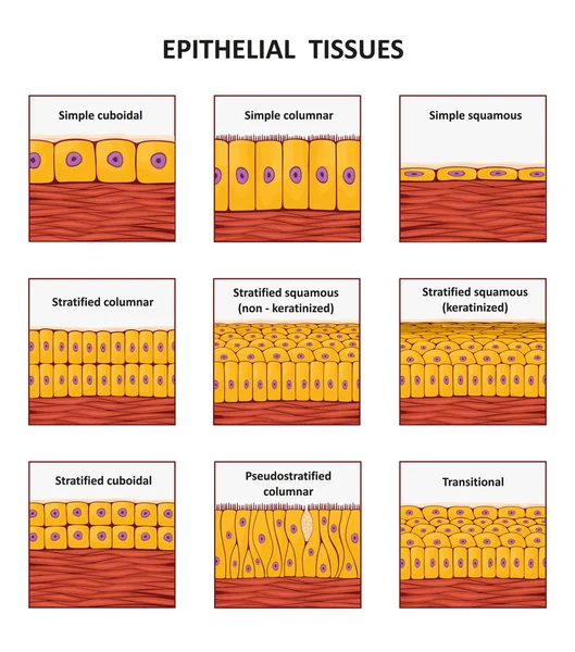
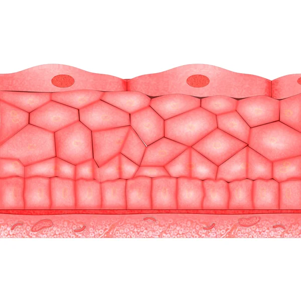
Cells Of Epithelial Tissue: Squamous (flattened And Thin), Cuboidal (boxy, As Wide As It Is Tall), Columnar (rectangular, Taller Than It Is Wide), Pseudostratified.
Vector, 6.24MB, 5209 × 3125 eps
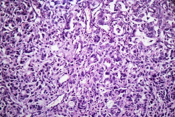
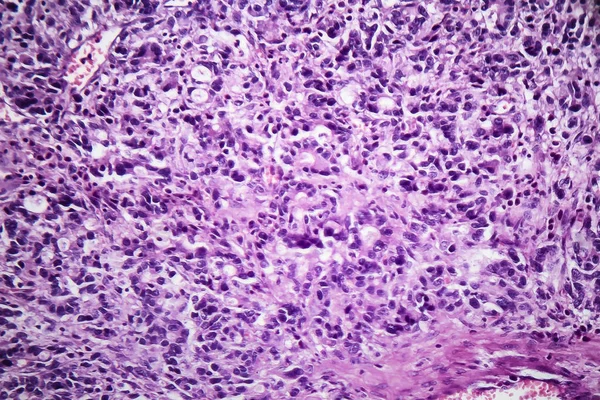
Backgrounds Of Characteristics Tissue Of Stomach Human, Small Intestine Human, Pancreas Human And Large Intestine Human Under The Microscope In Lab.
Image, 20.75MB, 6720 × 4480 jpg

Page 1 >> Next