photo free
Home Images Vectors Videos

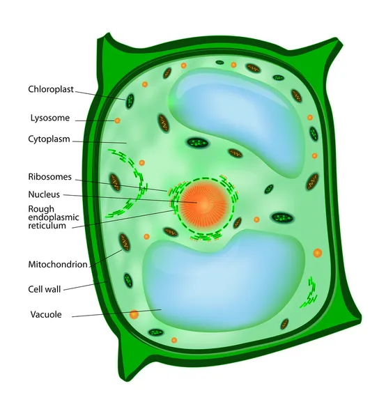
Stock image Eukaryote

stock image Scientist researching in laboratory, pipetting cell culture medium samples in laminar flow

Scientist Researching In Laboratory, Pipetting Cell Culture Medium Samples In Laminar Flow

Image, 3.79MB, 4000 × 2667 jpg
stock image Internal parts of bacteria

Internal Parts Of Bacteria

Image, 5.54MB, 3840 × 2160 jpg
stock image Laboratory work with plasmid DNA extraction

Laboratory Work With Plasmid DNA Extraction

Image, 6.04MB, 4000 × 2667 jpg
stock image Scientist researching in laboratory, pipetting cell culture medium samples in laminar flow

Scientist Researching In Laboratory, Pipetting Cell Culture Medium Samples In Laminar Flow

Image, 3.77MB, 4000 × 2667 jpg
stock image Cells divide by osmosis

Cells Divide By Osmosis

Image, 0.41MB, 2000 × 2000 jpg
stock vector Plant cell

Plant Cell

Vector, 4.07MB, 3870 × 4189 eps
stock vector Types of RNA: messenger RNA (mRNA), transport RNA (tRNA), ribosomal RNA (rRNA). Vector illustration.

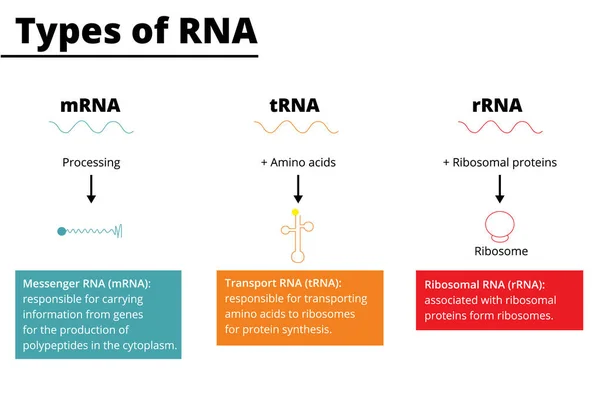
Types Of RNA: Messenger RNA (mRNA), Transport RNA (tRNA), Ribosomal RNA (rRNA). Vector Illustration.

Vector, 0.73MB, 5000 × 3500 ai
stock image Parasites, human pathogenic microbes

Parasites, Human Pathogenic Microbes

Image, 2.49MB, 4500 × 3000 jpg
stock image Parasites, pathogenic microbes

Parasites, Pathogenic Microbes

Image, 5.58MB, 4500 × 3000 jpg
stock vector Set of different single-celled eukaryote  Protozoas, Vector illustration

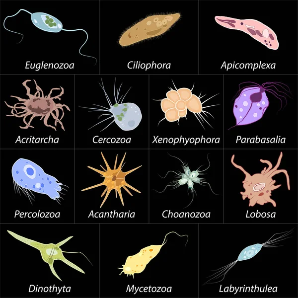
Set Of Different Single-celled Eukaryote Protozoas, Vector Illustration

Vector, 9.57MB, 4167 × 4167 eps
stock image Human forefinger touches cell dividing. Nebula dust in infinite space. Mixed media.

Human Forefinger Touches Cell Dividing. Nebula Dust In Infinite Space. Mixed Media.

Image, 9.78MB, 5736 × 3824 jpg
stock image Entamoeba histolytica protozoan

Entamoeba Histolytica Protozoan

Image, 1.53MB, 4500 × 3000 jpg
stock image cell culture at the medicine  medical and cell culture laboratory

Cell Culture At The Medicine Medical And Cell Culture Laboratory

Image, 7.21MB, 5472 × 3648 jpg
stock image Organic cell. Virus, germ or bacteria. Isolated on white background. 3d render

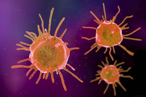
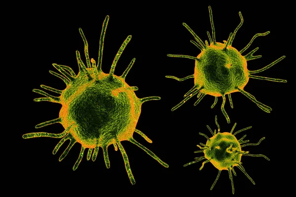
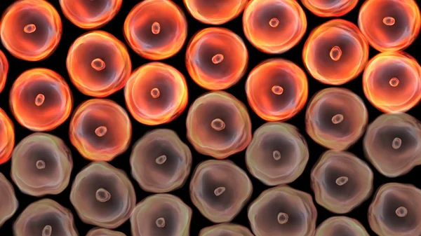
Organic Cell. Virus, Germ Or Bacteria. Isolated On White Background. 3d Render

Image, 2.96MB, 4244 × 3750 jpg
stock image Structure of a Eukaryotic Ribosome

Structure Of A Eukaryotic Ribosome

Image, 4MB, 8234 × 4232 jpg
stock vector mitosis cell division. Vector diagram. Poster for education

Mitosis Cell Division. Vector Diagram. Poster For Education

Vector, 10.78MB, 5000 × 3211 eps
stock image 3d Rendering of Mitochondria - realistic illustration on red background

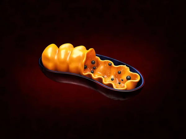
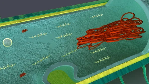
3d Rendering Of Mitochondria - Realistic Illustration On Red Background

Image, 10.21MB, 8000 × 6000 jpg
stock image Saccharomyces cerevisiae yeasts, hand drawn watercolor illustration. Baker's or brewer's yeast, probiotics restoring normal flora of intestine.

Saccharomyces Cerevisiae Yeasts, Hand Drawn Watercolor Illustration. Baker's Or Brewer's Yeast, Probiotics Restoring Normal Flora Of Intestine.

Image, 14.78MB, 4846 × 3403 jpg
stock vector Bacteria cell structure

Bacteria Cell Structure

Vector, 2.57MB, 6000 × 4212 eps
stock image Blue glowing cell mitosis fractal

Blue Glowing Cell Mitosis Fractal

Image, 7.08MB, 4000 × 3000 jpg
stock image Process division of cell

Process Division Of Cell

Image, 5.3MB, 5000 × 3750 jpg
stock image bad cells turn better

Bad Cells Turn Better

Image, 3.82MB, 4500 × 2532 jpg
stock image Meiosis and formation of zygote

Meiosis And Formation Of Zygote

Image, 8.32MB, 11811 × 7907 jpg
stock image Mitosis, a process of cell duplication.

Mitosis, A Process Of Cell Duplication.

Image, 0.58MB, 2500 × 2500 jpg
stock image Process division of cell

Process Division Of Cell

Image, 6.51MB, 5000 × 3750 jpg
stock vector organism blue cells

Organism Blue Cells

Vector, 3.6MB, 6944 × 6944 ai
stock image Human Cheek Epithelial Cells. The tissue that lines the inside of the mouth is known as the basal mucosa and is composed of squamous epithelial cells. Education pathology.

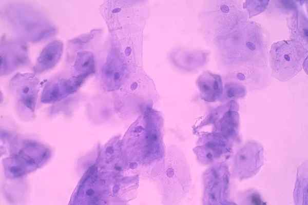
Human Cheek Epithelial Cells. The Tissue That Lines The Inside Of The Mouth Is Known As The Basal Mucosa And Is Composed Of Squamous Epithelial Cells. Education Pathology.

Image, 14.21MB, 6000 × 4000 jpg
stock image Cell division on blue background. 3d illustration

Cell Division On Blue Background. 3d Illustration

Image, 4.68MB, 4500 × 2700 jpg
stock vector Cell division. Mitosis

Cell Division. Mitosis

Vector, 1.93MB, 3860 × 3875 eps
stock image Human hand holding cell dividing. nebula dust. Mixed media.

Human Hand Holding Cell Dividing. Nebula Dust. Mixed Media.

Image, 6.9MB, 6000 × 4000 jpg
stock image Old brown toadstool in the forest in autumn. Natural backgrounds and textures.

Old Brown Toadstool In The Forest In Autumn. Natural Backgrounds And Textures.

Image, 4.11MB, 4080 × 3072 jpg
stock image Brown algae, fucus

Brown Algae, Fucus

Image, 9.71MB, 5184 × 3456 jpg
stock image Process division of cell

Process Division Of Cell

Image, 4.4MB, 3343 × 5879 jpg
stock vector Vector virus

Vector Virus

Vector, 1.27MB, 5669 × 5669 eps
stock vector Osteoblast

Osteoblast

Vector, 5.24MB, 4000 × 4000 eps
stock image The most beautiful bird in the world is a peacock from the chicken family with a large and bright tail like a chic fan of feathers with eyes for protection. An animal from the pheasant family.

The Most Beautiful Bird In The World Is A Peacock From The Chicken Family With A Large And Bright Tail Like A Chic Fan Of Feathers With Eyes For Protection. An Animal From The Pheasant Family.

Image, 25.58MB, 6000 × 4000 jpg
stock image Earthworm in the sun

Earthworm In The Sun

Image, 7.76MB, 4430 × 2652 jpg
stock image cells large size

Cells Large Size

Image, 4.17MB, 4500 × 2532 jpg
stock image Scientist researching in laboratory, pipetting cell culture medium samples in laminar flow

Scientist Researching In Laboratory, Pipetting Cell Culture Medium Samples In Laminar Flow

Image, 3.76MB, 4000 × 2667 jpg
stock image Process division of cell

Process Division Of Cell

Image, 4.67MB, 5000 × 3142 jpg
stock image Dividing cells on colorful background

Dividing Cells On Colorful Background

Image, 1.01MB, 4500 × 3000 jpg
stock image Illustration of cells in blue

Illustration Of Cells In Blue

Image, 3.23MB, 5700 × 4331 jpg
stock vector Process of division of organic cell

Process Of Division Of Organic Cell

Vector, 15.76MB, 6124 × 4082 eps
stock image color cells focus

Color Cells Focus

Image, 2.99MB, 4500 × 2532 jpg
stock image Cell division happens when a parent cell divides into two or more cells called daughter cells 3D rendering

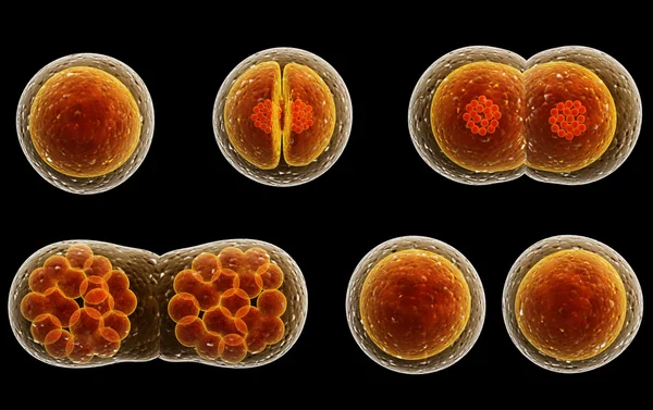
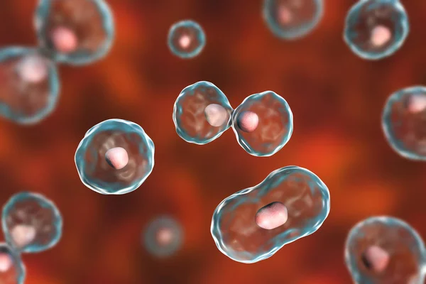
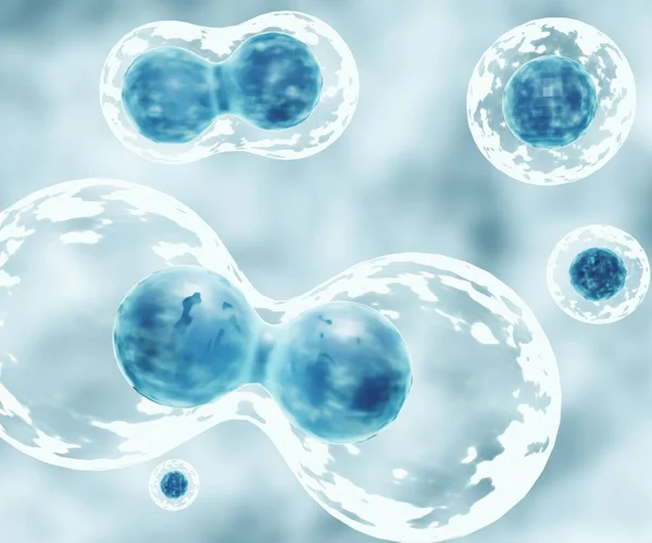
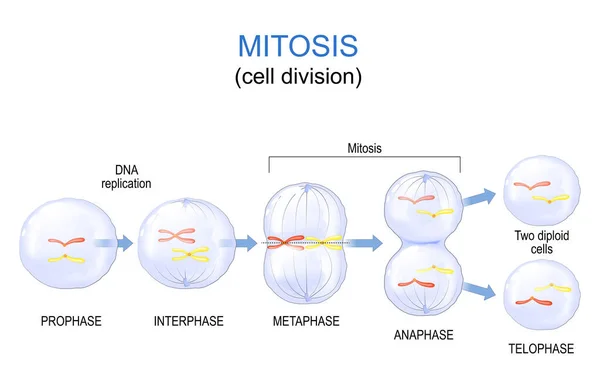
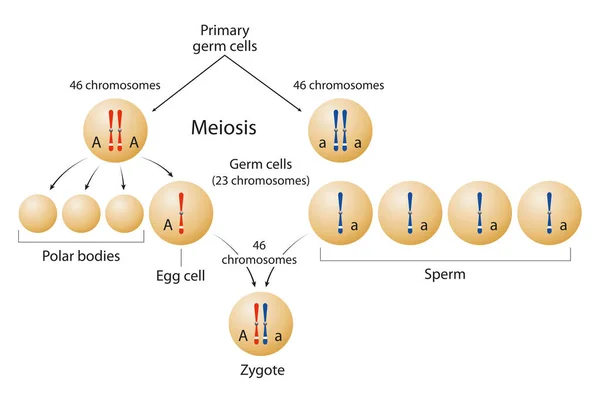
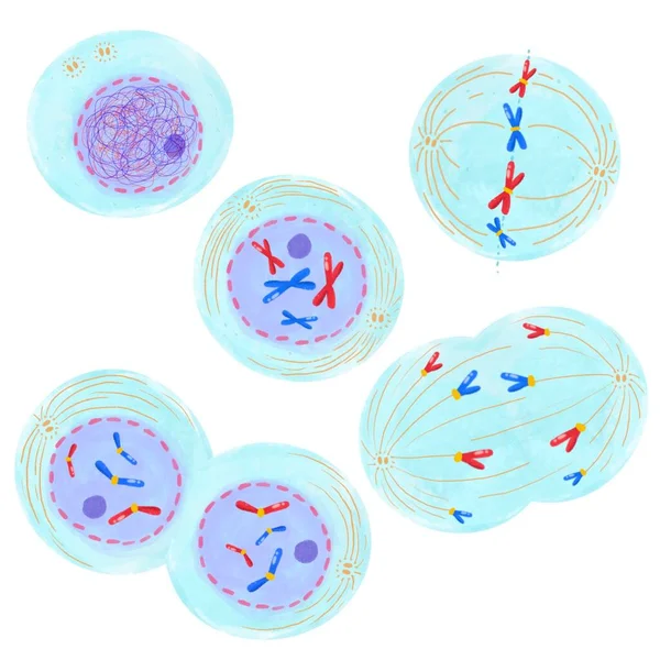
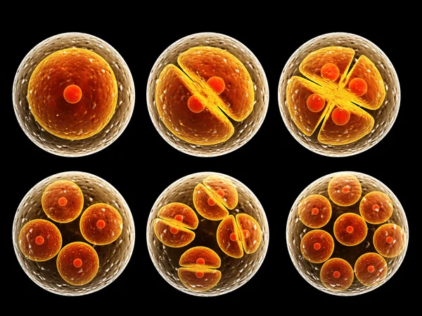
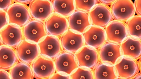
Cell Division Happens When A Parent Cell Divides Into Two Or More Cells Called Daughter Cells 3D Rendering

Image, 0.25MB, 2400 × 2000 jpg
stock image Human forefinger touches cell dividing. Mixed media.

Human Forefinger Touches Cell Dividing. Mixed Media.

Image, 5.78MB, 6000 × 4000 jpg
stock image Human hand holding with cell dividing. nebula dust. Mixed media.

Human Hand Holding With Cell Dividing. Nebula Dust. Mixed Media.

Image, 10.3MB, 6000 × 4000 jpg
stock image Human hand holding with cell dividing. nebula dust. Mixed media.

Human Hand Holding With Cell Dividing. Nebula Dust. Mixed Media.

Image, 10.49MB, 6000 × 4000 jpg
stock vector Eukaryotic vs Prokaryotic cells, educational biology vector illustration diagram

Eukaryotic Vs Prokaryotic Cells, Educational Biology Vector Illustration Diagram

Vector, 6.95MB, 3333 × 5000 eps
stock vector Human mitochondria structure diagram schematic vector illustration. Medical science educational illustration

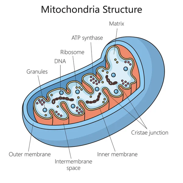
Human Mitochondria Structure Diagram Schematic Vector Illustration. Medical Science Educational Illustration

Vector, 0.64MB, 4000 × 4000 eps
Page 1 >> Next