
Stock image Extracellular


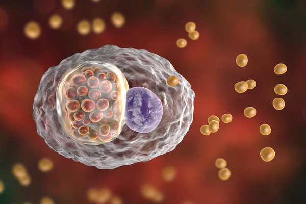
Chlamydophila Psittaci, Intracellular Bacteria That Cause Psittacosis
Image, 1.33MB, 4500 × 3000 jpg
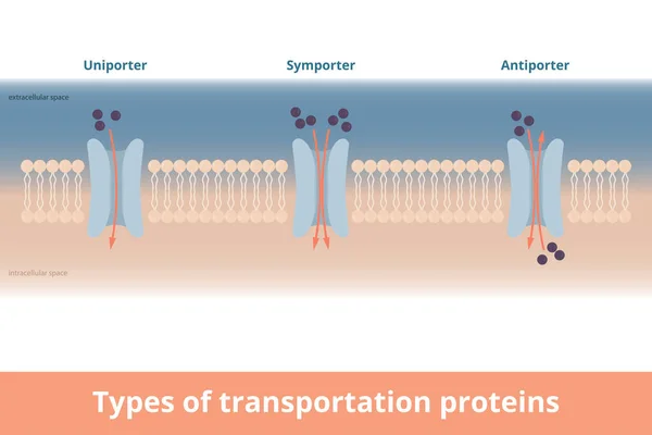
Types Of Cell Membrane Transportation Proteins. Visualization Of Uniporter (one Molecule, One Direction), Symporter (two Molecules, Same Directions), Antiporter (two Molecules, Different Directions).
Vector, 7.12MB, 6250 × 4167 eps
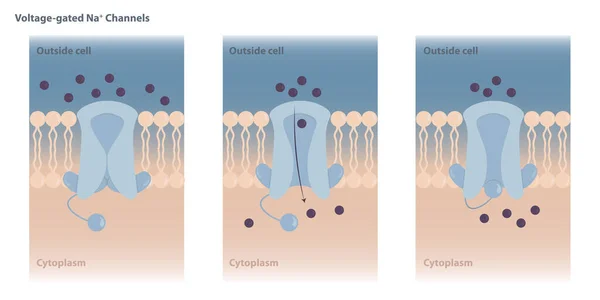
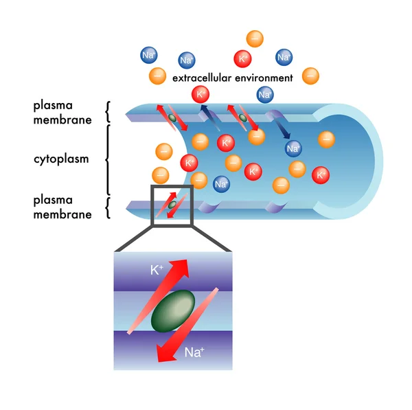
Neuronal Charged Membranes. Voltage-gated Ion Channels Are Closed At The Resting Potential And Open In Response To Changes In Membrane Voltage.
Vector, 7.67MB, 8334 × 4167 eps

Biofilm Formation. Stages Of Biofilm Development. Life Cycle Of Staphylococcus Aureus. Adherent Cells Of Bacteria Become Embedded Within A Slimy Extracellular Matrix. Vector Illustration For Science And Education Use
Vector, 6.69MB, 5000 × 3427 eps

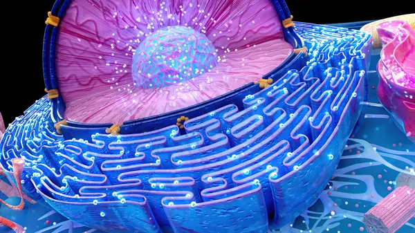
3d Rendering Of Molecules Passing Through Carbon Nanotube Porins On Lipid Bilayer Membrane
Image, 0.81MB, 3840 × 2160 jpg

3d Rendering Of Molecules Passing Through Carbon Nanotube Porins On Lipid Bilayer Membrane
Image, 0.57MB, 3840 × 2160 jpg

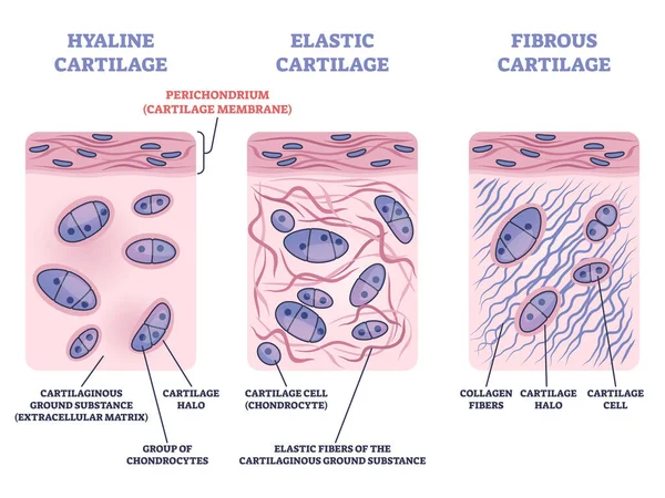
Perichondrium As Hyaline And Elastic Cartilage Membrane Outline Diagram
Vector, 6.99MB, 4700 × 3525 eps

Tight Junction As Intercellular Barrier Between Epithelial Cells Outline Diagram. Labeled Educational Scheme With Microbiological Protein Location To Separate Bowel Tissue Spaces Vector Illustration.
Vector, 6.52MB, 5200 × 3536 eps


3D Rendering Of Carbon Nanotube Porins, Short Pieces Of Carbon Nanotubes Capable Of Self-inserting Into A Lipid Bilayer, Model Of Biological Membrane Channels.
Image, 0.81MB, 3840 × 2160 jpg


Synapse Types Axo-somatic, Axo-axonic, Axoextracellular, Dendritic Spine, And Axo-dendritic Connections Diagram Hand Drawn Schematic Raster Illustration. Medical Science Educational Illustration
Image, 3.07MB, 6696 × 3803 jpg
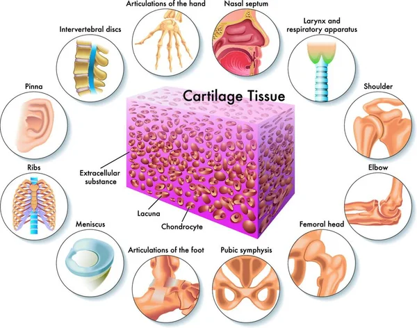
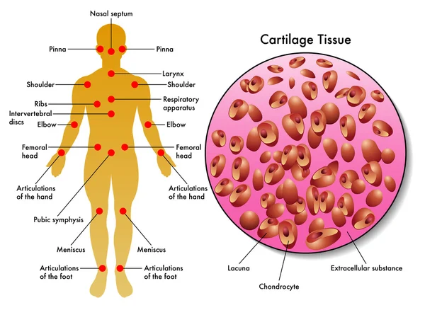
Medical Illustration Of Cartilage Tissue And Its Position In The Human Body
Vector, 0MB, 5144 × 4048 zip

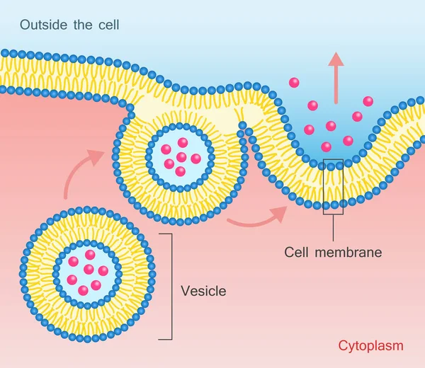
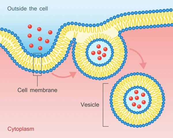
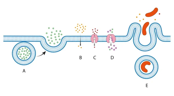
Exocytosis Process Explanation As Proteins Release Mechanism Outline Diagram
Vector, 5.5MB, 4500 × 3750 eps
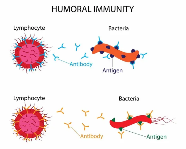
Illustration Of Biology, Humoral Immunity Is Also Referred To As Antibody Mediated Immunity, Humoral Immunity Is The Aspect Of Immunity That Is Mediated By Macromolecules, Cellular Immune Elements
Vector, 5.54MB, 2346 × 1885 eps
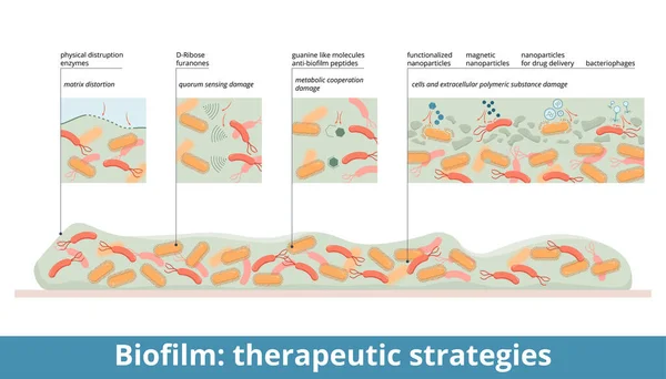
Biofilm: Therapeutic Strategies. Biofilm Treatment: Physical Disruption (enzymes), Quorum Sensing Damage (D-ribose), Metabolic Cooperation Damage, Magnetic And Functionalized Nanoparticles, Bacteriophages.
Vector, 10.78MB, 7292 × 4167 eps
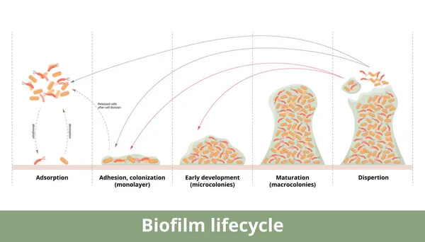
Biofilm Formation. Process Of Biofilm Formation With Mechanics Of Its Development And Growth. Stages Include First Contact, Strong Adhesion, Formation Of Monolayer, Colonies And Dispertion.
Vector, 22.62MB, 7292 × 4167 eps
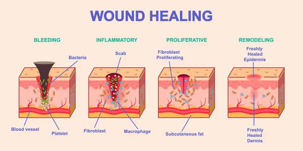
Wound Healing Process. Medical Infographics Or Diagram With Stages Or Phases Of Skin Regeneration After Injury. Bleeding, Inflammation, Proliferation And Remodeling. Cartoon Flat Vector Illustration
Vector, 9.06MB, 5831 × 2915 eps
Collagen Fibers Regeneration In The Skin Tissues. Wrinkled Skin Before And Smooth Skin After Anti-aging Treatment Or Cosmetics Action. Skin Layers, Matrix, Collagen, Elastin Fibers, Fibroblasts. Comparison Of 3D Illustrations Of Young And Aged Skin
Image, 9.87MB, 3840 × 4380 jpg
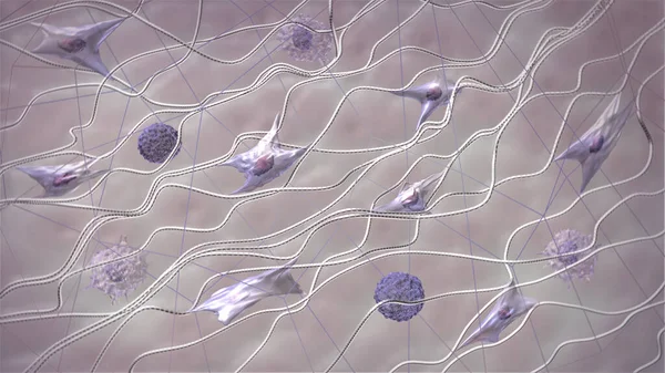
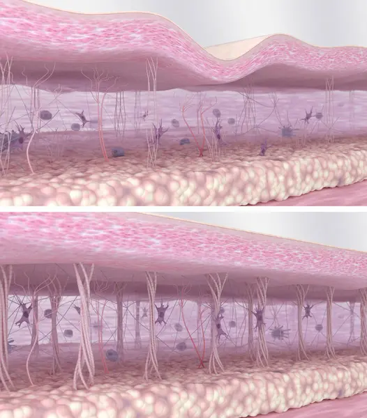
Skin Extracellular Matrix Structure. Fibroblasts, Collagen, And Elastic Fibers 3D Model. Skin In Macro Zoom. Scientific Medical Anatomical 3d Illustration
Image, 2.44MB, 3840 × 2160 jpg


ECF _ Extra Cellular Fluid, Letters And Icons, And Vector Illustration.
Vector, 1.25MB, 3402 × 3402 eps

3D Rendering Of Carbon Nanotube Porins, Short Pieces Of Carbon Nanotubes Capable Of Self-inserting Into A Lipid Bilayer, Model Of Biological Membrane Channels.
Image, 0.43MB, 3840 × 2160 jpg

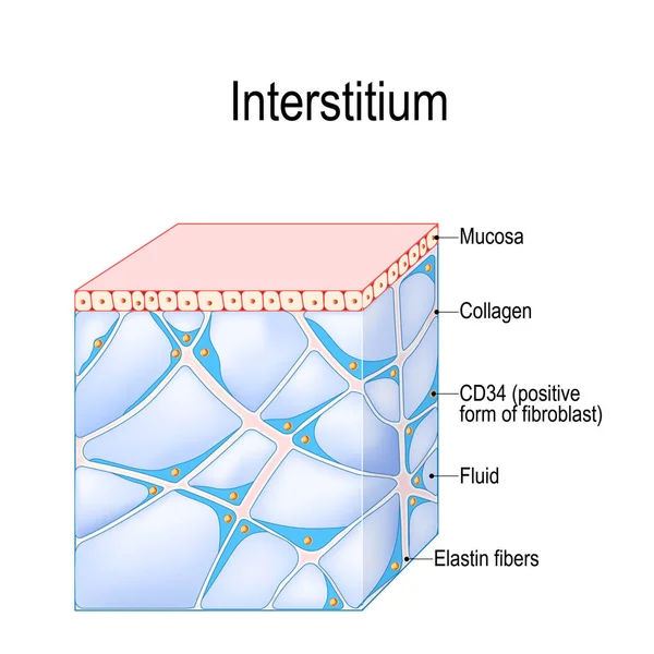
Structure Of Interstitium. New Organ. Interstitial Is A Reservoir And Transportation System For Nutrients And Solutes Distributing Among Organs, And Cells. Immune Regulation. Human Tissues
Vector, 2.83MB, 4372 × 4373 eps
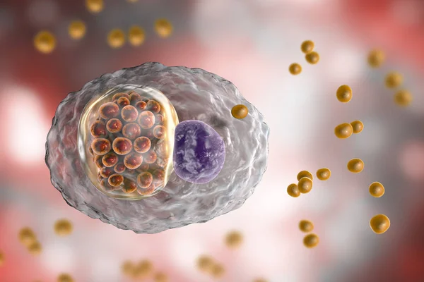
Chlamydophila Psittaci, Intracellular Bacteria That Cause Psittacosis
Image, 1.34MB, 4500 × 3000 jpg

Biofilm Structure. Bacterial Cell Colony: Protein, Polysaccharide, Extracellular DNA, Horizontal Gene Transfer Between Bacteria, Resistant Bacterium, And Other Bacterial Cell. Vector Poster
Vector, 1.28MB, 4444 × 4444 eps
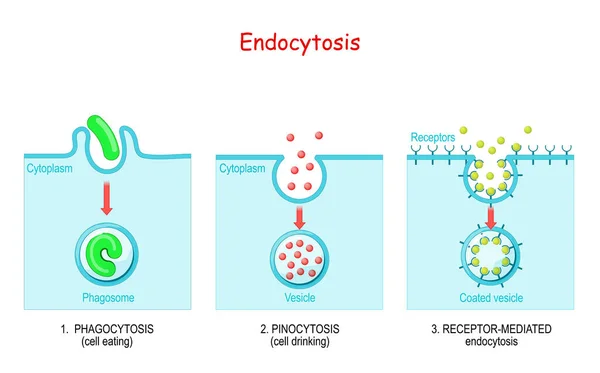
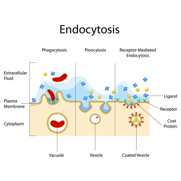
Endocytosis. Phagocytosis Is Cell Eating, Pinocytosis Is A Cell Drinking, Receptor-mediated Endocytosis - When Cells Absorb Metabolites, Hormones, Proteins And Viruses By Receptors On The Surface Of The Cell.
Vector, 1.03MB, 5357 × 3333 eps

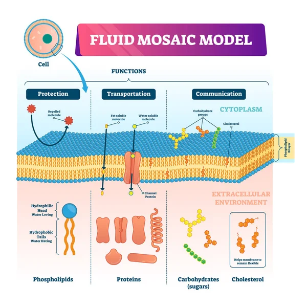
Fluid Mosaic Model Vector Illustration. Cell Membrane Structure Infographic
Vector, 9.21MB, 4000 × 4000 eps
Page 1 >> Next