
Stock image Femur Neck

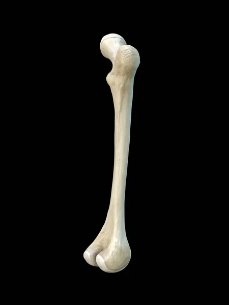
Right Human Femur Bone, Posterior View, Bone Anatomy, Black Background, 3d Rendering
Image, 1.48MB, 3375 × 4500 jpg
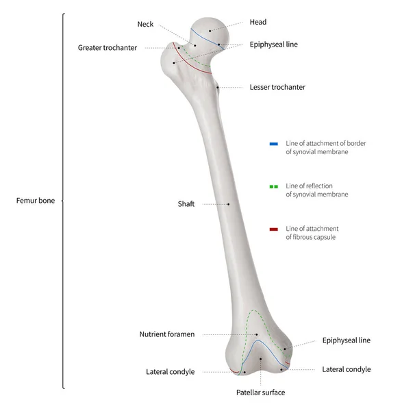
Infographic Diagram Of Human Femur Bone Or Leg Bone Anatomy System Anterior View- 3D- Human Anatomy- Medical Diagram- Educational And Human Body Concept- Isolated On White Background
Image, 4.11MB, 9797 × 10000 jpg
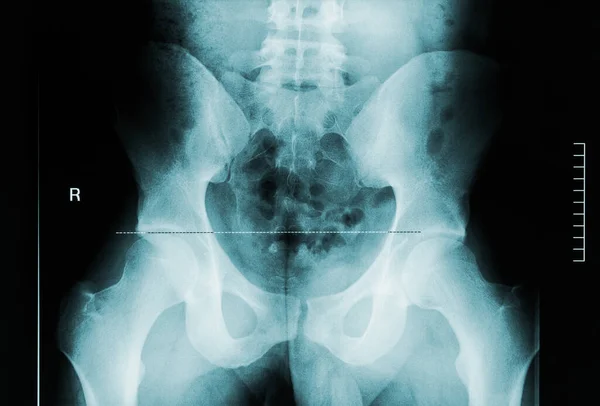
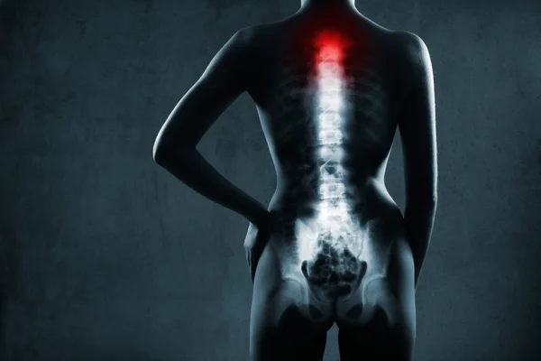
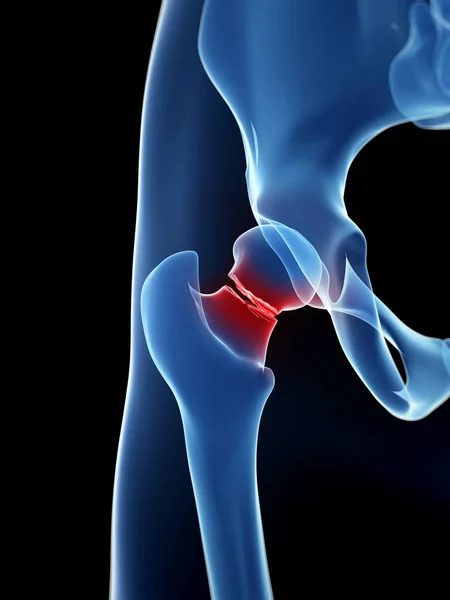
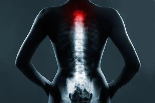
Back View X-ray Image Of Male Pelvis, Femoral Neck And Lumbar Vertebrae. Medical And Human Anatomy Imagery.
Image, 3.3MB, 3000 × 2033 jpg
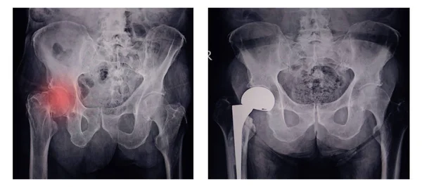
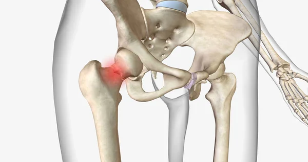
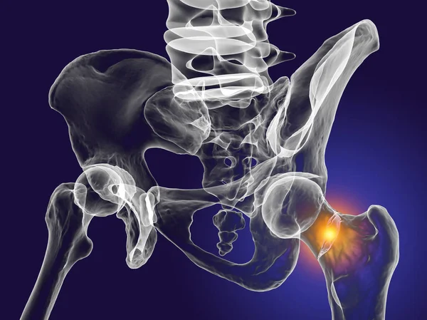
X--ray Image Of Painful Hip In Woman Present Fracture Right Hip Joint At Red Area Mark With Orthopedic Hip Joint Replacement Implant Head And Screws In Human Skeleton In Blue Gray Tones.
Image, 7.69MB, 6296 × 2800 jpg

Human Skeleton Vertebral Column Cervical Vertebrae Anatomy 3D Illustration
Image, 1.73MB, 3840 × 2160 jpg
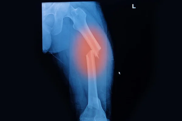
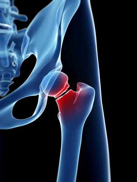
A Femoral Neck Fracture Is A Type Of Hip Fracture That Occurs In The Section Of The Femur Closest To The Pelvis.3D Rendering
Image, 2.83MB, 7340 × 3884 jpg
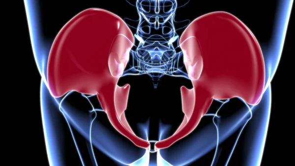
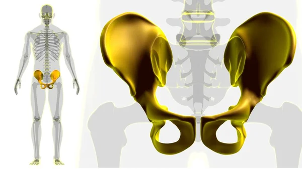
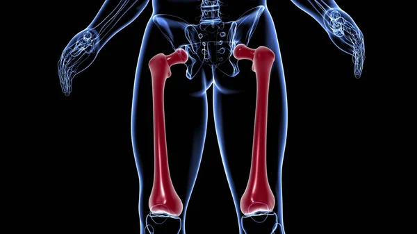
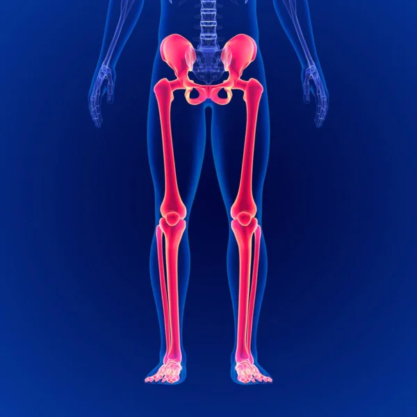
Human Skeleton Hip Or Pelvic Bone Anatomy For Medical Concept 3D Illustration
Image, 2.3MB, 3840 × 2160 jpg

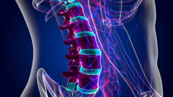
Thoracic Spine Joint Pain Anatomy For Medical Concept 3D Illustration
Image, 2.59MB, 3840 × 2160 jpg
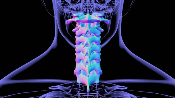
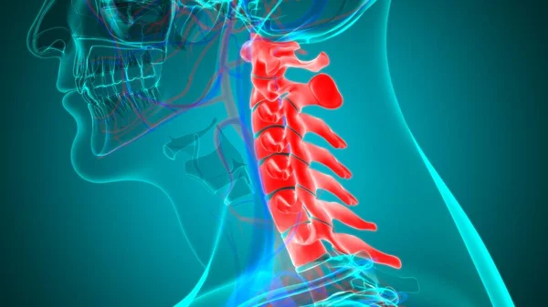
Human Skeleton Vertebral Column Cervical Vertebrae Anatomy 3D Illustration
Image, 1.88MB, 3840 × 2160 jpg

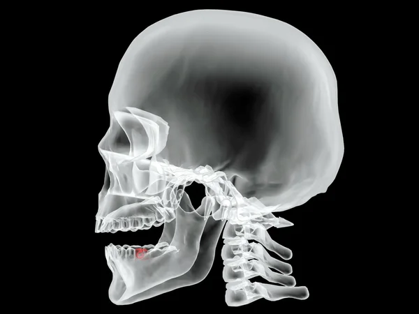
Human Skeleton Skull Temporal Bone Anatomy For Medical Concept 3D Illustration
Image, 0.82MB, 3840 × 2160 jpg
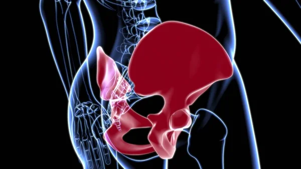
Human Skeleton Hip Or Pelvic Bone Anatomy For Medical Concept 3D Illustration
Image, 1.96MB, 3840 × 2160 jpg


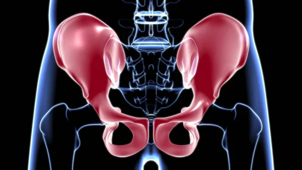
Human Skeleton Hip Or Pelvic Bone Anatomy For Medical Concept 3D Illustration
Image, 1.98MB, 3840 × 2160 jpg
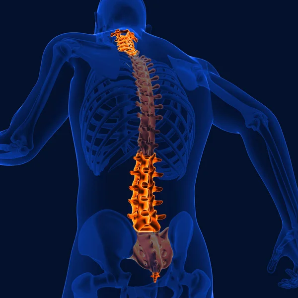
Human Skeleton Vertebral Column Lumbar Vertebrae Anatomy 3D Illustration
Image, 3.51MB, 3840 × 2160 jpg
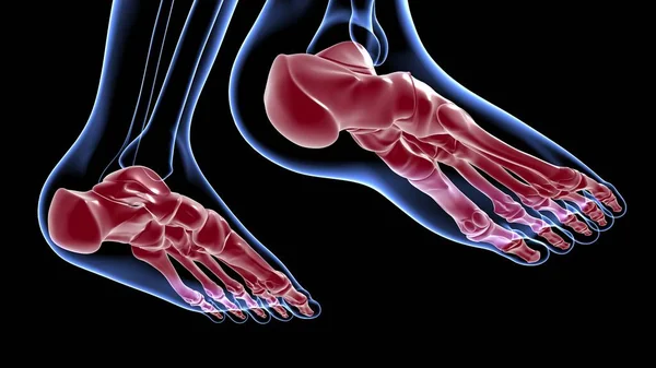
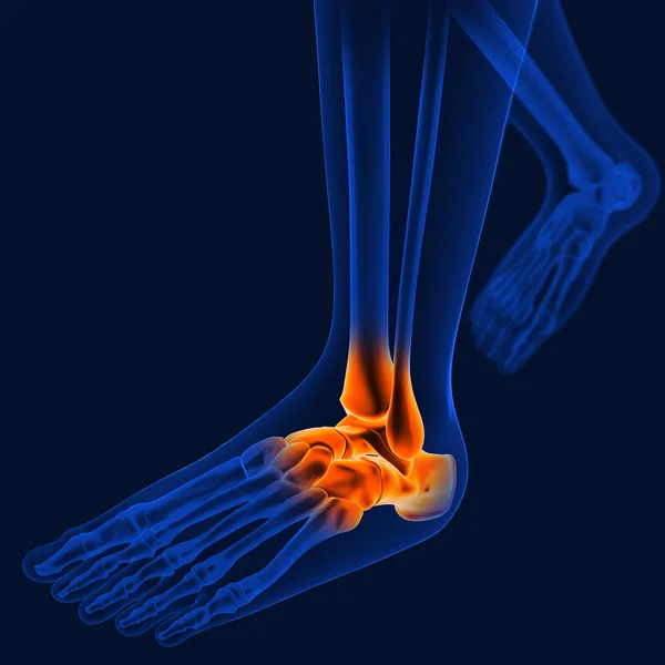
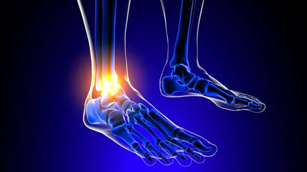
Human Skeleton Foot Bones Anatomy For Medical Concept 3D Illustration
Image, 1.64MB, 3840 × 2160 jpg
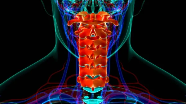
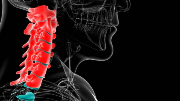
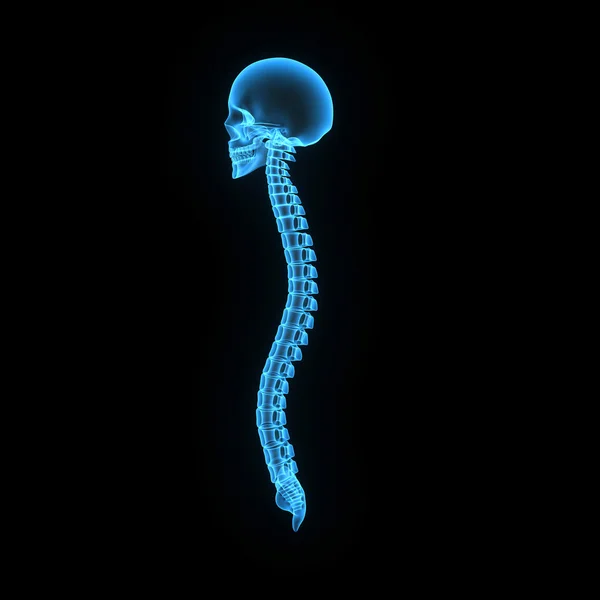
Human Skeleton Vertebral Column Cervical Vertebrae Anatomy 3D Illustration
Image, 2.78MB, 3840 × 2160 jpg

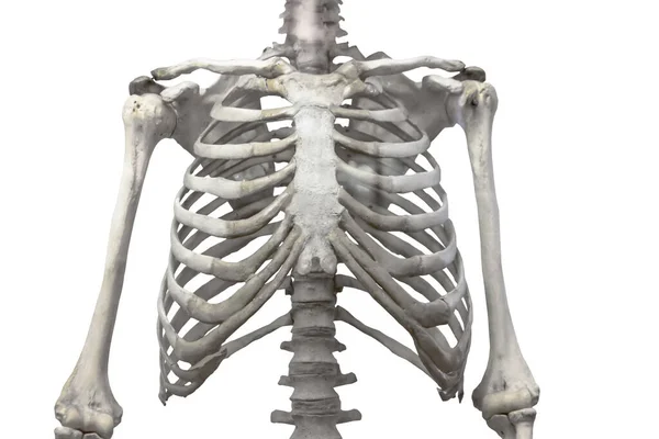
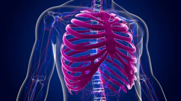
Thoracic Spine, Chest And Ribs Of Bone With Arms And Shoulders Isolated On A White Background.
Image, 2.94MB, 5250 × 3500 jpg
Thoracic Spine, Chest And Ribs Of Bone With Arms And Shoulders Isolated On A White Background.
Image, 2.94MB, 5250 × 3500 jpg

Human Skeleton Hip Or Pelvic Bone Anatomy For Medical Concept 3D Illustration
Image, 2.25MB, 3840 × 2160 jpg
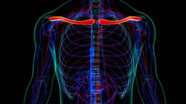
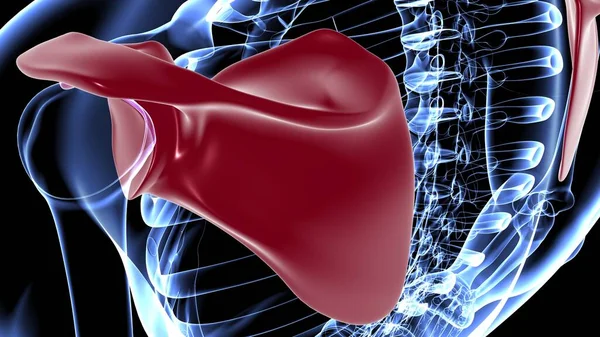
Human Skeleton Anatomy Clavicle Bones 3D Rendering For Medical Concept
Image, 3.11MB, 3840 × 2160 jpg

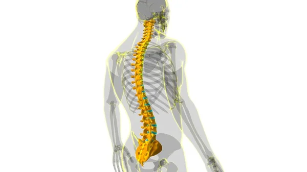
Human Skeleton Vertebral Column Lumbar Vertebrae Anatomy 3D Illustration
Image, 0.96MB, 3840 × 2160 jpg
Joint Pain Can Be Caused By Injury Affecting Any Of The Ligaments, Bursae, Or Tendons Surrounding The Joint.
Image, 7.88MB, 8192 × 8192 jpg
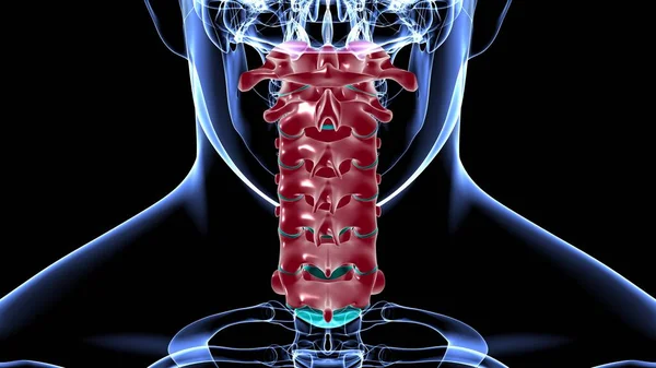
Human Skeleton Vertebral Column Cervical Vertebrae Anatomy 3D Illustration
Image, 1.77MB, 3840 × 2160 jpg
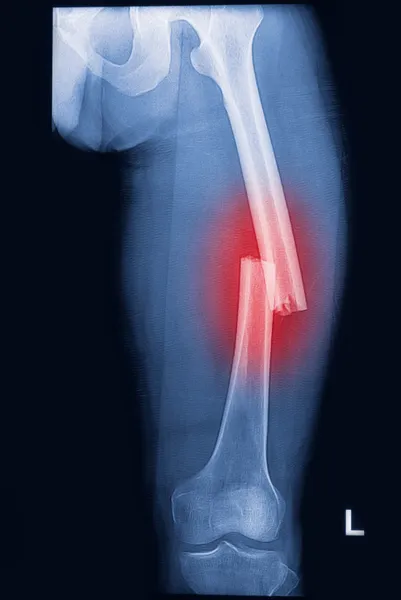
A Fracture Of The Femur Neck, A Common Type Of Hip Fracture That Typically Occurs In Older Adults And Can Lead To Mobility Issues And Other Complications, Isolated On White Background, 3D Illustration
Image, 8.22MB, 7000 × 5243 jpg
Joint Pain Can Be Caused By Injury Affecting Any Of The Ligaments, Bursae, Or Tendons Surrounding The Joint.
Image, 10.11MB, 8192 × 8192 jpg

Human Skeleton Vertebral Column Thoracic Vertebrae Anatomy 3D Illustration
Image, 1.39MB, 3840 × 2160 jpg
Page 1 >> Next