
Stock image Fibroblasts
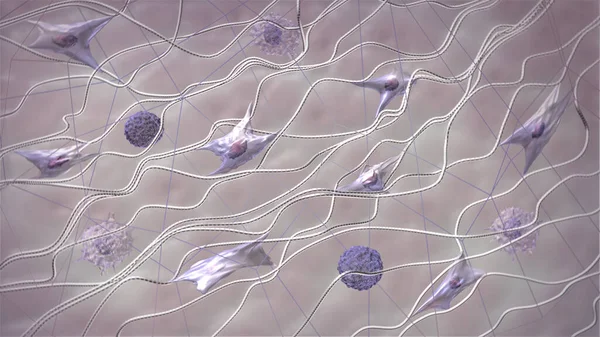
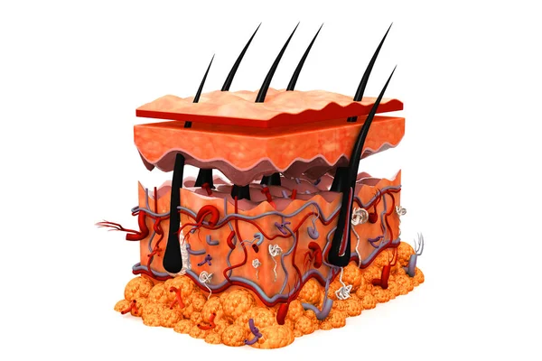
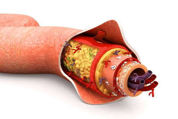
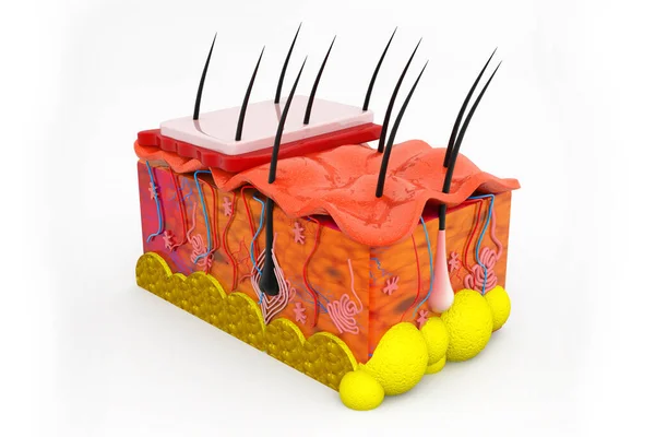
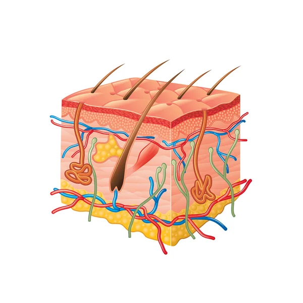
Skin Extracellular Matrix Structure. Fibroblasts, Collagen, And Elastic Fibers 3D Model. Skin In Macro Zoom. Scientific Medical Anatomical 3d Illustration
Image, 2.44MB, 3840 × 2160 jpg
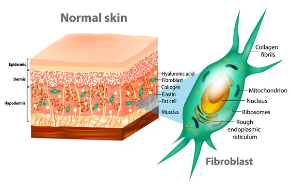
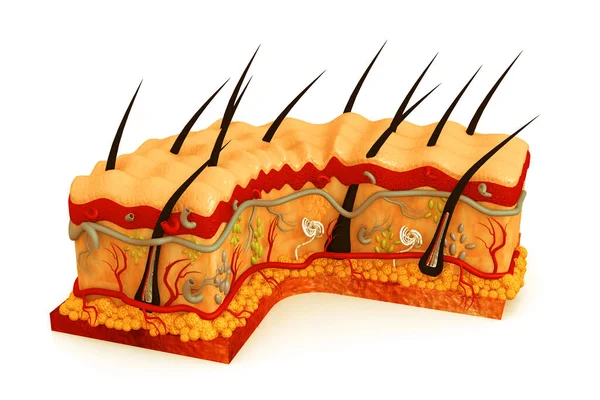
Fibroblast And Human Skin Structure (Muscles, Fat Cell, Hyaluronic Acid, Elastin, Collagen, Fibroblast).
Vector, 2.8MB, 5000 × 3166 eps
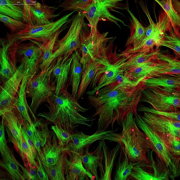
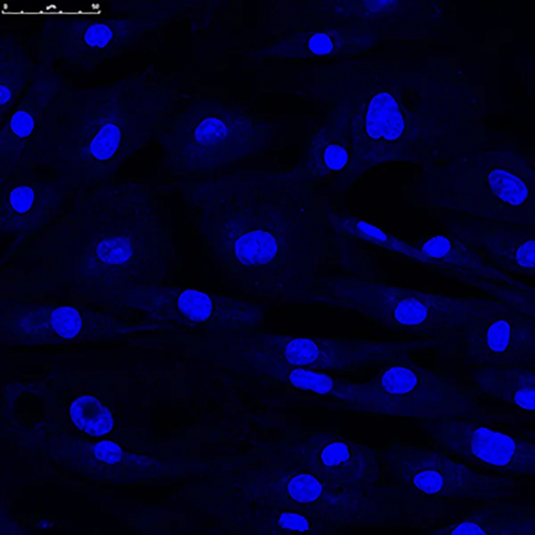
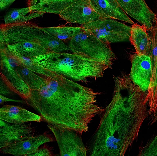
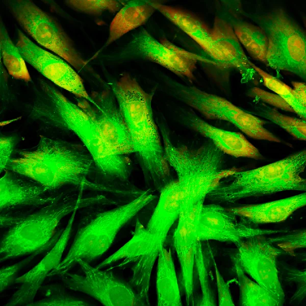
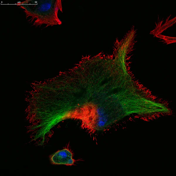
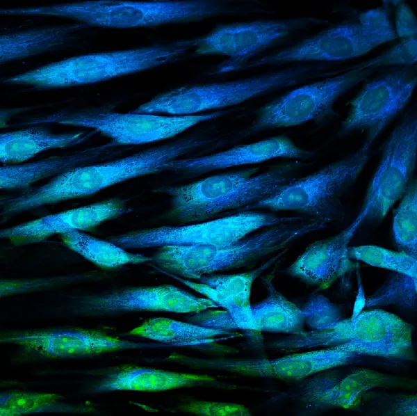
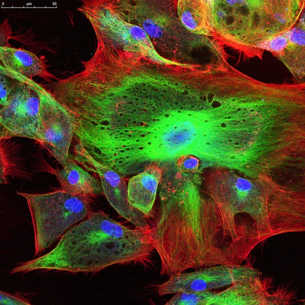
Real Fluorescence Microscopic View Of Human Skin Cells In Culture. Nucleus Are In Blue, Actin Filaments Are In Pink, Tubulin Was Labeled With Green
Image, 16.31MB, 4000 × 4000 jpg

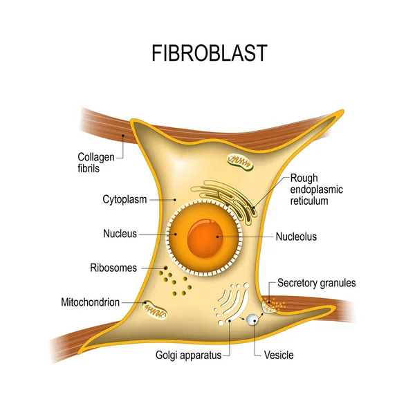
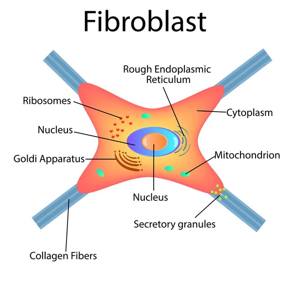
Fibroblast Anatomy. Structure Of Cell. Diagram With Golgi Apparatus, Nucleus, Mitochondrion And Ribosomes. Vector Illustration. Poster
Vector, 14.88MB, 4444 × 4444 eps
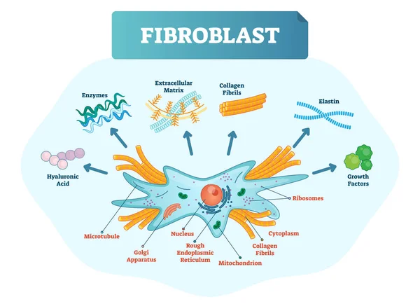
Fibroblast Vector Illustration. Scheme With Extracellular, Collagen Fibrils, Elastin, Hyaluronic Acid, Microtubule, Golgi Apparatus, Nucleus And Ribosomes.
Vector, 8.13MB, 5877 × 4252 eps
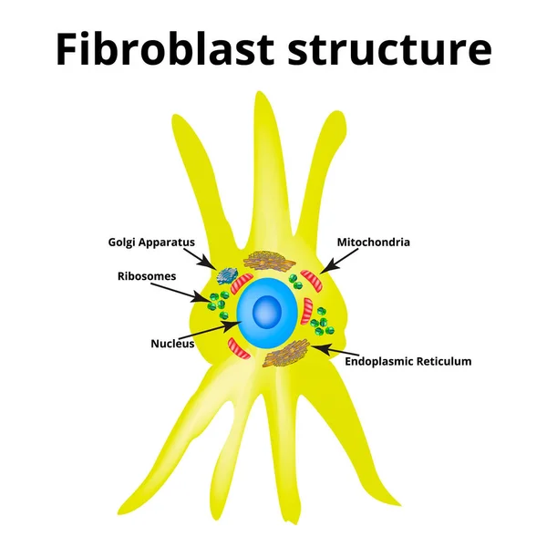
Fibroblast Structure. Fibroblast Cell. Vector Illustration On Isolated Background
Vector, 19.29MB, 5000 × 5000 eps

Skin Icon In Flat Style Isolated On White Background. Organs Symbol Stock Vector Illustration.
Vector, 1.15MB, 5000 × 5000 eps
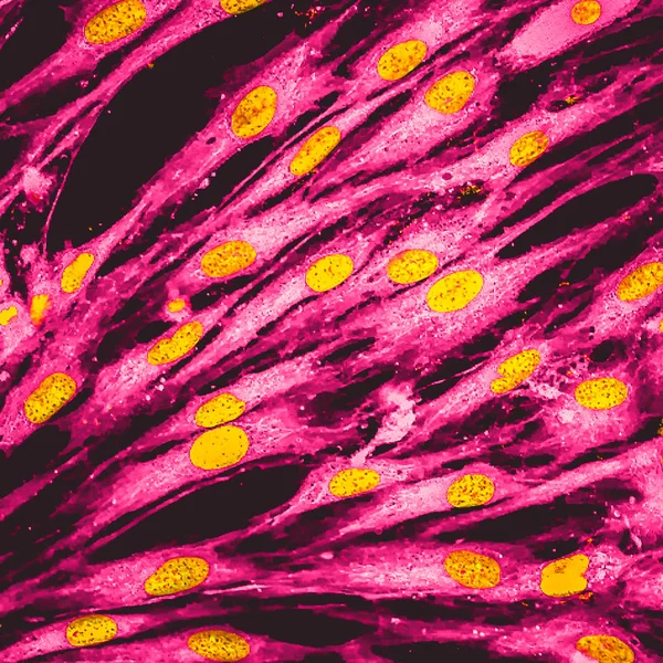
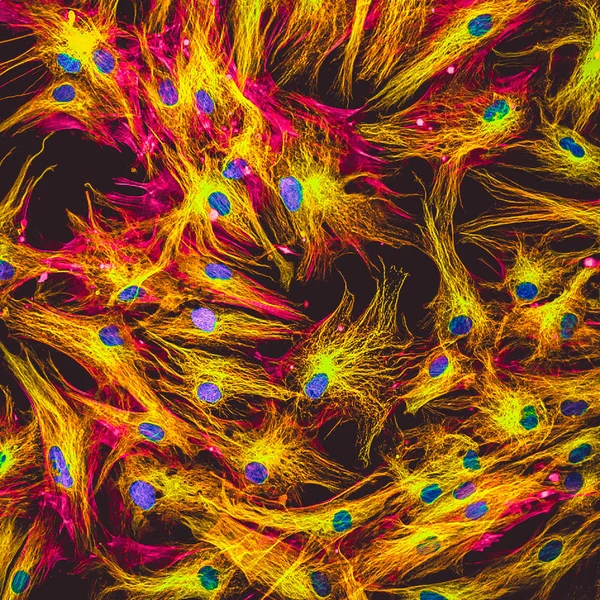
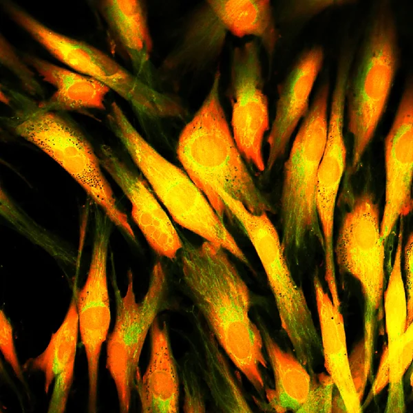
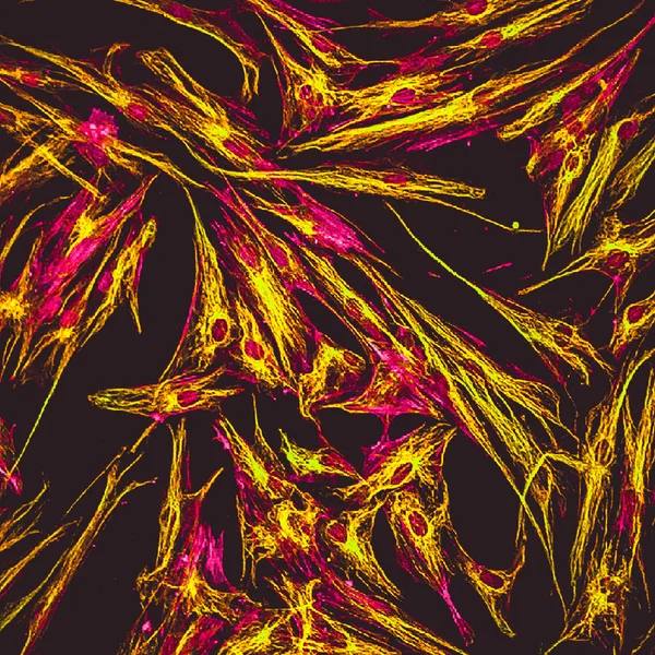
Real Fluorescence Microscopic View Of Human Skin Cells In Culture. Nucleus Are In Yellow, Cell Membranes Were Labeled With Pink
Image, 13.66MB, 4000 × 4000 jpg
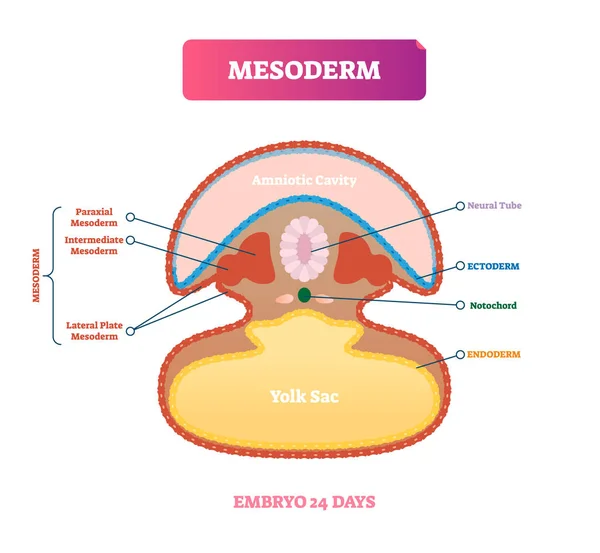
Mesoderm Vector Illustration. Labeled Medical Diagram With Embryo Structure
Vector, 5.53MB, 5000 × 4609 eps

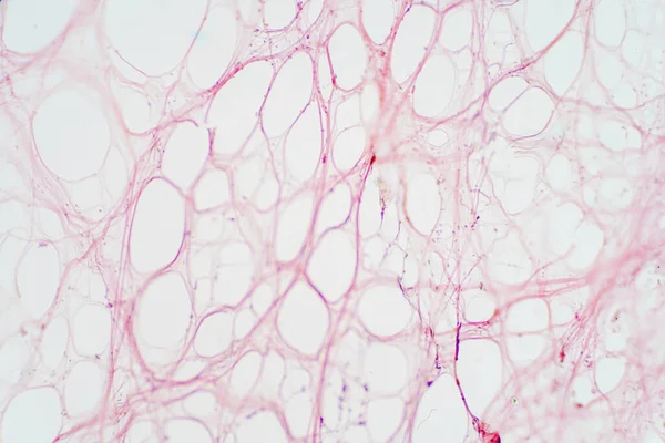
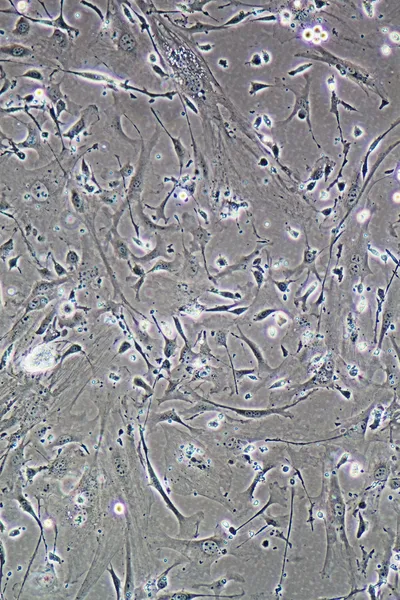
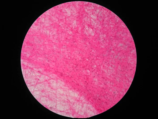
Areolar Connective Tissue Under The Microscope View. Histological For Human Physiology.
Image, 9.51MB, 6000 × 4000 jpg
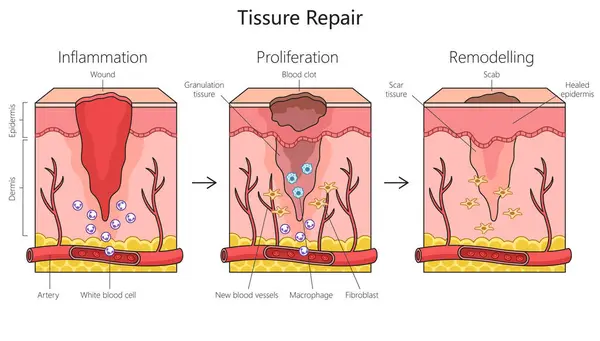
Tissue Repair Structure Diagram Hand Drawn Schematic Raster Illustration. Medical Science Educational Illustration
Image, 4.77MB, 7304 × 4148 jpg
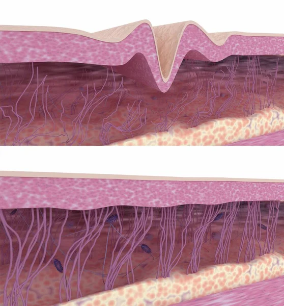
Skin Rejuvenation. Collagen And Elastin Fibers Rebuilding. 3D Rendered Illustration.
Image, 7.83MB, 3840 × 4126 jpg

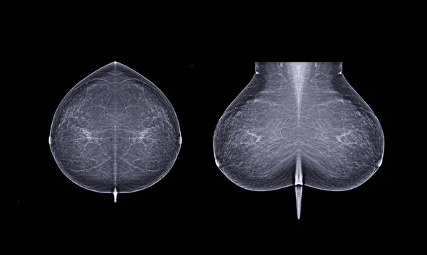
Basic Position X-ray Digital Mammogram Both Side Name Is CC View And MLO . Mammography Or Breast Scan For Breast Cancer.
Image, 4.67MB, 5472 × 3280 jpg

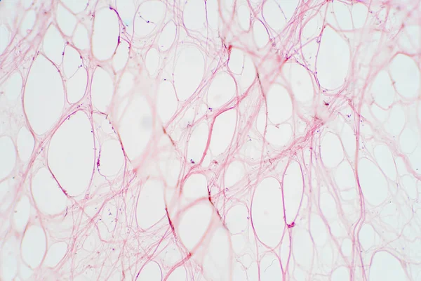
Areolar Connective Tissue Under The Microscope View. Histological For Human Physiology.
Image, 10.06MB, 6000 × 4000 jpg
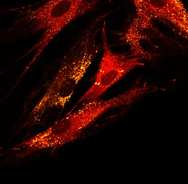
Real Fluorescence Microscopic View Of Human Skin Cells In Culture. Actin Filaments Are In Pink, Tubulin Was Labeled With Green
Image, 11.97MB, 4000 × 4000 jpg

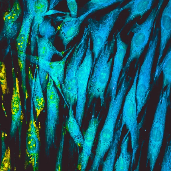
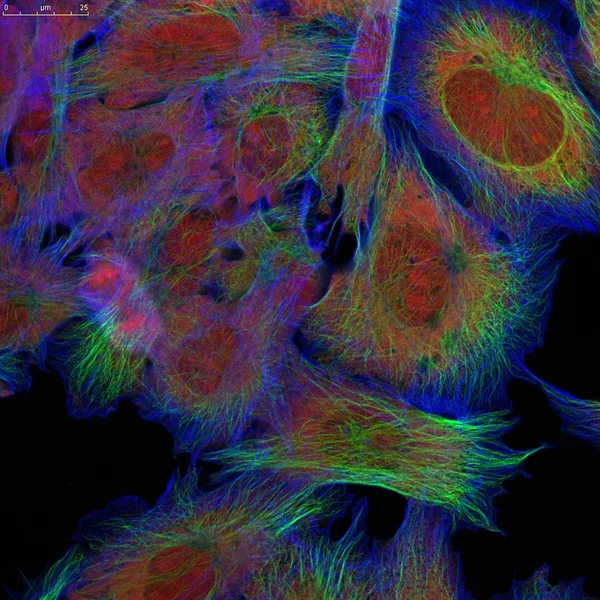
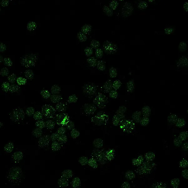
Real Fluorescence Microscopic View Of Pseudocolored Human Skin Cells In Culture
Image, 11.22MB, 4000 × 4000 jpg


Fibroblast Structure. Fibroblast Cell. Vector Illustration Isolated
Vector, 19.48MB, 5000 × 5000 eps
Page 1 >> Next