
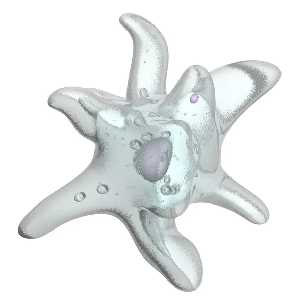
Stock image Food Vacuole


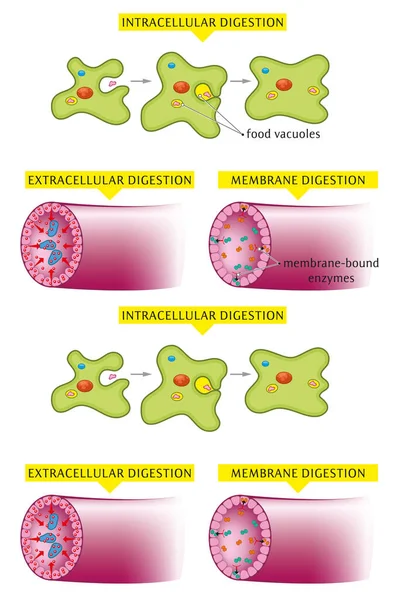
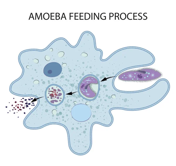
Illustrated Examples Of Intracellular Extracellular And Membrane Type Of Digestion.
Vector, 1.85MB, 3500 × 5267 eps
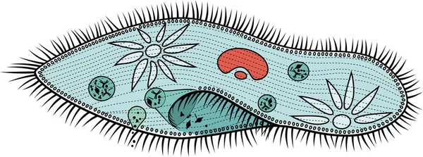
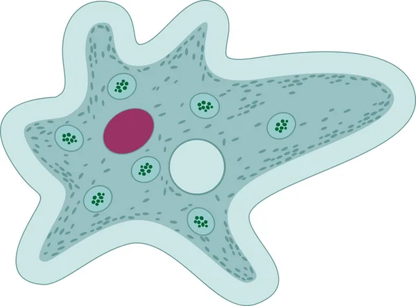
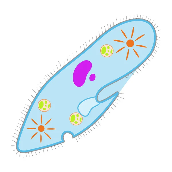
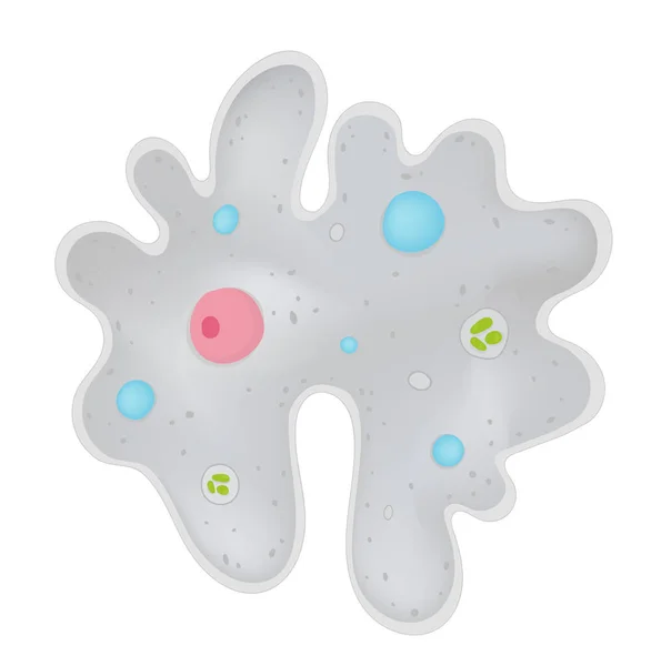
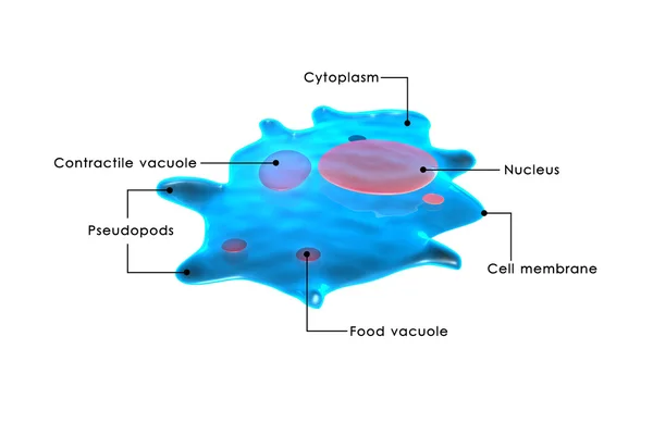
Amoeba Proteus With Red Nucleus, Contractile Vacuole And Other Organelles
Vector, 0.22MB, 5000 × 3685 ai

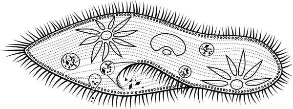
Coloring Page. Amoeba Proteus With Nucleus, Contractile Vacuole And Other Organelles
Vector, 0.14MB, 5000 × 3692 ai
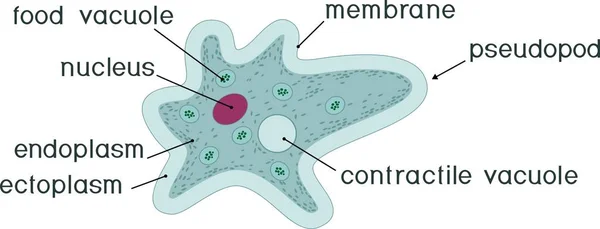
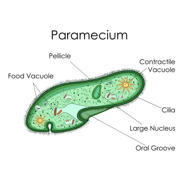
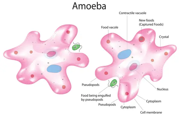
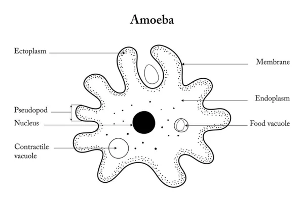
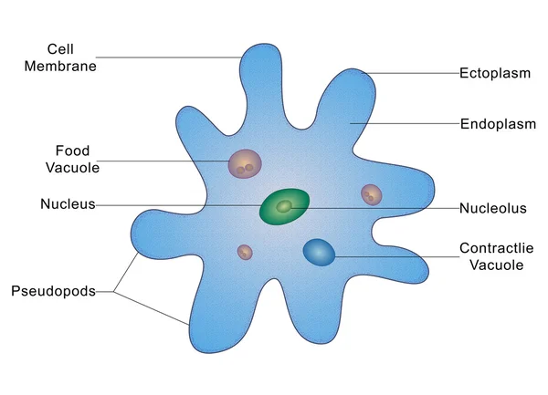
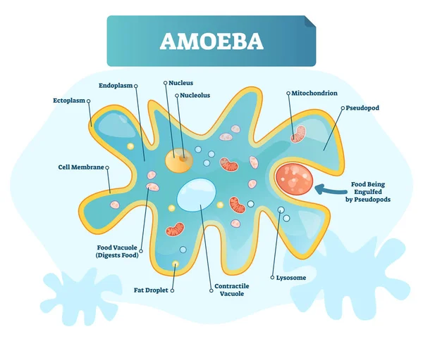
Amoeba Proteus With Nucleus, Contractile Vacuole, Other Organelles And Titles
Vector, 0.29MB, 7833 × 3000 ai
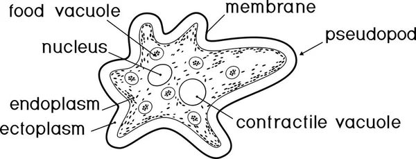
Coloring Page. Amoeba Proteus With Nucleus, Contractile Vacuole, Other Organelles And Titles
Vector, 0.22MB, 7854 × 3000 ai
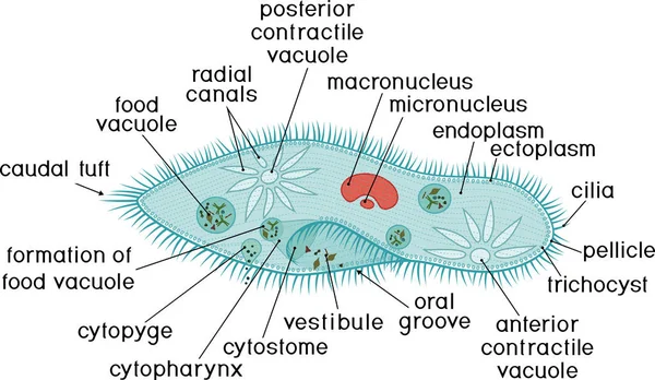
Vector Isolated Illustration Of Amoeba Proteus On A White Background. Studying The Structure Of An Amoeba.
Vector, 0.76MB, 5000 × 5000 eps

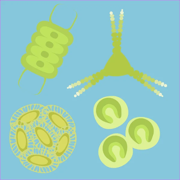
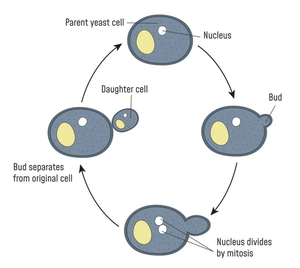
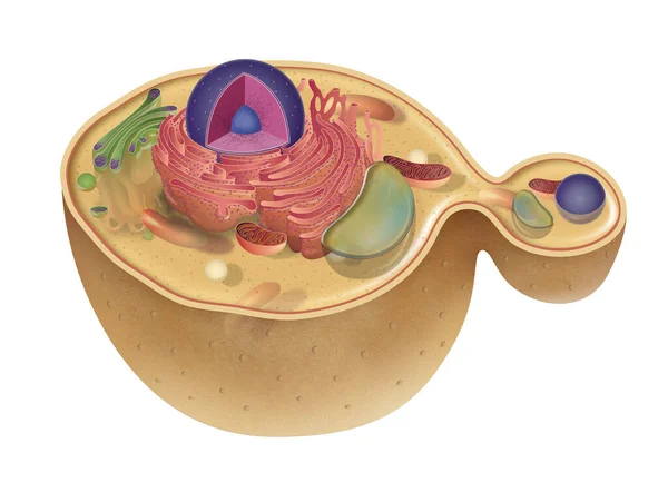
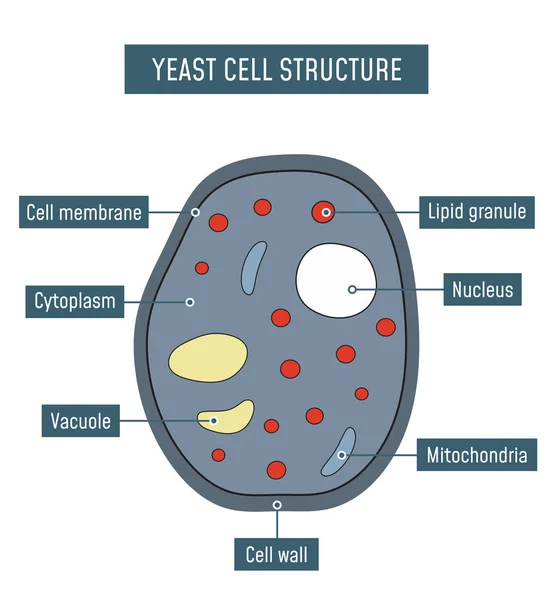
Yeast Cells Are Round To Long Cells That Reproduce Vegetatively By Budding Or Germinate To Produce A Mycelium
Image, 12.14MB, 8268 × 7234 jpg
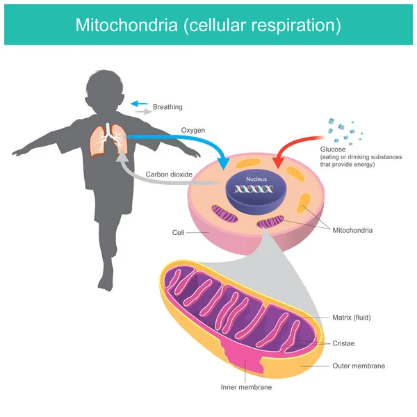
Mitochondria. Illustration Explain Human Body Received Glucose And Oxygen Such As Eating Or Drinking After That The Cell System Changes Glucose In A Fluid Matrix From Mitochondria To Energy Stored And Release Carbon Dioxide Gas Out
Vector, 7.33MB, 5000 × 5000 eps


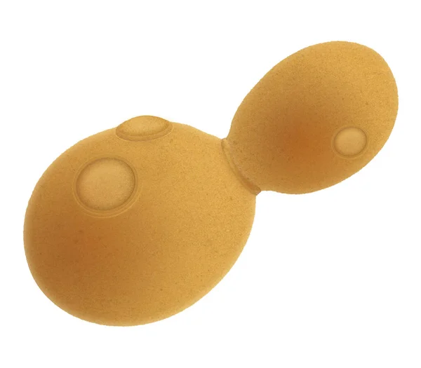
Pichia Is A Genus Of Yeasts In The Family Saccharomycetaceae Under The Microscope For Education.
Image, 19.01MB, 6720 × 4480 jpg
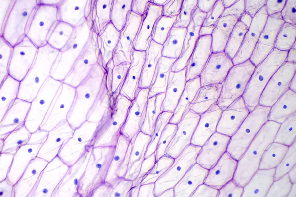
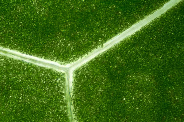
Onion Epidermis Under Light Microscope. Purple Colored, Large Epidermal Cells Of An Onion, Allium Cepa, In A Single Layer. Each Cell With Wall, Membrane, Cytoplasm, Nucleus And Large Vacuole.
Image, 3.62MB, 3786 × 2524 jpg

Scientists Characters Learning Plasmodium Parasites Landing Page Template. Tiny Microbiology Doctor At Huge Infographics
Vector, 2.64MB, 8331 × 5000 eps

Pichia Is A Genus Of Yeasts In The Family Saccharomycetaceae Under The Microscope For Education.
Image, 14.93MB, 6720 × 4480 jpg
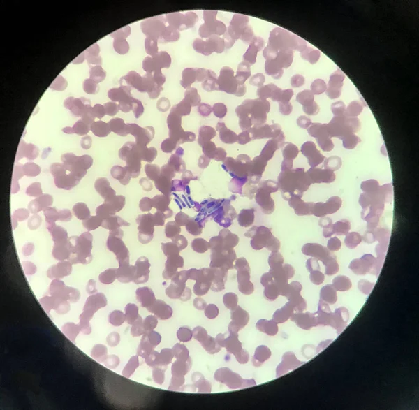
Yeast Cells Phagocytosis By White Blood Cell In Blood Smear.Fungus Blood Infection Medical Science Background.
Image, 3.3MB, 3030 × 2966 jpg

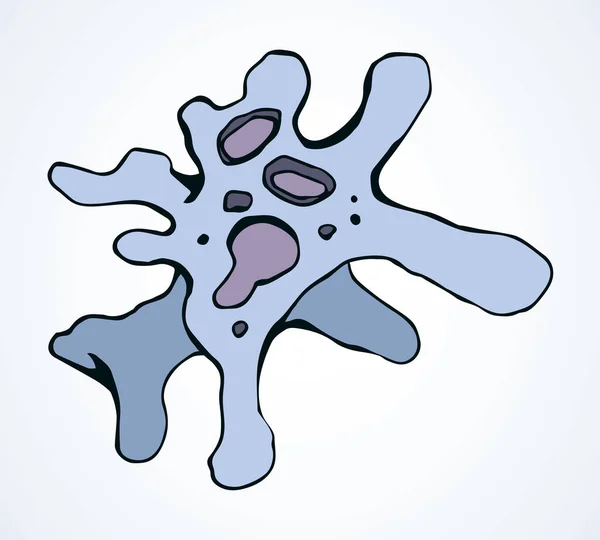
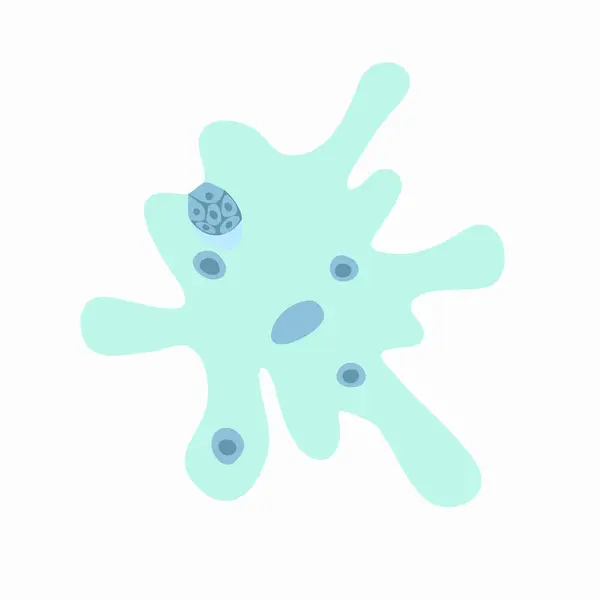
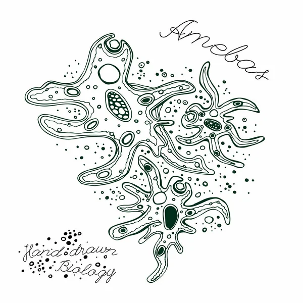
Abstract Chaos Shape Tiny Protist Element. Line Black Hand Drawn Water Nature Life Medicine School Study Icon Sign Symbol Pictogram Diagram Sketch. Art Doodle Cartoon Style Design. Closeup Macro View
Vector, 0.42MB, 5381 × 4846 eps
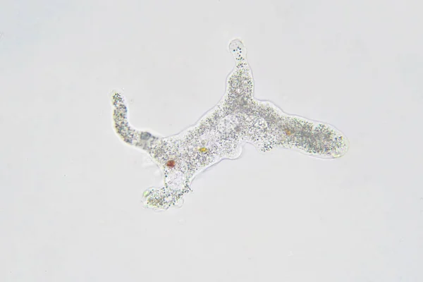
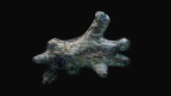
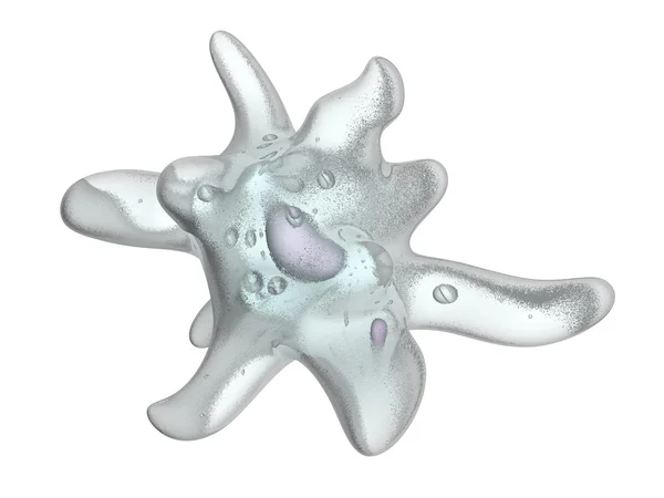
Amoeba Proteus This Small Protozoan Uses Tentacular Protuberances Called Pseudopodia To Move And Phagocytose Smaller Unicellular Organisms.
Image, 11.46MB, 6000 × 4000 jpg
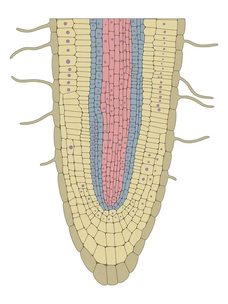
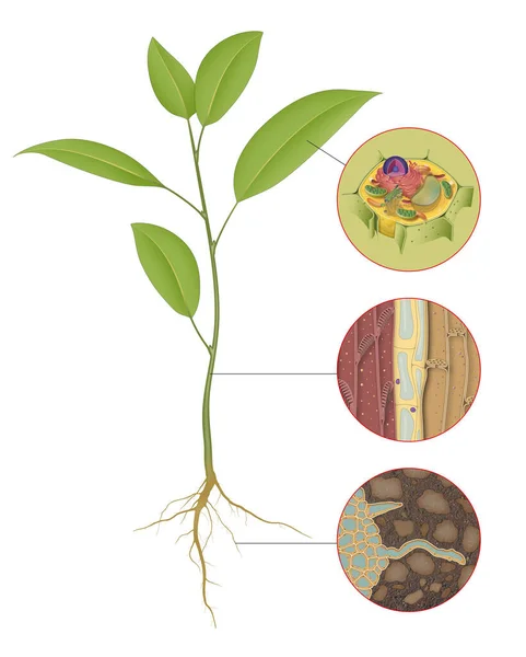
Typical Roots Contain Three Different Sections, Or Zones: The Meristematic Zone, The Zone Of Elongation, And The Zone Of Differentiation
Image, 9.29MB, 7087 × 9449 jpg
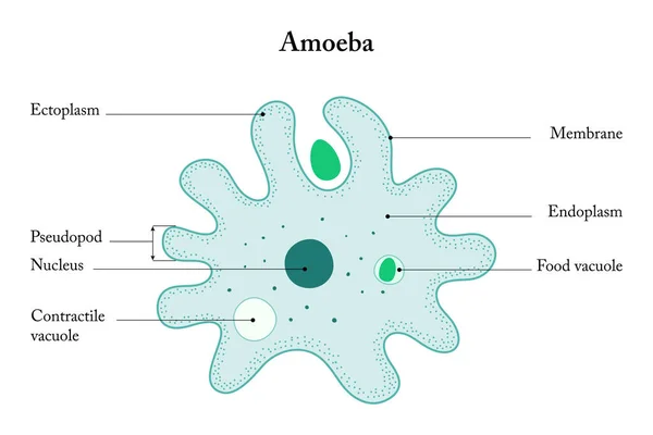
Amoeba Labeled Vector Illustration. Single Cell Animal Structure Scheme.
Vector, 7.2MB, 4500 × 3643 eps
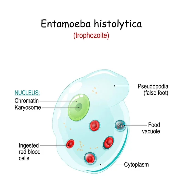
Entamoeba Histolytica. Anatomy Of Trophozoite. Entamoeba Is An Anaerobic Parasitic Amoeba That Cause Infection Disease Of Intestine And Gastrointestinal Tract, Liver And Other Internal Organs. Structure Of Unicellular Organism
Vector, 7.7MB, 4444 × 4444 eps


Abstract Chaos Shape Tiny Protist Element. Line Black Hand Drawn Water Nature Life Medicine School Study Icon Sign Symbol Pictogram Diagram Sketch. Art Doodle Cartoon Style Design. Closeup Macro View
Vector, 0.41MB, 5381 × 4846 eps

Pichia Is A Genus Of Yeasts In The Family Saccharomycetaceae Under The Microscope For Education.
Image, 15.37MB, 6720 × 4480 jpg
Page 1 >> Next