
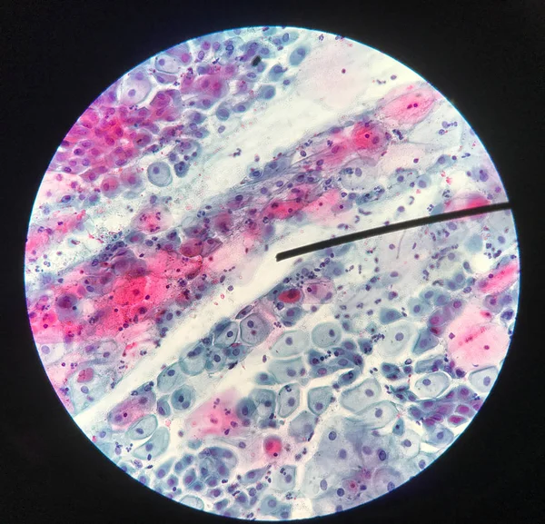
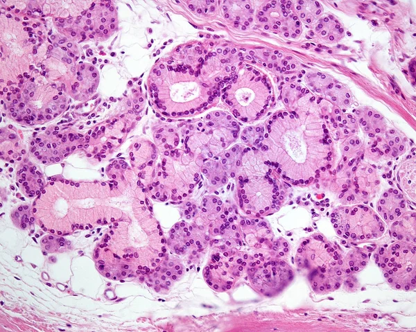
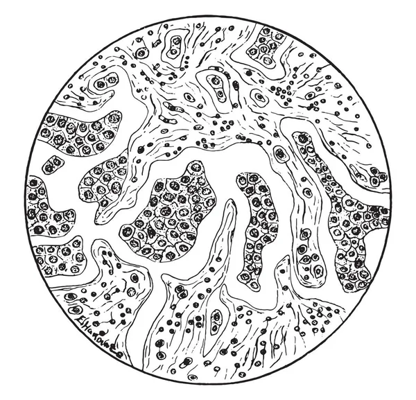
Stock image Glandular Cell


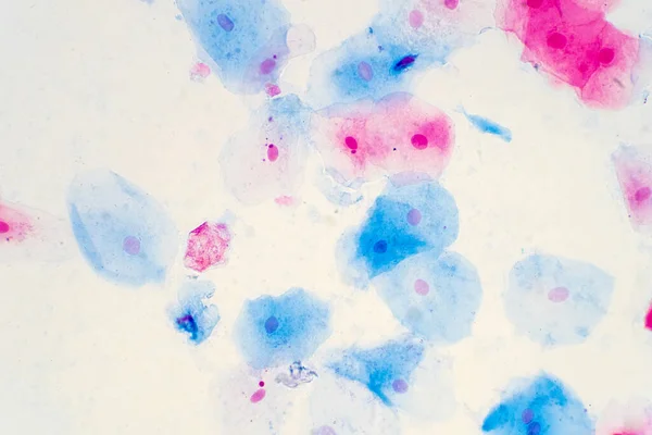
Squamous Epithelial Cells Of Human Cervix Under The Microscope View. Pap Smear Test Is A Procedure To Test For Cervical Cancer In Women.
Image, 14.68MB, 5385 × 3590 jpg

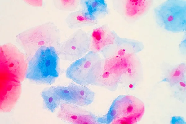
Squamous Epithelial Cells Of Human Cervix Under The Microscope View. Pap Smear Test Is A Procedure To Test For Cervical Cancer In Women.
Image, 17.23MB, 5883 × 3922 jpg

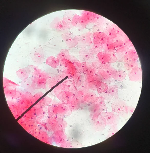
Squamous Epithelial Cells Of Human Cervix Under The Microscope View. Pap Smear Test Is A Procedure To Test For Cervical Cancer In Women.
Image, 18.53MB, 6000 × 4000 jpg
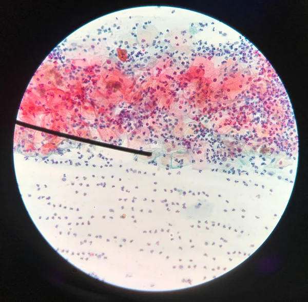
Squamous Epithelial Cells Of Human Cervix Under The Microscope View. Pap Smear Test Is A Procedure To Test For Cervical Cancer In Women.
Image, 11.03MB, 4506 × 3004 jpg
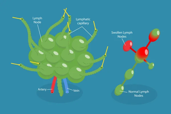
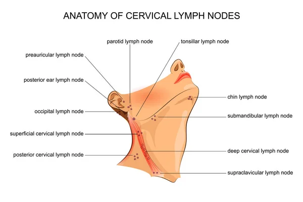
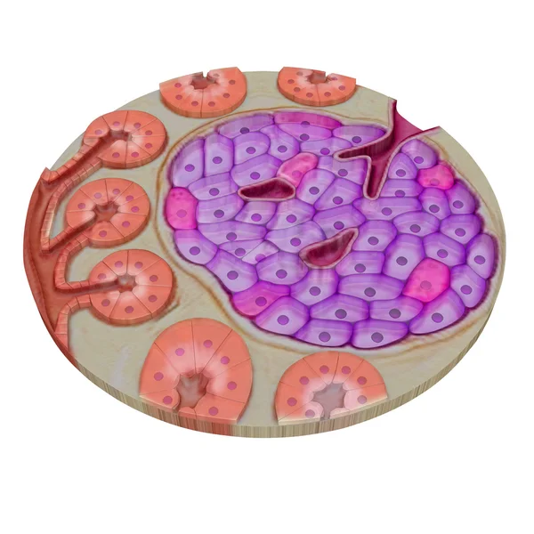
3D Isometric Flat Vector Conceptual Illustration Of Lymph Node, Labeled Diagram
Vector, 1.89MB, 6000 × 4000 eps


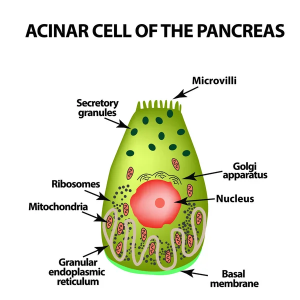
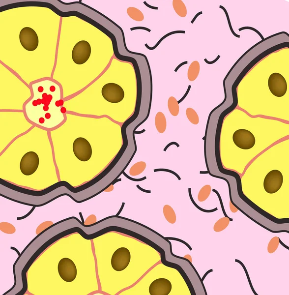
Acinar Cell Of The Pancreas. Acinus. Infographics. Vector Illustration On Isolated Background
Vector, 1.08MB, 5000 × 5000 eps

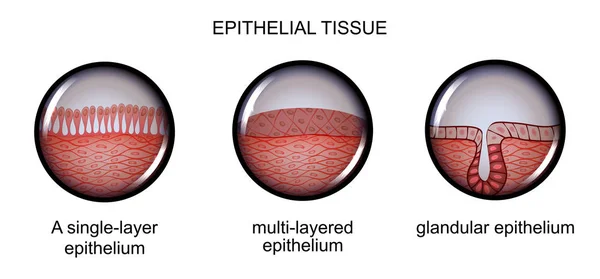

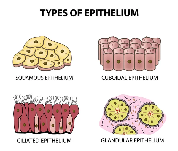
Types Of Epithelium. Squamous, Cubic, Ciliated, Glandular. Set. Infographics. Vector Illustration On Isolated Background
Vector, 2.27MB, 5000 × 4253 eps

Epithelium. Squamous, Cubic, Ciliated, Glandular. Set. Infographics. Vector Illustration.
Vector, 1.56MB, 5000 × 5093 eps


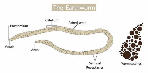
Illustration Of Biology And Animal, Anatomy Of Earthworms, Earthworm Structure, Form And Function, The Basic Shape Of The Earthworm Is A Cylindrical Tube
Vector, 5.85MB, 3074 × 1521 eps
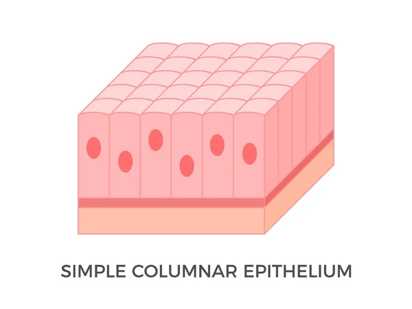
Simple Columnar Epithelium. Epithelial Tissue Types. Tall And Slender Cells With Oval-shaped Nuclei. Lines Most Organs Of The Digestive Tract Like Stomach, Intestines. Medical Illustration. Vector.
Vector, 4.96MB, 5000 × 3750 eps

Pharynx Cancer And Nasopharyngeal Cancer. Malignant Neoplasm Originating From Epithelial Cells. Medical Vector Illustration.
Vector, 0.34MB, 5615 × 3406 eps


Glandular Epithelium. Epithelial Tissue Types. Compound Tubular, Acinar And Tubuloacinar Glandular Epithelium. They Produce And Release Different Secretory Products In Glands. Vector Illustration.
Vector, 7.79MB, 6700 × 2251 eps
Page 1 >> Next