photo free
Home Images Vectors Videos

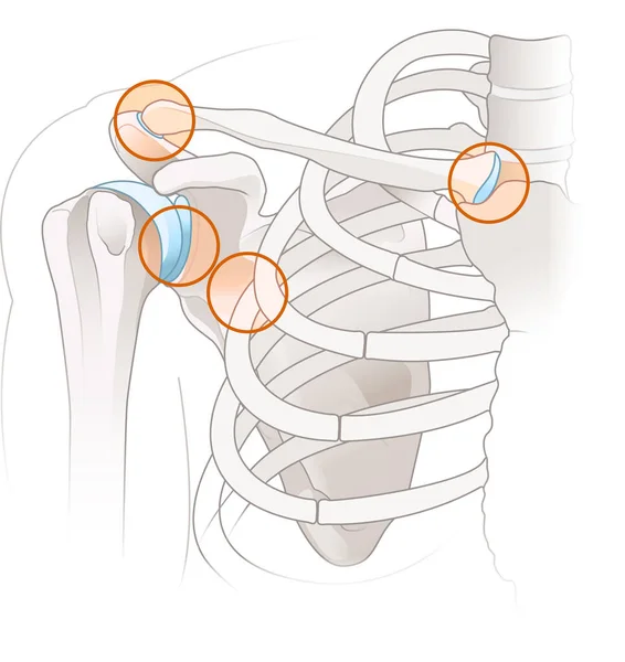
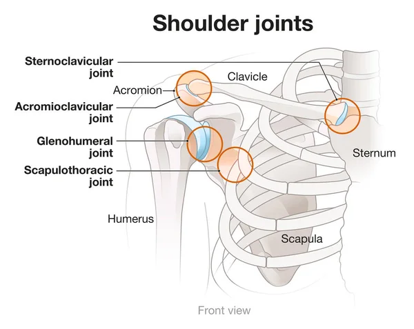
Stock image Glenohumeral Joint

stock vector vector illustration of shoulder joint anatomy. blade

Vector Illustration Of Shoulder Joint Anatomy. Blade

Vector, 2.38MB, 5500 × 4200 eps
stock vector vector illustration of anatomy of the elbow joint

Vector Illustration Of Anatomy Of The Elbow Joint

Vector, 3.55MB, 5787 × 4200 eps
stock vector vector illustration of shoulder replacement. surgery. traumatology

Vector Illustration Of Shoulder Replacement. Surgery. Traumatology

Vector, 6.12MB, 5501 × 2751 eps
stock image Bones of Arm and Hand

Bones Of Arm And Hand

Image, 2.25MB, 6480 × 4096 jpg
stock image 3D illustration showing human shoulder joint with bones and ligaments and capsule

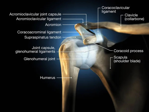
3D Illustration Showing Human Shoulder Joint With Bones And Ligaments And Capsule

Image, 5.68MB, 6653 × 5000 jpg
stock image Suffering patient

Suffering Patient

Image, 8.24MB, 3630 × 5446 jpg
stock image Shoulder anatomy. Four major joints of the shoulder.

Shoulder Anatomy. Four Major Joints Of The Shoulder.

Image, 2.69MB, 6595 × 6994 jpg
stock image Shoulder anatomy. Four major joints of the shoulder.

Shoulder Anatomy. Four Major Joints Of The Shoulder.

Image, 3.6MB, 9828 × 7819 jpg
stock vector Detailed diagram of SLAP tear in shoulder joint

Detailed Diagram Of SLAP Tear In Shoulder Joint

Vector, 1.4MB, 4990 × 4452 eps
stock vector vector illustration of the types of prosthesis of shoulder

Vector Illustration Of The Types Of Prosthesis Of Shoulder

Vector, 5.33MB, 5847 × 3999 eps
stock vector Shoulder joint isolated on white vector

Shoulder Joint Isolated On White Vector

Vector, 2.96MB, 4313 × 4313 eps
stock image 3d illustration of the endoprosthesis of the elbow joint.

3d Illustration Of The Endoprosthesis Of The Elbow Joint.

Image, 2.61MB, 6000 × 3264 jpg
stock image Patient and doctor in surgery

Patient And Doctor In Surgery

Image, 8.25MB, 3679 × 5518 jpg
stock vector vector illustration of the endoprosthesis of the elbow joint

Vector Illustration Of The Endoprosthesis Of The Elbow Joint

Vector, 4.71MB, 6125 × 3894 eps
stock image Sprain of glenohumeral joint

Sprain Of Glenohumeral Joint

Image, 8.31MB, 3669 × 5503 jpg
stock image Doctor diagnosing broken arm

Doctor Diagnosing Broken Arm

Image, 7.05MB, 5228 × 3485 jpg
stock image Synovial joint anatomy

Synovial Joint Anatomy

Image, 3.24MB, 4096 × 4096 jpg
stock vector vector illustration of the endoprosthesis of the elbow joint

Vector Illustration Of The Endoprosthesis Of The Elbow Joint

Vector, 13.7MB, 5501 × 4200 eps
stock image Doctor putting on sling

Doctor Putting On Sling

Image, 9.16MB, 5616 × 3744 jpg
stock image Bones of shoulder

Bones Of Shoulder

Image, 6.03MB, 8120 × 8372 jpg
stock image 3d illustration of the endoprosthesis of the elbow joint.

3d Illustration Of The Endoprosthesis Of The Elbow Joint.

Image, 3.48MB, 6000 × 4500 jpg
stock image A Bankart lesion occurs as the result of a forward shoulder dislocation.3D rendering

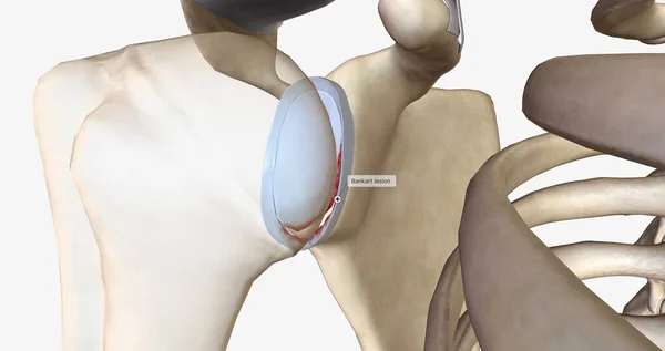
A Bankart Lesion Occurs As The Result Of A Forward Shoulder Dislocation.3D Rendering

Image, 2.77MB, 7340 × 3884 jpg
stock vector Vector illustration of the spinal implant medical

Vector Illustration Of The Spinal Implant Medical

Vector, 2.12MB, 4500 × 3376 eps
stock image A Bony Bankart lesion in the shoulder involves a fracture of the anterior glenoid rim, often resulting from dislocation, leading to instability and requiring surgical repair for stability restoration.

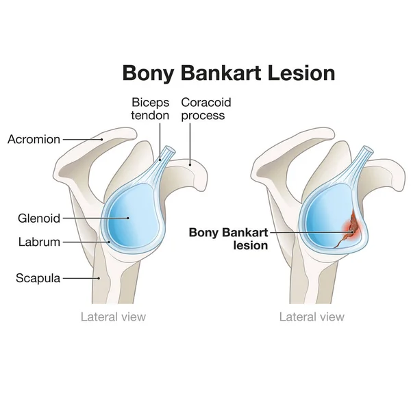
A Bony Bankart Lesion In The Shoulder Involves A Fracture Of The Anterior Glenoid Rim, Often Resulting From Dislocation, Leading To Instability And Requiring Surgical Repair For Stability Restoration.

Image, 2.88MB, 5000 × 5000 jpg
stock image A SLAP lesion in the shoulder refers to an injury to the superior labrum, often caused by trauma or overuse, resulting in pain, instability, and reduced shoulder function. Treatment may involve arthroscopy or rehabilitation.

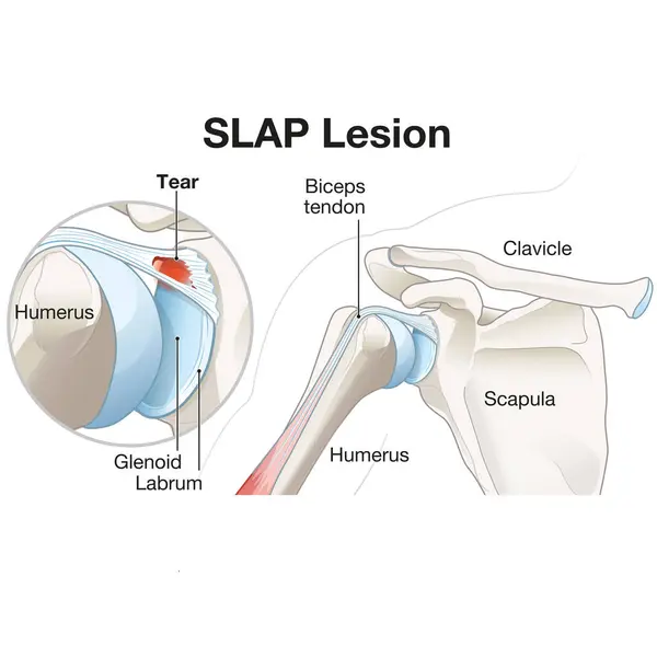
A SLAP Lesion In The Shoulder Refers To An Injury To The Superior Labrum, Often Caused By Trauma Or Overuse, Resulting In Pain, Instability, And Reduced Shoulder Function. Treatment May Involve Arthroscopy Or Rehabilitation.

Image, 3.08MB, 5000 × 5000 jpg
stock image A Hill-Sachs lesion is a divot-like defect on the humeral head, often resulting from shoulder dislocation. It can contribute to instability and limited range of motion in the joint.

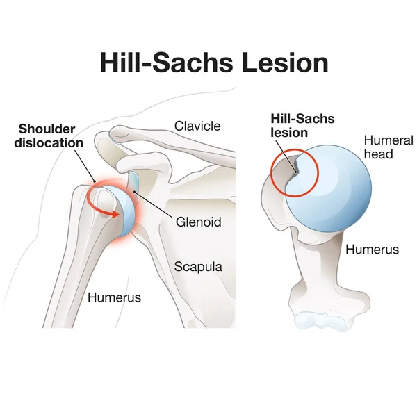
A Hill-Sachs Lesion Is A Divot-like Defect On The Humeral Head, Often Resulting From Shoulder Dislocation. It Can Contribute To Instability And Limited Range Of Motion In The Joint.

Image, 2.83MB, 5000 × 5000 jpg
stock image Asian woman suffering from frozen shoulder with pain and stiffness, Rotator cuff tear concept

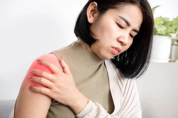
Asian Woman Suffering From Frozen Shoulder With Pain And Stiffness, Rotator Cuff Tear Concept

Image, 9.76MB, 6000 × 4000 jpg
stock image Surgeon inserting syringe needle through skin into female patient glenohumeral joint under ultrasound guidance supervised by colleague

Surgeon Inserting Syringe Needle Through Skin Into Female Patient Glenohumeral Joint Under Ultrasound Guidance Supervised By Colleague

Image, 15.23MB, 7008 × 4672 jpg
stock vector Joint stability or body mobility skeletal bone division outline diagram. Labeled educational anatomical scheme with skeleton functions as spinal, chest, hip, knee or ankle purpose vector illustration

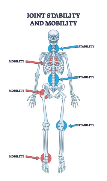
Joint Stability Or Body Mobility Skeletal Bone Division Outline Diagram. Labeled Educational Anatomical Scheme With Skeleton Functions As Spinal, Chest, Hip, Knee Or Ankle Purpose Vector Illustration

Vector, 10.45MB, 3300 × 5544 eps
stock image Surgeon inserting syringe needle into deltoid muscle on female patient upper arm under ultrasound guidance supervised by ultrasonographer

Surgeon Inserting Syringe Needle Into Deltoid Muscle On Female Patient Upper Arm Under Ultrasound Guidance Supervised By Ultrasonographer

Image, 15.73MB, 7008 × 4672 jpg
stock image Closeup of doctor hands in nitrile gloves inserting syringe needle into patient glenohumeral joint under ultrasound guidance

Closeup Of Doctor Hands In Nitrile Gloves Inserting Syringe Needle Into Patient Glenohumeral Joint Under Ultrasound Guidance

Image, 14.87MB, 6812 × 4541 jpg
Page 1 >> Next