
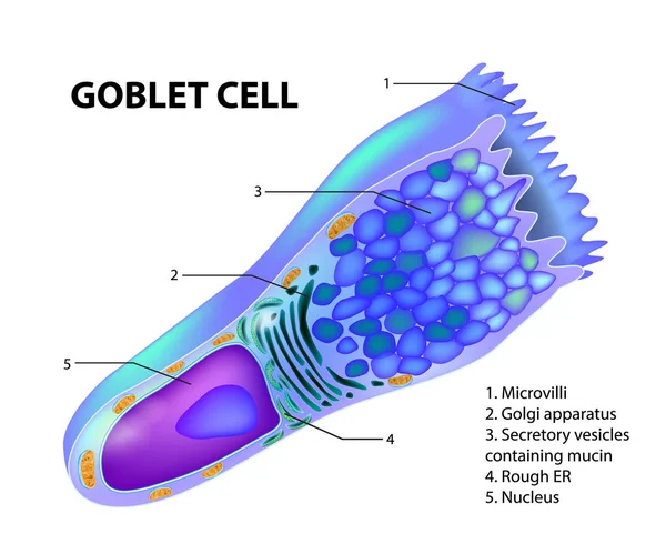
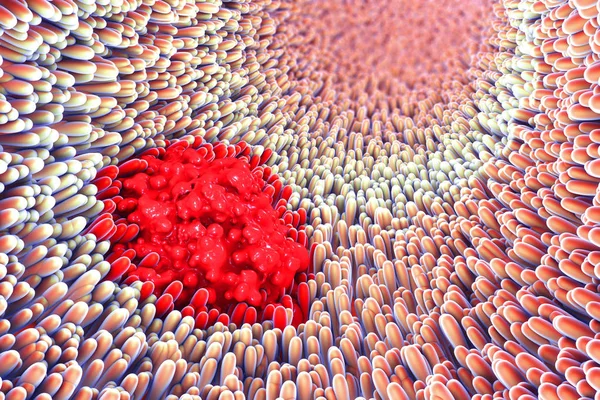
Stock image Goblet Cell
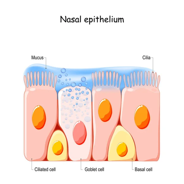
Nasal Mucosa Cells. Nasal Secretions. Ciliated, Basal And Goblet Cells. Olfactory Epithelium. Cells Act As A Low Resistance Filter. Vector Illustration
Vector, 11.58MB, 4444 × 4444 eps
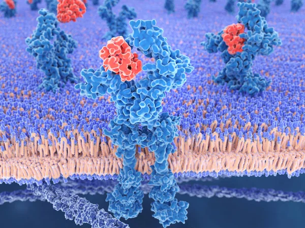
3d Computer Illustration Of Interleukin 13 And Its Receptor.IL-13 Is A Cytokine That Plays A Central Regulator Role In IgE Synthesis, Goblet Cell Hyperplasia, Mucus Hypersecretion, Airway Hyperresponsiveness, Fibrosis, It Is A Mediator Of Allergic I
Image, 5.59MB, 8000 × 6000 jpg

Glandular Epithelium. Epithelial Tissue Types. Compound Tubular, Acinar And Tubuloacinar Glandular Epithelium. They Produce And Release Different Secretory Products In Glands. Vector Illustration.
Vector, 7.79MB, 6700 × 2251 eps
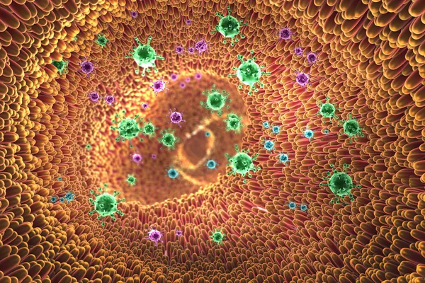
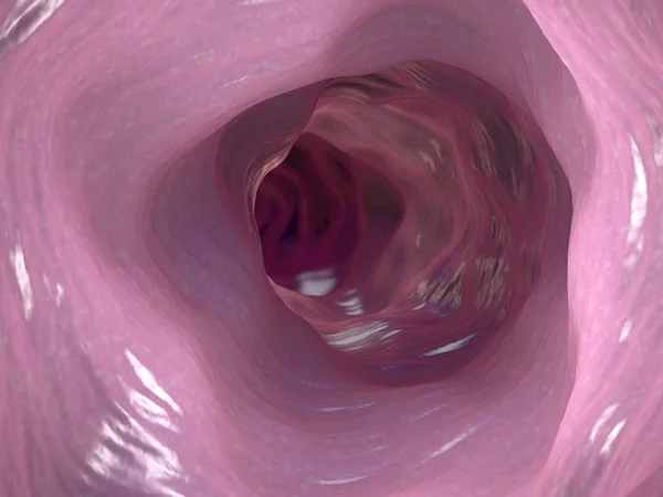
3d Illustration Of Microscopic Closeup Showing Viruses And Intestine Villus Into Digestive Tract
Image, 38.78MB, 9500 × 6333 jpg
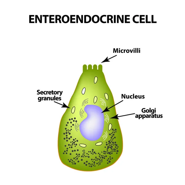
Enteroendocrine Cell. Cell Of The Intestines. Vector Illustration On Isolated Background
Vector, 0.97MB, 5000 × 5000 eps
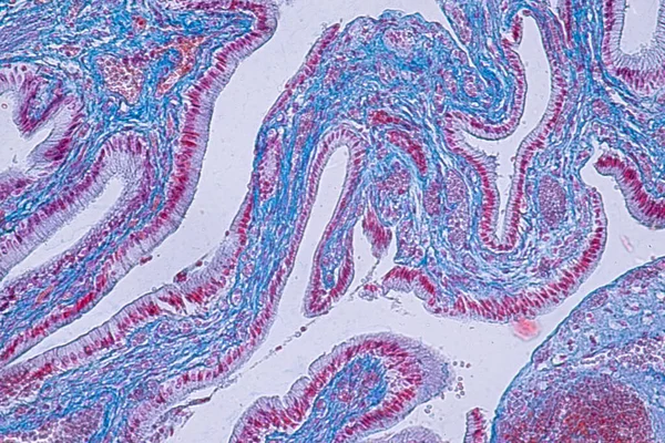
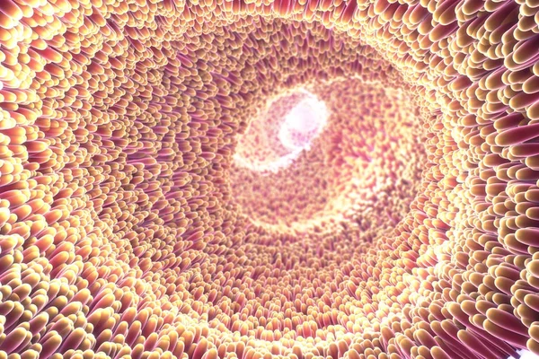
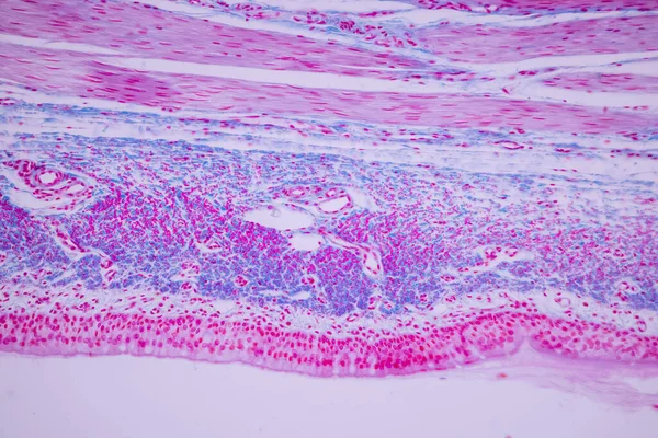
Cross Section Of Ciliated Epithelium Under The Microscope For Education Histology. Human Tissue.
Image, 14.75MB, 5168 × 3448 jpg


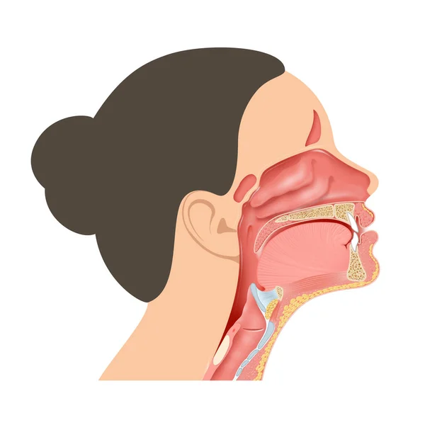
Gastric Glands And Cell Types. Sectional View Of Stomach Mucosa. Stomach Anatomy
Image, 10.89MB, 8858 × 7806 jpg
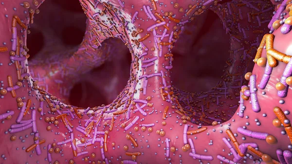
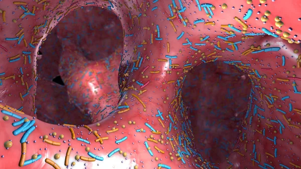
Different Germs In The Human Intestines Called Microbiota - 3d Illustration
Image, 17.83MB, 7500 × 4218 jpg
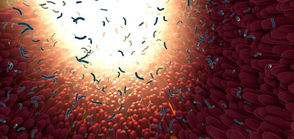
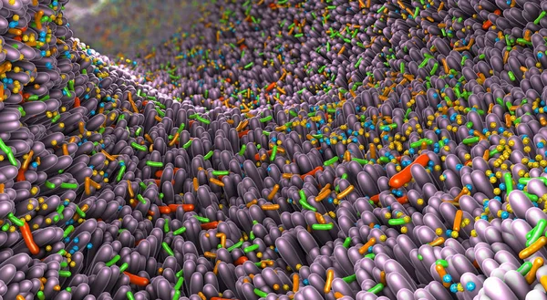
Bacteria As Part Of The Intestinal Microbiome In The Digestive Tract - 3d Illustration
Image, 9.16MB, 7500 × 3550 jpg

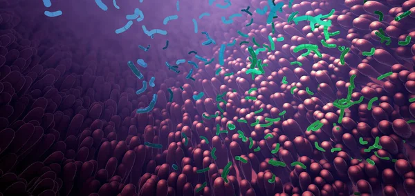
Bacteria As Part Of The Intestinal Microbiome In The Digestive Tract - 3d Illustration
Image, 15.08MB, 7500 × 3550 jpg
3d Illustration Of Microscopic Closeup Of Intestine Villus And Stomach Ulcer
Image, 32.13MB, 9500 × 6333 jpg

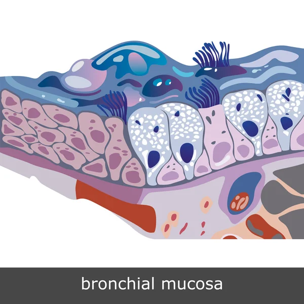
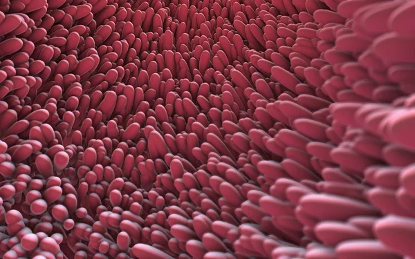
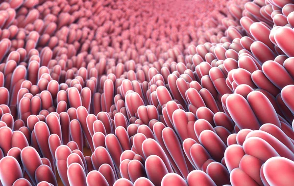
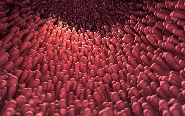
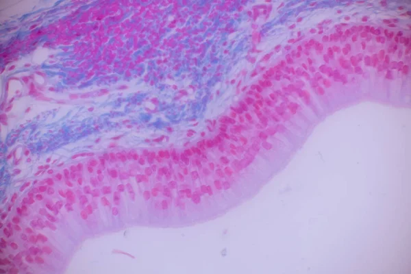
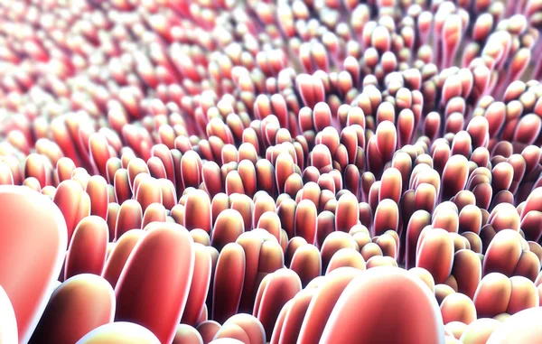
Pseudostratified Epithelium Is A Type Of Epithelium That, Though Comprising Only A Single Layer Of Cells.
Image, 11.24MB, 5840 × 3893 jpg

Smell (olfactory) Receptor Field In Nasal Lining - Isometric View 3d Illustration
Image, 12.55MB, 10000 × 6600 jpg

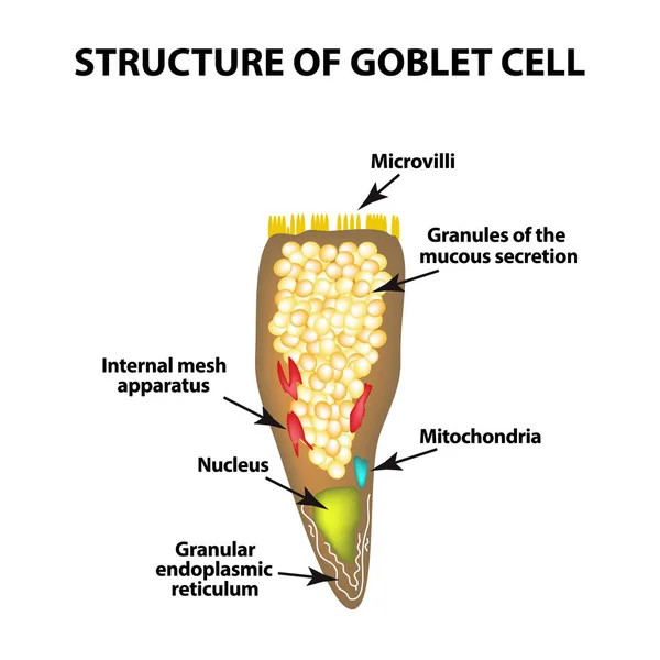
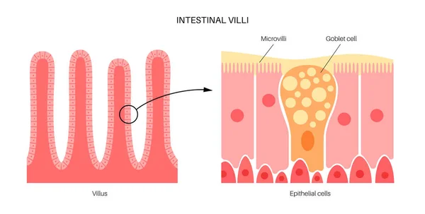
Structure Goblet Cells Of The Intestine. Infographics. Vector Illustration On Isolated Background
Vector, 3.04MB, 5000 × 5000 eps

Enteroendocrine Cell. Cell Of The Intestines. Vector Illustration On Isolated Background
Vector, 0.97MB, 5000 × 5000 eps
Structure Goblet Cells Of The Intestine. Infographics. Vector Illustration On Isolated Background
Vector, 3.04MB, 5000 × 5000 eps

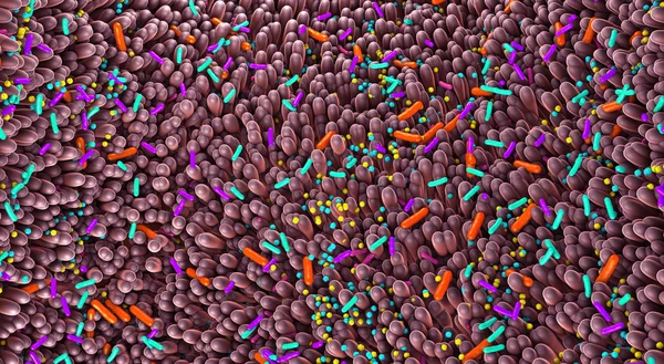
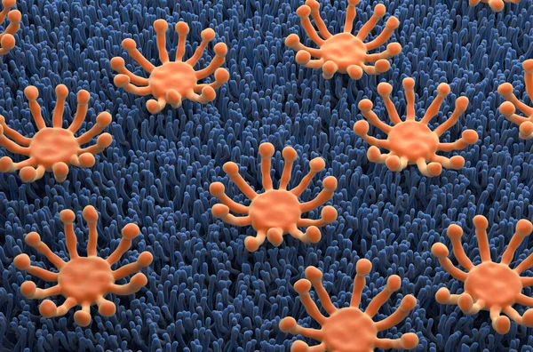
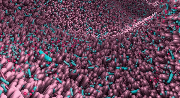
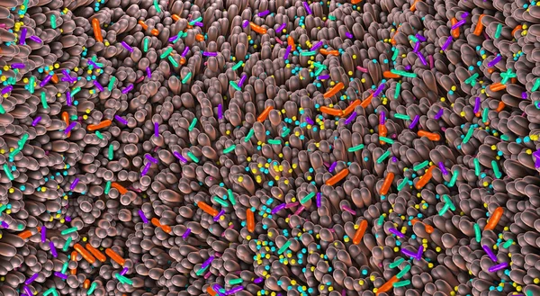
Different Germs In The Human Intestines Called Microbiome - 3d Illustration
Image, 13.37MB, 10000 × 5500 jpg
Different Germs In The Human Intestines Called Microbiome - 3d Illustration
Image, 20MB, 10000 × 5500 jpg
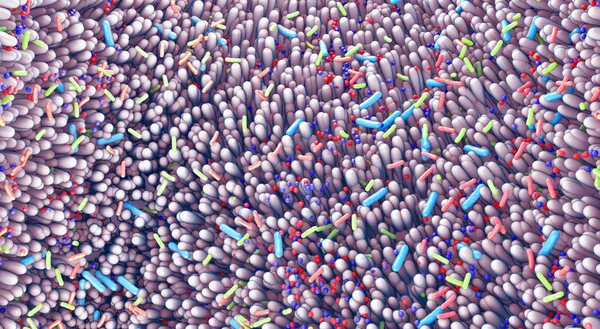
Different Germs In The Human Intestines Called Microbiota - 3d Illustration
Image, 16.45MB, 7500 × 4218 jpg
Different Germs In The Human Intestines Called Microbiome - 3d Illustration
Image, 20.35MB, 10000 × 5500 jpg


Different Germs In The Human Intestines Called Microbiome - 3d Illustration
Image, 45.22MB, 10000 × 5500 jpg

A Trophy With Gifts And A Heart As A Sign. A Mobile Phone With A Blank Screen. The Concept For The Winner. 3d Rendering
Image, 5.39MB, 7451 × 4000 jpg
Different Germs In The Human Intestines Called Microbiome - 3d Illustration
Image, 21.03MB, 10000 × 5500 jpg
Page 1 >> Next