
Stock image Granulosum
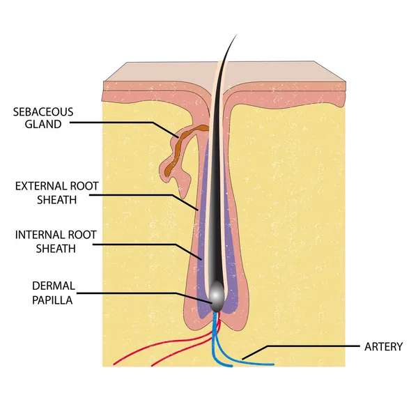


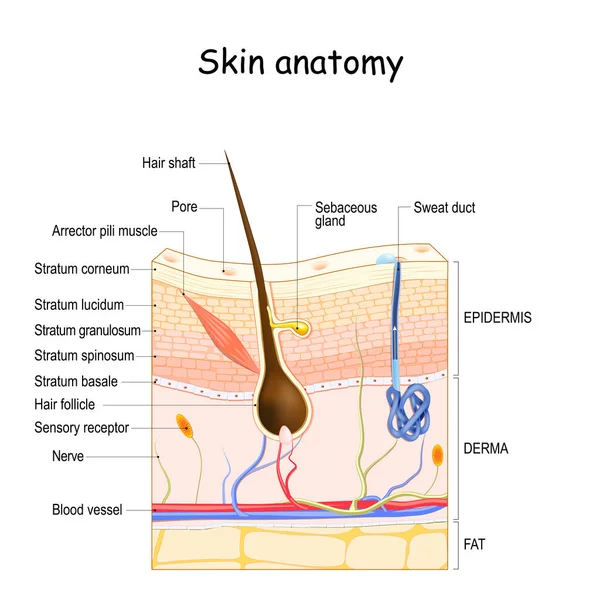
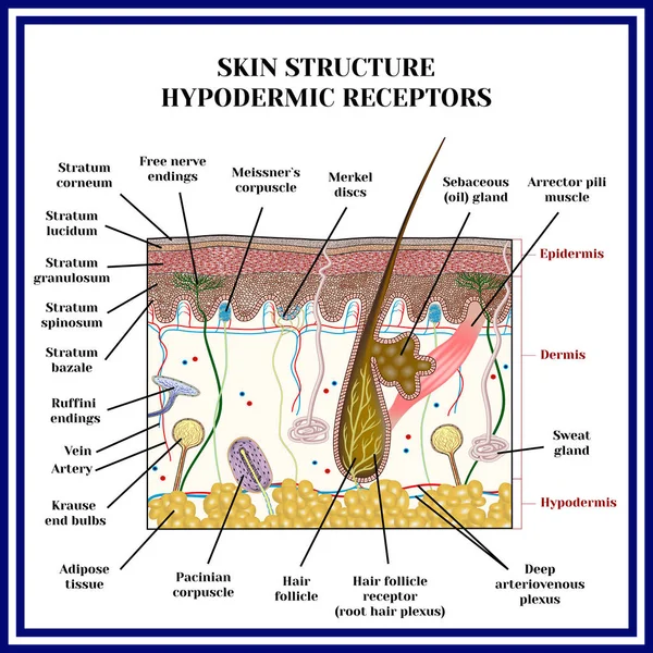
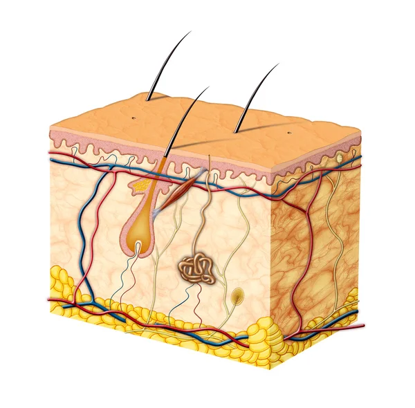
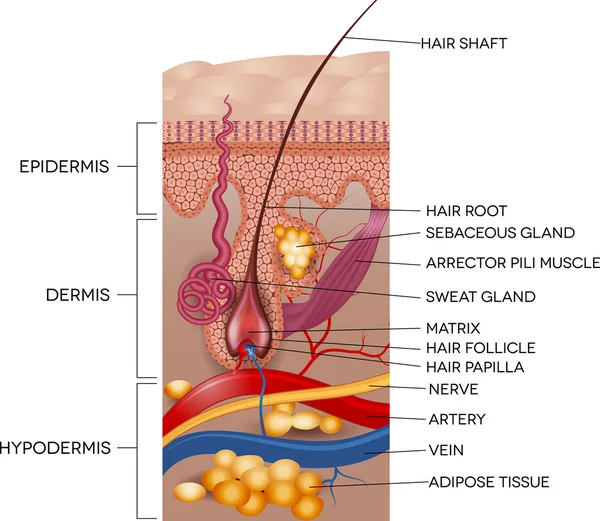
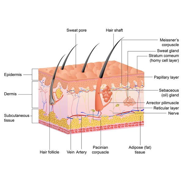
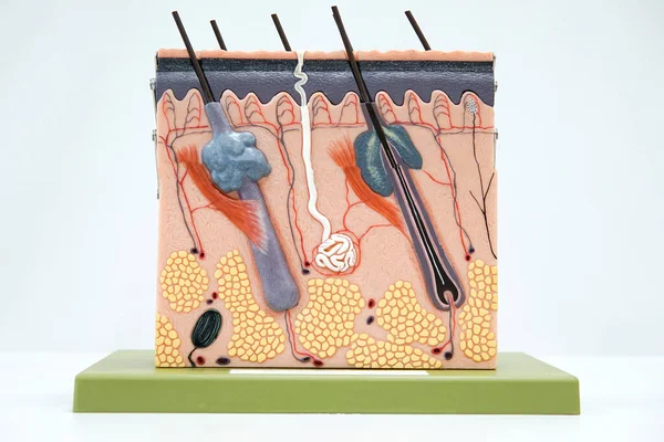
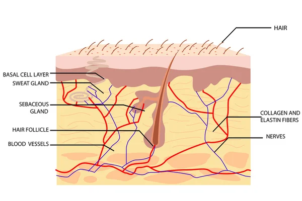
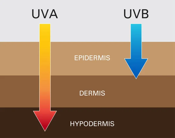
Skin Anatomy. Cross Section Of The Human Skin. Layers Of The Human Skin (epidermis, Dermis, Fat), Hair Follicle, Sensory Receptor, Sweat And Sebaceous Glands.
Vector, 7.34MB, 4444 × 4444 eps

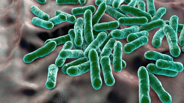
Cutibacterium Acnes, Formely Propionibacterium Acnes, 3D Illustration. Bacteria Found In Hair Follicles And Associated With Development Of Acne, Chronic Blepharitis And Endophthalmitis
Image, 17.5MB, 7200 × 4050 jpg


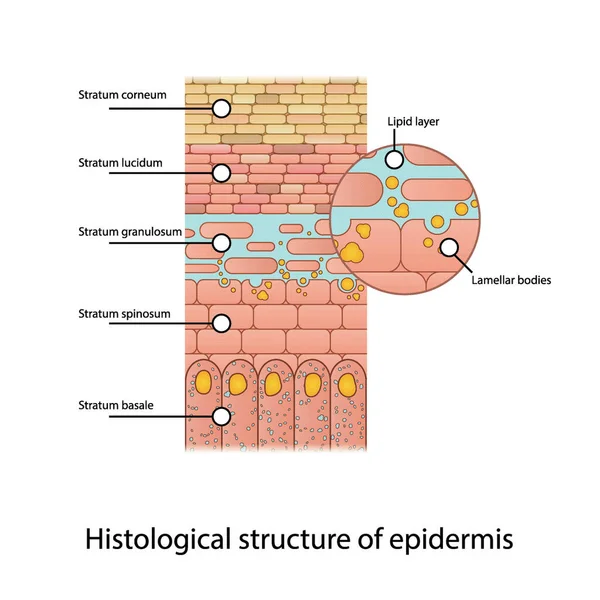
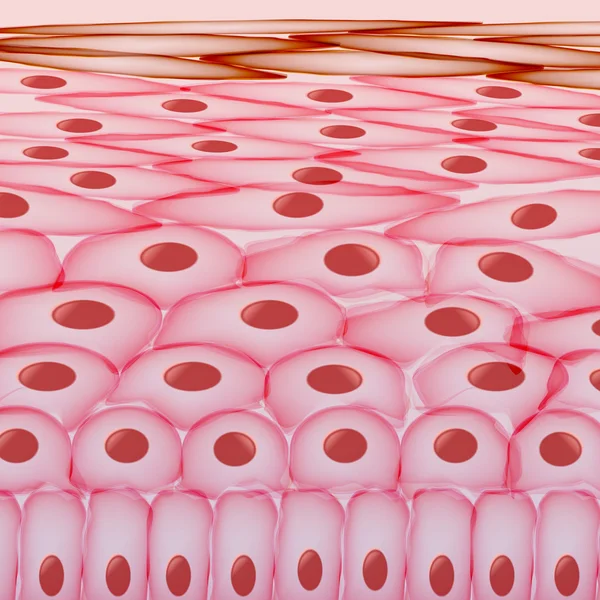
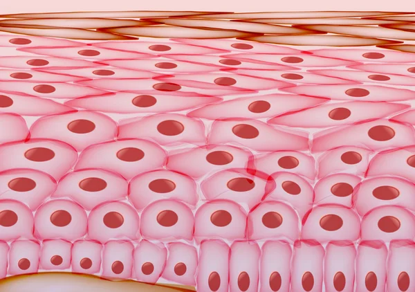
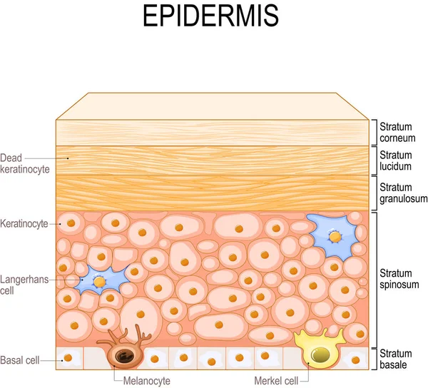
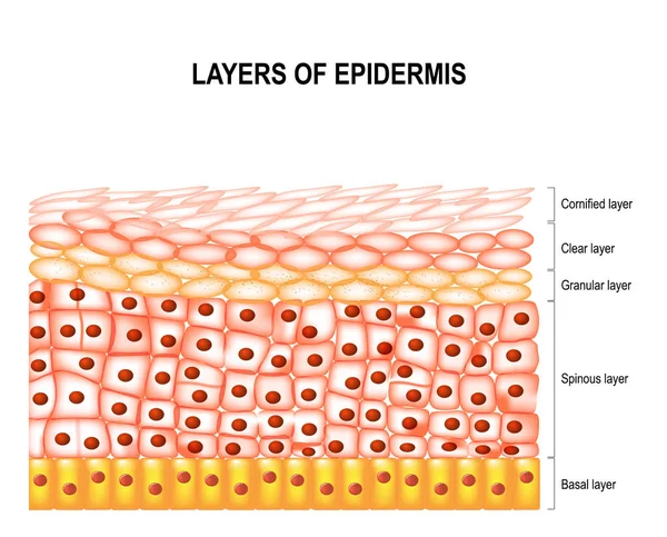
Histological Structure Of Epidermis - Skin Layers Shcematic Vector Illustration Showing Stratum Basale, Spinosum, Granulosum, Lucidum And Corneum And Lamellar Bodies
Vector, 7.07MB, 3090 × 3090 eps
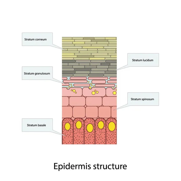
Histological Structure Of Epidermis - Skin Layers Shcematic Vector Illustration Showing Stratum Basale, Spinosum, Granulosum, Lucidum And Corneum
Vector, 5.89MB, 3090 × 3090 eps

Epidermis Anatomy. Layers And Cell Structure Of The Human Skin. Close-up Of Epidermis. Vector Illustration
Vector, 2.34MB, 4444 × 4444 eps
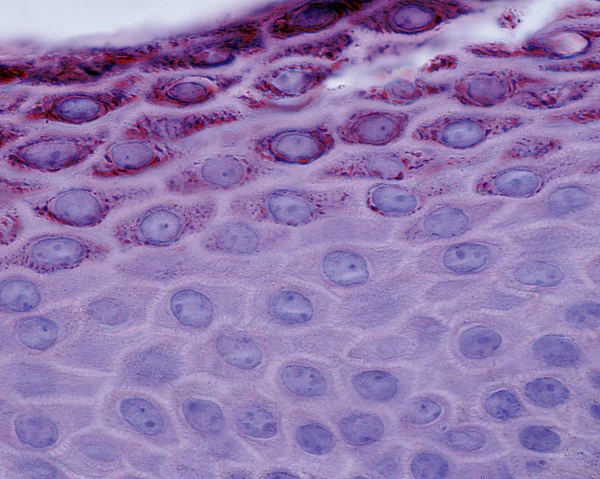

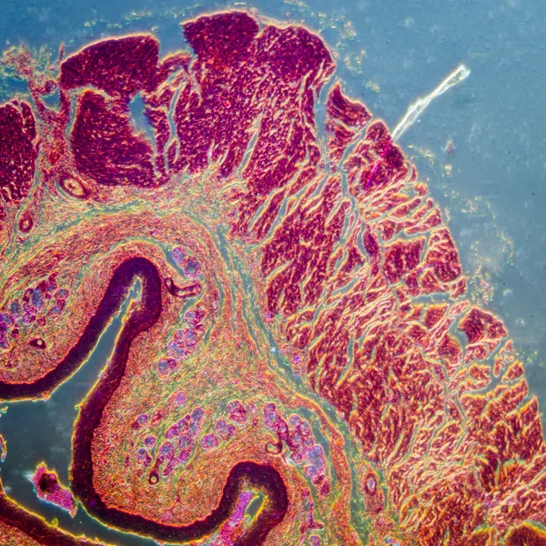

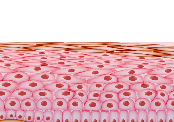
Cross Section Human Skin Tissue Under Microscope View For Physiology Education.
Image, 17.18MB, 6000 × 4000 jpg
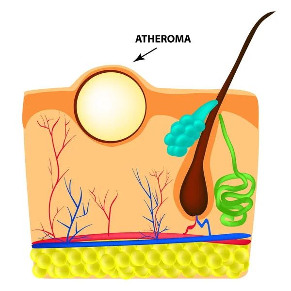
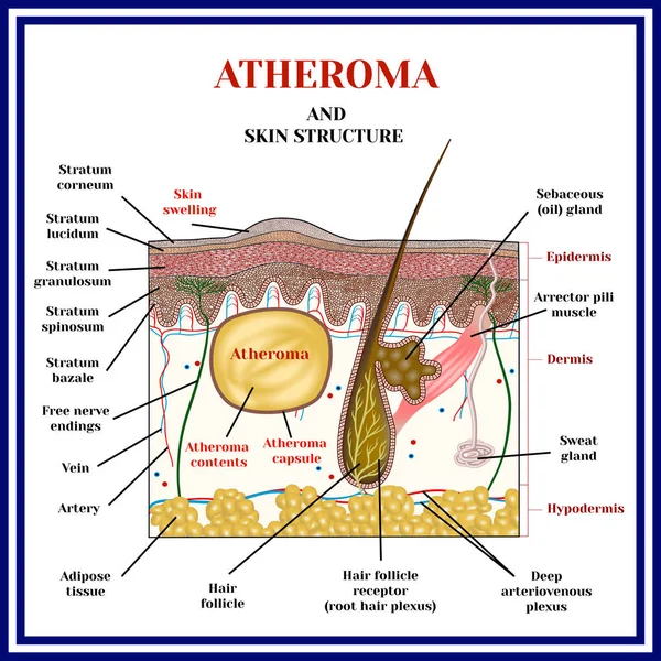
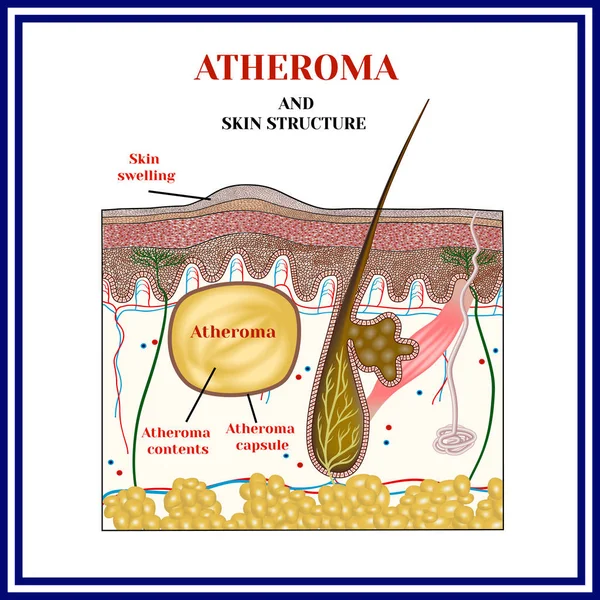
Atheroma Structure. The Structure Of Moles On The Skin. Infographics. Vector Illustration On Isolated Background
Vector, 3.4MB, 5000 × 5000 eps
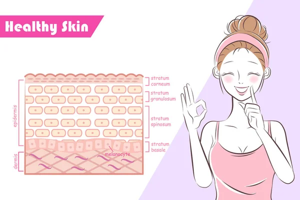
Epidermis Structure. Skin Anatomy. Cell, And Layers Of A Human Skin. Cross Section Of The Epidermis. Skin Care. Vector Illustration.
Vector, 2.11MB, 4444 × 4444 eps

Histological Structure Of Epidermis - Skin Layers Shcematic Vector Illustration Showing Stratum Basale, Spinosum, Granulosum, Lucidum And Corneum
Vector, 7.1MB, 3090 × 3090 eps
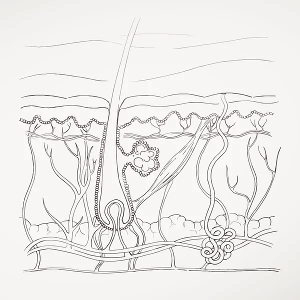
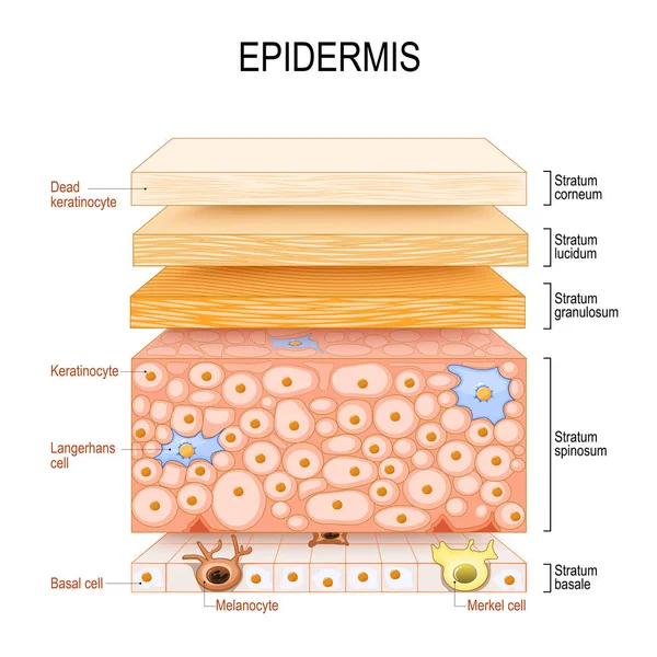
Layers Of Epidermis. Epithelial Cells: Keratinocytes, Melanocyte, Langerhans, Merkel And Basal Cells. Poster For Medical And Educational Use. Structure Of The Humans Skin. Vector Illustration
Vector, 1.67MB, 8717 × 7916 eps
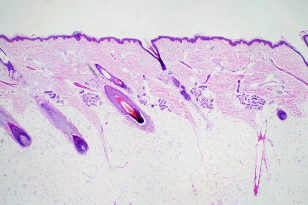
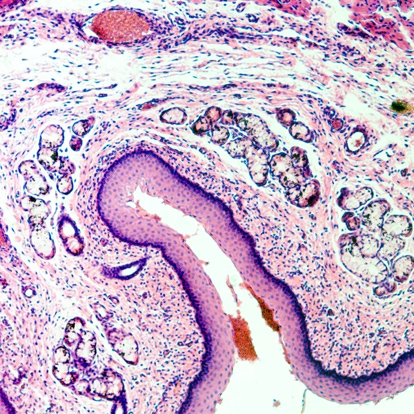
Cross Section Human Skin Head Under Microscope View For Education Histology. Histological For Human Physiology.
Image, 11.8MB, 6000 × 4000 jpg
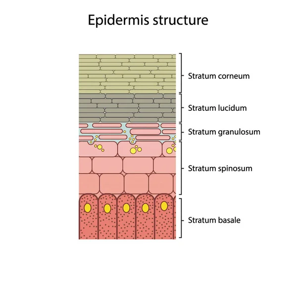
Histological Structure Of Epidermis - Skin Layers Shcematic Vector Illustration Showing Stratum Basale, Spinosum, Granulosum, Lucidum And Corneum
Vector, 5.89MB, 3090 × 3090 eps
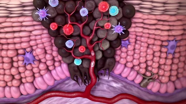
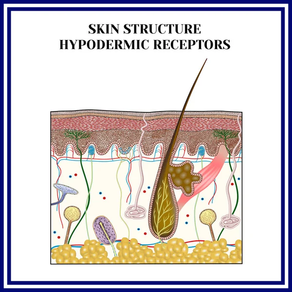
Natural-killer Receptor Group 2, Member D (NKG2D) Is A Well Characterized Natural Killer (NK) Cell Activating Receptor That Recognizes Several Ligands Poorly Expressed On Healthy Cells But Up-regulated Upon Stressing Stimuli In The Context Of Cancer
Image, 1.12MB, 3840 × 2160 jpg

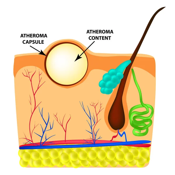
Atheroma Structure. The Structure Of Moles On The Skin. Infographics. Vector Illustration On Isolated Background
Vector, 3.41MB, 5000 × 5000 eps

Cross Section Human Skin Head Under Microscope View For Education Histology. Histological For Human Physiology.
Image, 12.94MB, 6000 × 4000 jpg
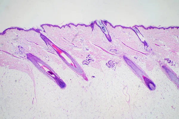
Cross Section Human Skin Head Under Microscope View For Education Histology. Histological For Human Physiology.
Image, 11.9MB, 6000 × 4000 jpg

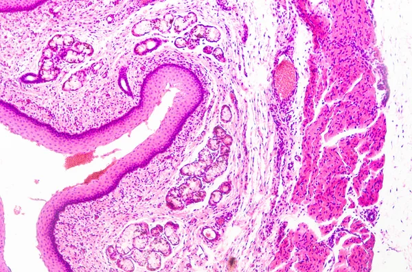
Cross Section Human Skin Head Under Microscope View For Education Pathology. Histological For Human Physiology.
Image, 10.09MB, 6000 × 4000 jpg

Cross Section Human Skin Tissue Under Microscope View For Physiology Education.
Image, 15.25MB, 6000 × 4000 jpg

Epidermis Anatomy. Skin Structure. Cell, And Layers Of A Human Skin. Cross Section Of The Epidermis. Vector Poster. Isometric Flat Illustration.
Vector, 2.09MB, 5000 × 3498 eps
Page 1 >> Next