
Stock image Hematoxylin And Eosin Staining

Buccal Mucosa Cell Is The Lining Of The Cheeks And The Back Of The Lips Under The Light Microscope.
Image, 27.23MB, 8182 × 5434 jpg


Photomicrograph Of Nasal Polyps, Displaying Abnormal Tissue Growth In The Nasal Passages Often Causing Congestion And Discomfort.
Image, 16.6MB, 6150 × 4100 jpg


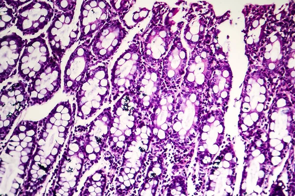
Bacillary Dysentery, Light Micrograph, Photo Under Microscope Showing Presence Of Bacteria And Accumulation Of Inflammatory Cells In Intestinal Epithelium
Image, 4.64MB, 3375 × 2250 jpg

Photomicrograph Of Nasal Polyps, Displaying Abnormal Tissue Growth In The Nasal Passages Often Causing Congestion And Discomfort.
Image, 18.16MB, 6363 × 4242 jpg

An Interesting Photo Taken With A Microscope. Unmyelinated Fibers In Peripheral Nerves. Longitudinal Section. Hematoxylin And Eosin Stainit.
Image, 2.42MB, 3000 × 2248 jpg

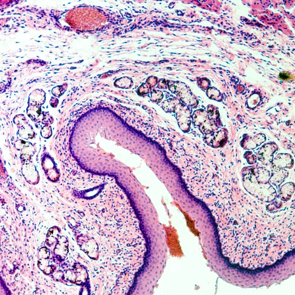
Follicular Adenoma Of Thyroid Gland, Light Micrograph. Histopathology Of Thydoid Adenoma. Photo Under Microscope
Image, 4.28MB, 4087 × 2725 jpg

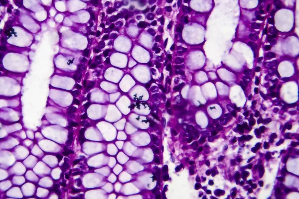
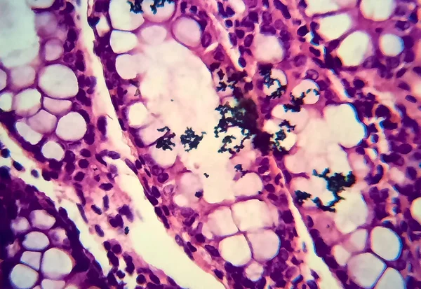
Bacillary Dysentery, Light Micrograph, Photo Under Microscope Showing Presence Of Bacteria And Accumulation Of Inflammatory Cells In Intestinal Epithelium. High Magnification
Image, 4.63MB, 4000 × 2755 jpg
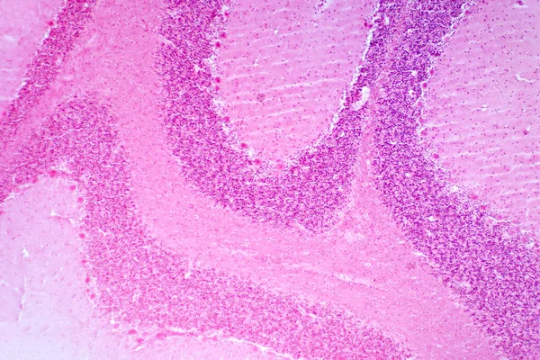
Cerebellum Cross Section Tissue Under The Light Microscope For Pathology Education. Haematoxylin And Eosin Staining Technique For Human Tissue.
Image, 22.42MB, 8192 × 5464 jpg

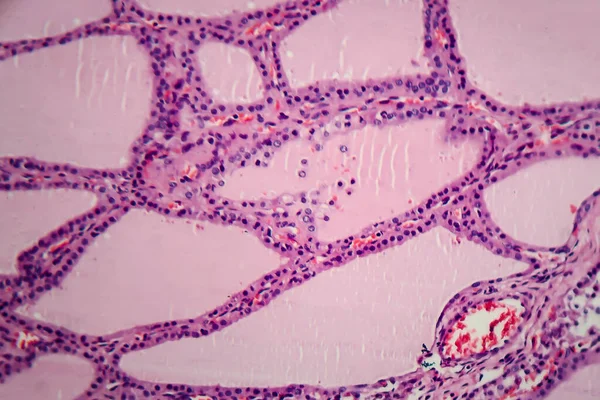
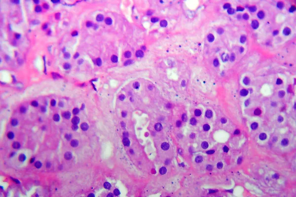
Endemic Goiter, Light Micrograph, Abnormal Enlargement Of The Thyroid Gland Due To Dietary Iodine Deficiency. Photomicrograph Shows Follicles Of Varying Size, Abundant Colloid, Lymphocytic Infiltrate
Image, 6.63MB, 4197 × 2798 jpg
Page 1 >> Next