
Stock image Hip Necrosis

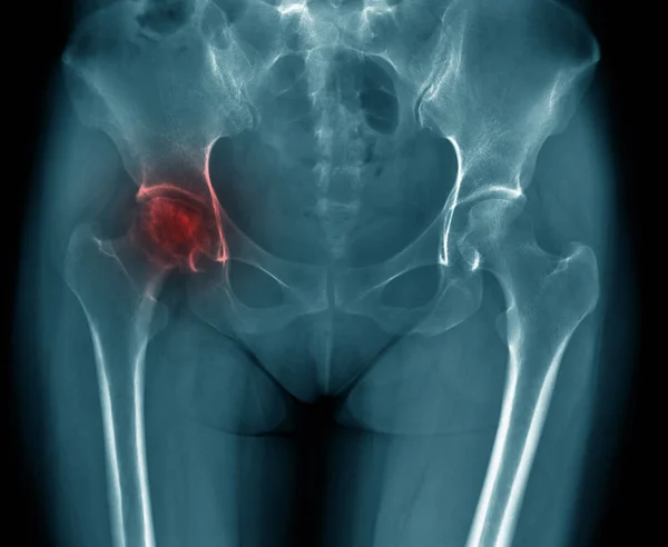
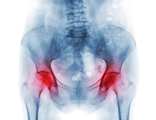
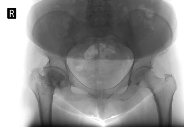
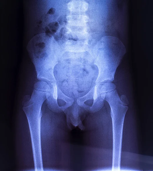
X-ray Image Of Old Woman Show Degenerative Change Of Hip Joint, Hip Avascular Necrosis Right Side Of Alderly
Image, 1.57MB, 2268 × 1862 jpg

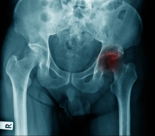
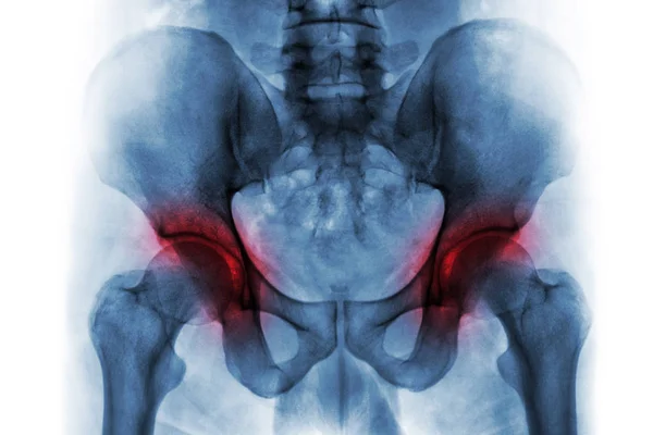
X-ray Image Of Old Woman Show Degenerative Change Of Hip Joint, Hip Avascular Necrosis Right Side Of Alderly
Image, 2.24MB, 2448 × 2010 jpg
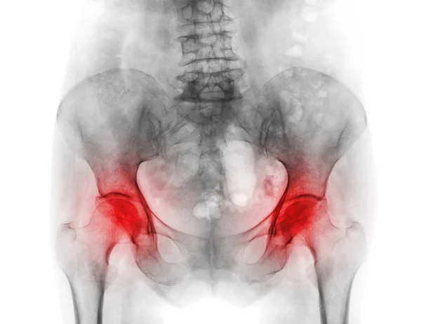
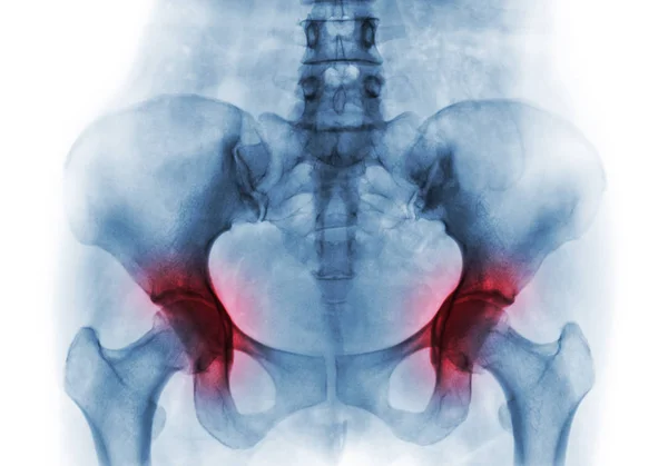
X-ray Image Of Old Woman Show Degenerative Change Of Hip Joint, Hip Avascular Necrosis Right Side Of Alderly
Image, 1.58MB, 2268 × 1862 jpg
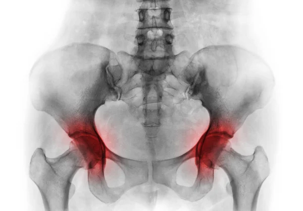
X-ray Image Of Old Woman Show Degenerative Change Of Hip Joint, Hip Avascular Necrosis Right Side Of Alderly
Image, 1.92MB, 2276 × 1994 jpg

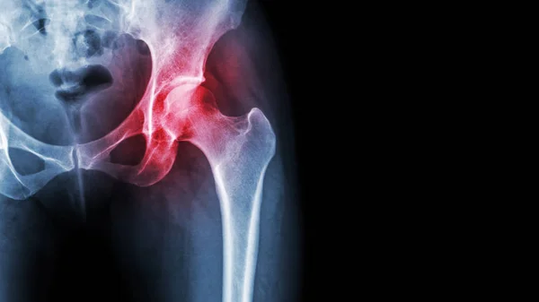
Arthritis At Hip Joint . Film X-ray Show Inflamed Of Hip Joint And Blank Area At Right Side . Avascular Necrosis Concept
Image, 4.8MB, 5815 × 3271 jpg
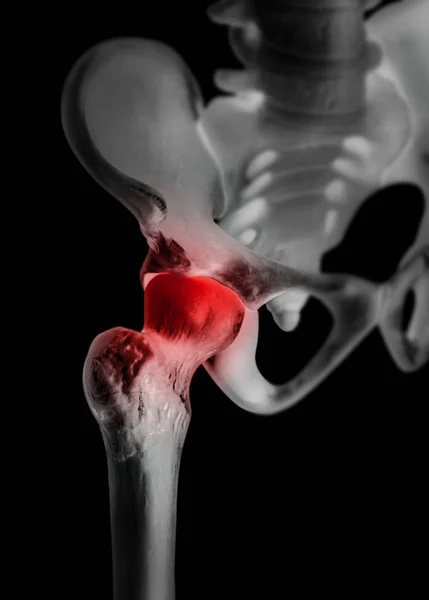
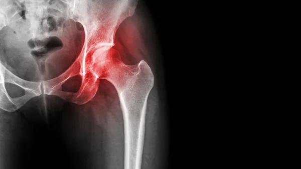
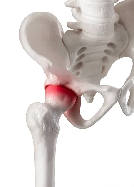
Human Hip Joint With Red Highlight On Pain Area - X Ray Film-Healthcare-Human Anatomy And Medical Concept-Isolated On Black Background.
Image, 7.01MB, 4856 × 6789 jpg
Arthritis At Hip Joint . Film X-ray Show Inflamed Of Hip Joint And Blank Area At Right Side . Avascular Necrosis Concept
Image, 5.61MB, 5815 × 3271 jpg
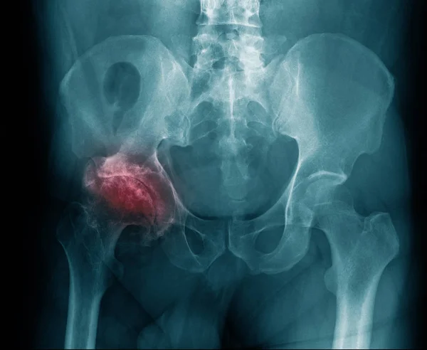


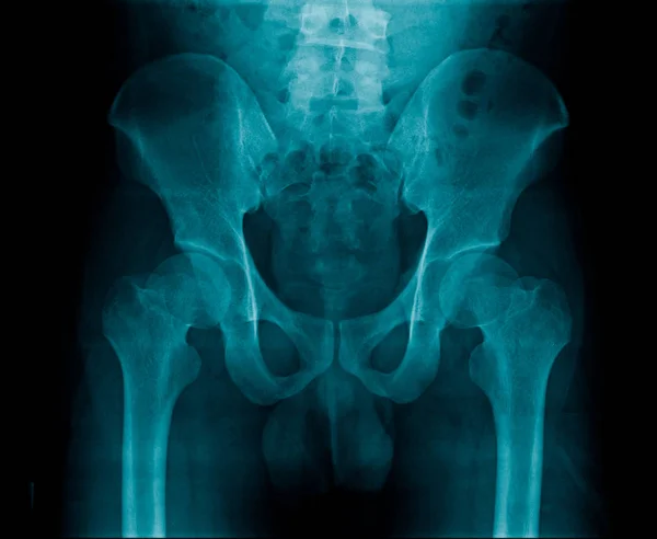
X-ray Of The Pelvis. Deforming Arthrosis Of The Right Hip Joint. Aseptic Necrosis Of The Femoral Head. Negative.
Image, 2.61MB, 3345 × 2317 jpg
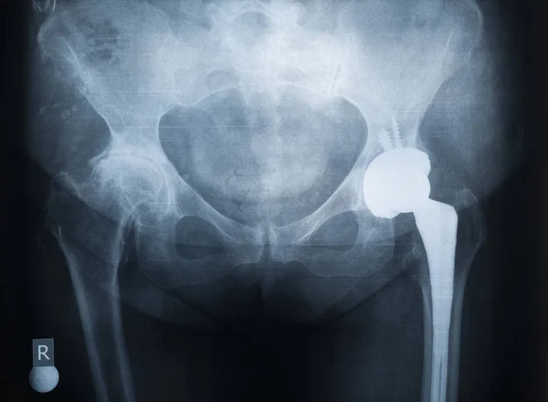
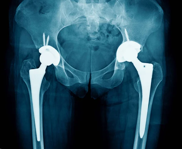
X-ray Of The Prosthesis Of The Left Hip Joint. The Right Joint Is Affected By Rheumatoid Arthritis
Image, 6.48MB, 4282 × 3144 jpg
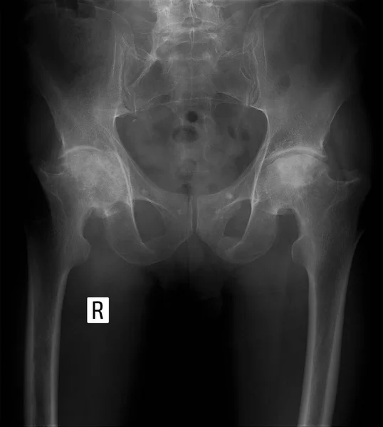
Degenerative Change Of Hip Joint, Old Man Pain At Hip Joint, X-ray Pelvic Bone And Hip Jiont For Visible Diagnosis Hip Joint Pain Cause
Image, 2.83MB, 2448 × 2010 jpg
Human Hip Joint With Red Highlight On Pain Area-Healthcare-Human Anatomy And Medical Concept-Isolated On White Background.
Image, 8.9MB, 4856 × 6789 jpg
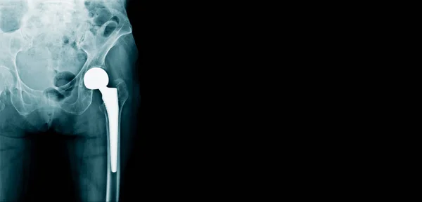
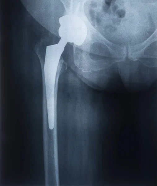
X-ray Image Of Hip Replacement On Black Background, Banner Design For Webpace And Copy Space For Text
Image, 1.49MB, 5041 × 2428 jpg
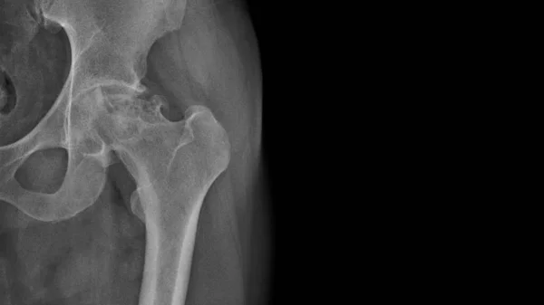
X-ray Of A Painful Hip In A Man With Osteoarthritis Of The Left Hip Joint In The Red Area, Very Painful, Difficult To Walk, Worn Out Joint, Endoprosthetics. Surgical Work Required
Image, 1.12MB, 2767 × 1557 jpg
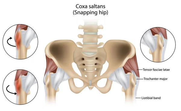
Coxa Saltans Or Snapping Hip Snapping Hip Syndrome Also Referred To As Dancer Hip. Trochanter Major, Tensor Fasciae Latae And Liotibial Band
Vector, 3.84MB, 6240 × 3840 eps
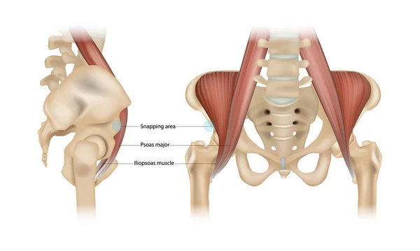
Internal Snapping Hip Syndrome. Psoas Major, Iliopsoas Muscle And Snapping Area. Vector
Vector, 30.04MB, 6000 × 3600 eps
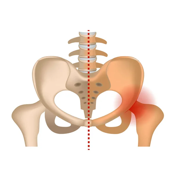
Aseptic Necrosis. Hip Bone With Damaged Femoral Head. Infographic With Axis Of Symmetry. Vector Illustration
Vector, 10.33MB, 2000 × 2000 eps
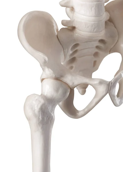
Human Hip Joint-Bone-Healthcare-Human Anatomy And Medical Concept-Isolated On White Background.
Image, 7.41MB, 4856 × 6789 jpg
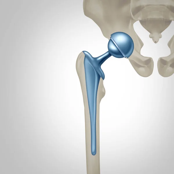
Hip Replacement Surgery Concept As An Artificial Joint Or Prosthesis With Orthopedic Surgery Inserting A Metal Ball And Socket To Replace A Damaged By Disease Femure Joint In A 3D Illustration Style.
Image, 5.18MB, 5100 × 5100 jpg

Doctor Surgeon With A Scalpel Makes An Incision To The Patient To Replace An Artificial Hip Joint, Hip Fracture, Endoprosthetic Replacement, Copy Space, Aseptic Necrosis Of The Femoral Head
Image, 2.89MB, 5306 × 3474 jpg
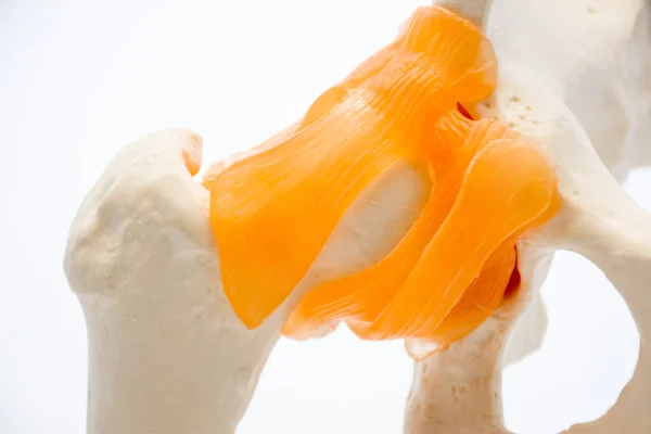
Photo Close-up Anatomy And Hip Structure With Ligaments And Tendons, Which, Together With Pelvic Bones: Ilium, Ischium, Pubis And Thighs Form Acetabulum. For Use In Medicine, Orthopedics And Trauma
Image, 5.61MB, 6000 × 4000 jpg


MRI Scan Of Both Hip Joints Reveals Avascular Necrosis Of The Femoral Head.
Image, 3.01MB, 4112 × 3286 jpg
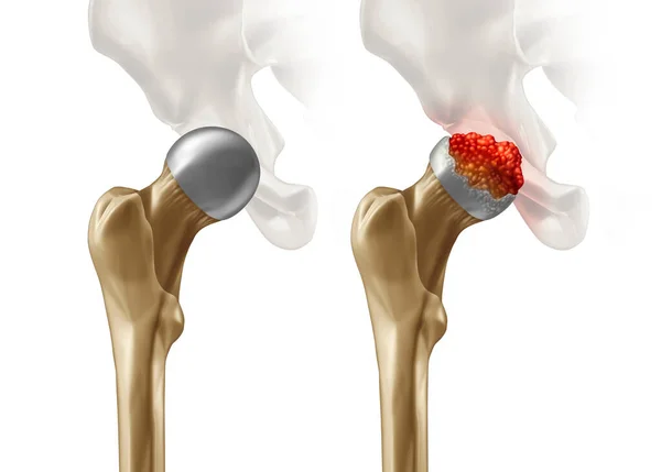
Femoral Head Disease And Osteonecrosis Or Avascular Necrosis And Aseptic Necrosis With A Healthy Hip Compared To An Osteoarthritis Damaged Pelvic Joint In A 3D Illustration Style.
Image, 3.44MB, 6053 × 4330 jpg

Medical Illustration Of Human Male Skeleton With Osteoarthritis Hip Joint Injury On Femur And Pelvic Joint
Image, 2.93MB, 5000 × 3300 jpg
Page 1 >> Next